Bio-Synthesis is a leading global manufacturer of high quality custom-synthesized peptides. We have successfully synthesized tens of thousands of synthetic peptides for the biomedical research community, biotechnology and pharmaceutical companies and have consistently met the highest peptide standards of quality, service and technical expertise. As a peptide manufacturer, we also have the capability to perform a wide variety of synthetic peptide chemistries.
We routinely perform difficult custom peptide synthesis such as synthesis of long peptides (>136 mers), peptides containing unusual modifications, cyclic peptides, cosmetic peptides and peptide oligonucleotide conjugates. In addition, Bio-Synthesis has the capacity for large-scale synthesis (>1 kg) or custom GMP peptide production. Our facility is equipped with state-of-the-art peptide synthesizers and, coupled with our experienced peptide chemists, we are able to deliver your custom peptide synthesis products quickly and with high quality (purity of 90-95% is typical).
Each custom peptide synthesis project is carefully analyzed using an in-house, proprietary PEPscan Identifier and is carefully examined by our chemists. Final synthetic peptide products are shipped with detailed quality control documentation including an HPLC trace, Mass Spec, and a Certificate of Analysis. Custom peptide synthesis is carried out using an assortment of state-of-the-art instrumentation in our 20,000 square foot facility. As a custom peptide manufacturer since 1989, we have earned a reputation that speaks for itself. Our turnaround for custom peptide production is rapid. Most custom peptide orders are shipped within 1 to 2 weeks!
Because Bio-Synthesis maintains its own facilities located in Lewisville, Texas, we are able to provide comprehensive oversight to ensure the highest standards of quality. All custom peptides are accompanied with MS and HPLC analyses. Additional analytical data (e.g., sequencing, amino acid analysis) are available upon request.
Peptide Synthesis Capabilities
Personalized consultation with experienced peptide and immunology experts
Peptides from 2 to 120 amino acids
Scales from 1mg to multi grams research grade to GMP production
Purities from crude to >98%
Solid and solution phase chemistry
Wide range of peptide modifications
Cyclic peptides
Special modifications including: phosphorylation, fluoroscein, glycosylation, PEGylation, rhodamine, AMC, pNA, biotin, boronic amino acids, d-amino acids, stable isotopes, EDANS, dabsyl, dansyl, Abz, thiolactic acids, and many others
High throughput synthesis - peptide library design tools
Specialized peptide synthesis: peptide nucleic acid conjugation
Fast, on-time delivery”
Monday, June 28, 2010
DFF40 Antibody
DFF40 Antibody
Catalog# : 2155
Apoptosis is related to many diseases and induced by a family of cell death receptors and their ligands. Cell death signals are transduced by death domain containing adapter molecules and members of the caspase family of proteases. These death signals finally cause the degradation of chromosomal DNA by activated DNase. A mouse DNase that causes DNA fragmentation was identified recently and designated CAD for caspase activated deoxyribonuclease (1,2). The human homologue of mouse CAD was more recently identified by three groups independently and termed CPAN, DFF40, and human CAD, respectively, (3-5). DFF45/ICAD is the inhibitory protein of DFF40/CAD (1,2,6) and forms complex with DFF40/CAD. Upon cleavage of DFF45/ICAD by activated caspase, DFF40/CAD is released and activated and eventually causes the degradation of DNA in the nuclei. Activation of DFF40/CAD, which causes DNA degradation, is the hallmark of apoptotic cell death.
Additional Names : DFF40 (IN), CAD
 Description
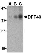
Left: Western blot analysis of DFF40 in Jurkat cell lysate with DFF40 antibody at (A) 0.5, (B) 1 and (C) 2 µg/ml.
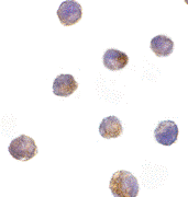
Below: Immunocytochemistry of DFF40 in K562 cells with DFF40 antibody at 10 µg/ml.
Other Product Images
 Source : DFF40 antibody was raised against a peptide corresponding to amino acids 203 to 218 of human DFF40 .
Purification : Affinity chromatography purified via peptide column
Clonality and Clone : This is a polyclonal antibody.
Host : DFF40 antibody was raised in rabbit. Please use anti-rabbit secondary antibodies.
Immunogen : Human DFF40 (Intermediate Domain) Peptide (Cat. No. 2155P)
Application : DFF40 antibody can be used for detection of DFF40 /CAD by Western blot at 1:500 dilution. A 40 kDa band can be detected. It is human, mouse, and rat reactive,
Tested Application(s) : E, WB, ICC
Buffer : Antibody is supplied in PBS containing 0.02% sodium azide.
Blocking Peptide : Cat. No. 2155P - DFF40 Peptide
Long-Term Storage : DFF40 antibody can be stored at 4ºC, stable for one year. As with all antibodies care should be taken to avoid repeated freeze thaw cycles. Antibodies should not be exposed to prolonged high temperatures.
Positive Control
1.Cat. No. 1204 - K562 Cell Lysate
2.Cat. No. 1205 Jurkat Whole Cell Lysate
Species Reactivity :H, M, R
GI Number : 4758150
Accession Number : NP_004393
Short Description : (IN) Caspase Activated DNase
References
1.Enari M, Sakahira H, Yokoyama H, Okawa K, Iwamatsu A, Nagata S. A caspase-activated DNase that degrades DNA during apoptosis, and its inhibitor ICAD. Nature 1998;391:43-50
2.Sakahira H, Enari M, Nagata S. Cleavage of CAD inhibitor in CAD activation and DNA degradation during apoptosis. Nature 1998;391:96-99
3.Halenbeck R, MacDonald H, Roulston A, Chen TT, Conroy L, Williams LT. CPAN, a human nuclease regulated by the caspase-sensitive inhibitor DFF45. Curr Biol 1998;8:537-40
4.Liu X, Li P, Widlak P, Zou H, Luo X, Garrard WT, Wang X The 40-kDa subunit of DNA fragmentation factor induces DNA fragmentation and chromatin condensation during apoptosis. Proc Natl Acad Sci USA 1998;95:8461-6
Catalog# : 2155
Apoptosis is related to many diseases and induced by a family of cell death receptors and their ligands. Cell death signals are transduced by death domain containing adapter molecules and members of the caspase family of proteases. These death signals finally cause the degradation of chromosomal DNA by activated DNase. A mouse DNase that causes DNA fragmentation was identified recently and designated CAD for caspase activated deoxyribonuclease (1,2). The human homologue of mouse CAD was more recently identified by three groups independently and termed CPAN, DFF40, and human CAD, respectively, (3-5). DFF45/ICAD is the inhibitory protein of DFF40/CAD (1,2,6) and forms complex with DFF40/CAD. Upon cleavage of DFF45/ICAD by activated caspase, DFF40/CAD is released and activated and eventually causes the degradation of DNA in the nuclei. Activation of DFF40/CAD, which causes DNA degradation, is the hallmark of apoptotic cell death.
Additional Names : DFF40 (IN), CAD
DescriptionLeft: Western blot analysis of DFF40 in Jurkat cell lysate with DFF40 antibody at (A) 0.5, (B) 1 and (C) 2 µg/ml.
Below: Immunocytochemistry of DFF40 in K562 cells with DFF40 antibody at 10 µg/ml.
Other Product Images
Source : DFF40 antibody was raised against a peptide corresponding to amino acids 203 to 218 of human DFF40 .Purification : Affinity chromatography purified via peptide column
Clonality and Clone : This is a polyclonal antibody.
Host : DFF40 antibody was raised in rabbit. Please use anti-rabbit secondary antibodies.
Immunogen : Human DFF40 (Intermediate Domain) Peptide (Cat. No. 2155P)
Application : DFF40 antibody can be used for detection of DFF40 /CAD by Western blot at 1:500 dilution. A 40 kDa band can be detected. It is human, mouse, and rat reactive,
Tested Application(s) : E, WB, ICC
Buffer : Antibody is supplied in PBS containing 0.02% sodium azide.
Blocking Peptide : Cat. No. 2155P - DFF40 Peptide
Long-Term Storage : DFF40 antibody can be stored at 4ºC, stable for one year. As with all antibodies care should be taken to avoid repeated freeze thaw cycles. Antibodies should not be exposed to prolonged high temperatures.
Positive Control
1.Cat. No. 1204 - K562 Cell Lysate
2.Cat. No. 1205 Jurkat Whole Cell Lysate
Species Reactivity :H, M, R
GI Number : 4758150
Accession Number : NP_004393
Short Description : (IN) Caspase Activated DNase
References
1.Enari M, Sakahira H, Yokoyama H, Okawa K, Iwamatsu A, Nagata S. A caspase-activated DNase that degrades DNA during apoptosis, and its inhibitor ICAD. Nature 1998;391:43-50
2.Sakahira H, Enari M, Nagata S. Cleavage of CAD inhibitor in CAD activation and DNA degradation during apoptosis. Nature 1998;391:96-99
3.Halenbeck R, MacDonald H, Roulston A, Chen TT, Conroy L, Williams LT. CPAN, a human nuclease regulated by the caspase-sensitive inhibitor DFF45. Curr Biol 1998;8:537-40
4.Liu X, Li P, Widlak P, Zou H, Luo X, Garrard WT, Wang X The 40-kDa subunit of DNA fragmentation factor induces DNA fragmentation and chromatin condensation during apoptosis. Proc Natl Acad Sci USA 1998;95:8461-6
Sunday, June 27, 2010
DEDD2 Antibody
DEDD2 Antibody
Catalog# : 3071
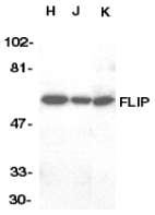
Apoptotic signals are often triggered by cell surface death receptors through protein-protein interactions mediated by conserved domains such as the death effector domain (1). A novel death effector domain (DED)-containing protein, DEDD2, has been recently identified and its over-expression in transfected cells induces moderate apoptosis and results in substantial sensitization to apoptosis induced by Fas, TRAIL, and FADD (2). More recently, work has shown that DEDD2 can bind caspase-8 and -10 in addition to FLIP but not FADD (2,3). Like the related protein DEDD, DEDD2 translocates from the cytosol to the nucleus upon induction of apoptosis, and it has been suggested that DEDD2 may target caspase-8 to the nucleus and that DEDD2 thus plays a critical role in death receptor-induced apoptosis (3). At least two alternatively spliced transcript variants encoding distinct isoforms have been found for DEDD2.
Additional Names : DEDD2, DNA-binding death effector domain-containing protein 2, FLAME-3
 Description
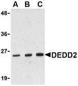
Left: Western blot analysis of DEDD2 in RAW264.7 cell lysate with DEDD2 antibody at (A) 0.5, (B) 1 and (C) 2 μg/ml.
Source : DEDD2 antibody was raised against a peptide corresponding to 11 amino acids near the amino-terminus of human DEDD2.
Purification : Immunoaffinity chromatography purified IgG
Clonality and Clone : This is a polyclonal antibody.
Host : DEDD2 antibody was raised in rabbit. Please use anti-rabbit secondary antibodies.
Immunogen : Human DEDD2 Peptide (Cat. No. 3071P)
Application : DEDD2 antibody can be used for detection of DEDD2 by Western blot at 0.5 to 1 µg/ml.
Tested Application(s) : E, WB
Buffer : Antibody is supplied in PBS containing 0.02% sodium azide.
Blocking Peptide : Cat. No. 3071P - DEDD2 Peptide
Long-Term Storage : DEDD2 antibody can be stored at 4ºC, stable for one year. As with all antibodies care should be taken to avoid repeated freeze thaw cycles. Antibodies should not be exposed to prolonged high temperatures.
Positive Control
1.Cat. No. 1283 - RAW264.7 Cell Lysate
Species Reactivity :H, M, R
GI Number : 19923050
Accession Number : NP_579874
Short Description : A novel death effector domain containing protein
References
1.Tibbetts MD, Zheng L, and Lenardo MJ. The death effector domain protein family: regulators of cellular homeostasis. Nat. Immunol. 2003; 4:404-9.
2.Roth W, Stenner-Liewen F, Pawlowski K, et al. Identification and characterization of DEDD2, a death effector domain-containing protein. J. Biol. Chem. 2002; 277:7501-8.
3.Alcivar A, Hu S, Tang J, et al. DEDD and DEDD2 associate with caspase-8/10 and signal cell death. Oncogene 2003; 22:291-7.
Catalog# : 3071
Apoptotic signals are often triggered by cell surface death receptors through protein-protein interactions mediated by conserved domains such as the death effector domain (1). A novel death effector domain (DED)-containing protein, DEDD2, has been recently identified and its over-expression in transfected cells induces moderate apoptosis and results in substantial sensitization to apoptosis induced by Fas, TRAIL, and FADD (2). More recently, work has shown that DEDD2 can bind caspase-8 and -10 in addition to FLIP but not FADD (2,3). Like the related protein DEDD, DEDD2 translocates from the cytosol to the nucleus upon induction of apoptosis, and it has been suggested that DEDD2 may target caspase-8 to the nucleus and that DEDD2 thus plays a critical role in death receptor-induced apoptosis (3). At least two alternatively spliced transcript variants encoding distinct isoforms have been found for DEDD2.
Additional Names : DEDD2, DNA-binding death effector domain-containing protein 2, FLAME-3
DescriptionLeft: Western blot analysis of DEDD2 in RAW264.7 cell lysate with DEDD2 antibody at (A) 0.5, (B) 1 and (C) 2 μg/ml.
Source : DEDD2 antibody was raised against a peptide corresponding to 11 amino acids near the amino-terminus of human DEDD2.
Purification : Immunoaffinity chromatography purified IgG
Clonality and Clone : This is a polyclonal antibody.
Host : DEDD2 antibody was raised in rabbit. Please use anti-rabbit secondary antibodies.
Immunogen : Human DEDD2 Peptide (Cat. No. 3071P)
Application : DEDD2 antibody can be used for detection of DEDD2 by Western blot at 0.5 to 1 µg/ml.
Tested Application(s) : E, WB
Buffer : Antibody is supplied in PBS containing 0.02% sodium azide.
Blocking Peptide : Cat. No. 3071P - DEDD2 Peptide
Long-Term Storage : DEDD2 antibody can be stored at 4ºC, stable for one year. As with all antibodies care should be taken to avoid repeated freeze thaw cycles. Antibodies should not be exposed to prolonged high temperatures.
Positive Control
1.Cat. No. 1283 - RAW264.7 Cell Lysate
Species Reactivity :H, M, R
GI Number : 19923050
Accession Number : NP_579874
Short Description : A novel death effector domain containing protein
References
1.Tibbetts MD, Zheng L, and Lenardo MJ. The death effector domain protein family: regulators of cellular homeostasis. Nat. Immunol. 2003; 4:404-9.
2.Roth W, Stenner-Liewen F, Pawlowski K, et al. Identification and characterization of DEDD2, a death effector domain-containing protein. J. Biol. Chem. 2002; 277:7501-8.
3.Alcivar A, Hu S, Tang J, et al. DEDD and DEDD2 associate with caspase-8/10 and signal cell death. Oncogene 2003; 22:291-7.
DEDAF Antibody
DEDAF Antibody
Catalog# : 2227
Apoptosis is related to many diseases and induced by a family of cell death receptors and their ligands. Cell death signals are transduced by death domain (DD) death effector domain (DED), and caspase recruitment domain (CARD) containing molecules. Several molecules including caspases and adaptor FADD contain DEDs. A novel protein that interacts with DED of caspase-8 and 10, and FADD was identified recently and designated DEDAF for DED associated factor . DEDAF is identical to the transcriptional repressor RYBP (2). DEDAF/RYBP is expressed in multiple tissues and cell lines. DEDAF interacts with FADD and augments the formation of CD95/FADD/capase-8 complexes at the cell membrane, and interacts with DED-containing DNA biding protein (DEDD) in the nucleus indicating it is involved in the regulation of both cytoplasmic and nuclear events of apoptosis.
Additional Names : DEDAF
 Description
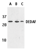
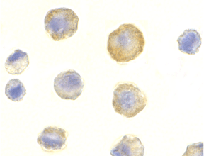
Left: Western blot analysis of DEDAF expression in human A549 (lane A), HepG2 (lane B), and mouse 3T3 (lane C) cell lysates with DEDAF antibody at 1 µg /ml.
Below: Immunohistochemistry of DEDAF in mouse liver tissue with DEDAF antibody at 10 µg/ml.
Other Product Images
 Source : DEDAF antibody was raised against a synthetic peptide corresponding to amino acids 215 to 228 of human DEDAF. The sequence is identical to that of mouse origin.
Purification : Affinity chromatography purified via peptide column
Clonality and Clone : This is a polyclonal antibody.
Host : DEDAF antibody was raised in rabbit. Please use anti-rabbit secondary antibodies.
Immunogen : Human DEDAF Peptide (Cat. No. 2227P)
Application : DEDAF can be used for detection of DEDAF by Western blot at 0.5 to 1 µg/ml.A band at approximately 32 kDa can be detected.
Tested Application(s) : E, WB, IHC
Buffer : Antibody is supplied in PBS containing 0.02% sodium azide.
Blocking Peptide : Cat. No. 2227P - DEDAF Peptide
Long-Term Storage : DEDAF antibody can be stored at 4ºC, stable for one year. As with all antibodies care should be taken to avoid repeated freeze thaw cycles. Antibodies should not be exposed to prolonged high temperatures.
Positive Control
1.Cat. No. 1203 - A549 Cell Lysate
2.Cat. No. 1211 - HepG2 Whole Cell Lysate
3.Cat. No. 1404 - Mouse Liver Tissue Lysate
Species Reactivity :H, M, R
GI Number : 5802963
Accession Number : AF179286
Short Description : A novel regulator of apoptosis
References
1.Zheng L, Schickling O, Peter ME, Lenardo MJ. The death effector domain-associated factor (DEDAF) plays distinct regulatory roles in the nucleus and cytoplasm. J Biol Chem. 2001;276(34):31945-52.
2.Garcia E, Marcos-Gutierrez C, del Mar Lorente M, Moreno JC, Vidal M. RYBP, a new repressor protein that interacts with components of the mammalian Polycomb complex, and with the transcription factor YY1. EMBO J. 1999;18(12):3404-18. (RD0201)
Catalog# : 2227
Apoptosis is related to many diseases and induced by a family of cell death receptors and their ligands. Cell death signals are transduced by death domain (DD) death effector domain (DED), and caspase recruitment domain (CARD) containing molecules. Several molecules including caspases and adaptor FADD contain DEDs. A novel protein that interacts with DED of caspase-8 and 10, and FADD was identified recently and designated DEDAF for DED associated factor . DEDAF is identical to the transcriptional repressor RYBP (2). DEDAF/RYBP is expressed in multiple tissues and cell lines. DEDAF interacts with FADD and augments the formation of CD95/FADD/capase-8 complexes at the cell membrane, and interacts with DED-containing DNA biding protein (DEDD) in the nucleus indicating it is involved in the regulation of both cytoplasmic and nuclear events of apoptosis.
Additional Names : DEDAF
DescriptionLeft: Western blot analysis of DEDAF expression in human A549 (lane A), HepG2 (lane B), and mouse 3T3 (lane C) cell lysates with DEDAF antibody at 1 µg /ml.
Below: Immunohistochemistry of DEDAF in mouse liver tissue with DEDAF antibody at 10 µg/ml.
Other Product Images
Source : DEDAF antibody was raised against a synthetic peptide corresponding to amino acids 215 to 228 of human DEDAF. The sequence is identical to that of mouse origin.Purification : Affinity chromatography purified via peptide column
Clonality and Clone : This is a polyclonal antibody.
Host : DEDAF antibody was raised in rabbit. Please use anti-rabbit secondary antibodies.
Immunogen : Human DEDAF Peptide (Cat. No. 2227P)
Application : DEDAF can be used for detection of DEDAF by Western blot at 0.5 to 1 µg/ml.A band at approximately 32 kDa can be detected.
Tested Application(s) : E, WB, IHC
Buffer : Antibody is supplied in PBS containing 0.02% sodium azide.
Blocking Peptide : Cat. No. 2227P - DEDAF Peptide
Long-Term Storage : DEDAF antibody can be stored at 4ºC, stable for one year. As with all antibodies care should be taken to avoid repeated freeze thaw cycles. Antibodies should not be exposed to prolonged high temperatures.
Positive Control
1.Cat. No. 1203 - A549 Cell Lysate
2.Cat. No. 1211 - HepG2 Whole Cell Lysate
3.Cat. No. 1404 - Mouse Liver Tissue Lysate
Species Reactivity :H, M, R
GI Number : 5802963
Accession Number : AF179286
Short Description : A novel regulator of apoptosis
References
1.Zheng L, Schickling O, Peter ME, Lenardo MJ. The death effector domain-associated factor (DEDAF) plays distinct regulatory roles in the nucleus and cytoplasm. J Biol Chem. 2001;276(34):31945-52.
2.Garcia E, Marcos-Gutierrez C, del Mar Lorente M, Moreno JC, Vidal M. RYBP, a new repressor protein that interacts with components of the mammalian Polycomb complex, and with the transcription factor YY1. EMBO J. 1999;18(12):3404-18. (RD0201)
DcR3 Antibody
DcR3 Antibody
Catalog# : 2140
Apoptosis is induced by certain cytokines including TNF and Fas ligand in the TNF family through their death domain containing receptors. Several novel members in the TNFR family including DR3, DR4, DR5, and DR6 were recently discovered and function as cell death receptors. Two decoy receptors, DcR1 and DcR2, were recently identified to compete with DR4 and DR5 for their ligand TRAIL binding. A novel decoy receptor was more recently discovered and designated DcR3 and TR6, respectively, (1,2). Unlike DcR1 and DcR2, DcR3 is a soluble rather than a membrane associated molecule. DcR3 binds to FasL and LIGHT and inhibits FasL and LIGHT induced apoptosis (1,2). DcR3 transcript is expressed in a number of lung and colon carcinomas and in some normal tissues.
Additional Names : DcR3, DCR3, TR6
 Description
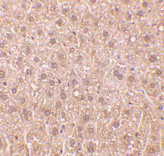
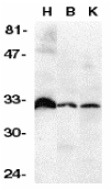
Left: Western blot analysis of DcR3 in human heart (H), brain (B), and kidney (K) tissue lysates with DcR3 antibody at 1:500 dilution.
Below: Immunohistochemistry of DcR3 in human heart tissue with DcR3 antibody at 1 µg/ml.
Other Product Images
 Source : DcR3 antibody was raised against a peptide corresponding to amino acids near the carboxy terminus of human DcR3 precursor.
Purification : Ion exchange chromatography purified
Clonality and Clone : This is a polyclonal antibody.
Host : DcR3 antibody was raised in rabbit. Please use anti-rabbit secondary antibodies.
Immunogen : Human DcR3 (N-Terminus) Peptide (Cat. No. 2140P)
Application : DcR3 antibody can be used for detection of DcR3 expression by Western blot at 1:500 to 1:1000 dilution. An approximately 33 kDa band can be detected.
Tested Application(s) : E, WB, IHC
Buffer : Antibody is supplied in PBS containing 0.02% sodium azide.
Blocking Peptide : Cat. No. 2140P - DcR3 Peptide
Long-Term Storage : DcR3 antibody can be stored at 4ºC, stable for one year. As with all antibodies care should be taken to avoid repeated freeze thaw cycles. Antibodies should not be exposed to prolonged high temperatures.
Positive Control
1.Cat. No. 1301 - Human Heart Tissue Lysate
Species Reactivity :H, M, R
GI Number : 4106877
Accession Number : AF104419
Short Description : Decoy Receptor for FASL and LIGHT
References
1.Pitti RM, Marsters SA, Lawrence DA, Roy M, Kischkel FC, Dowd P, Huang A, Donahue CJ, Sherwood SW, Baldwin DT, Godowski PJ, Wood WI, Gurney AL, Hillan KJ, Cohen RL, Goddard AD, Botstein D, Ashkenazi A. Genomic amplification of a decoy receptor for Fas ligand in lung and colon cancer. Nature 1998;396:699-703
2.Yu KY, Kwon B, Ni J, Zhai Y, Ebner R, Kwon BS. A newly identified member of tumor necrosis factor receptor superfamily (TR6) suppresses LIGHT-mediated apoptosis. J Biol Chem 1999;274:13733-6 (RD1299)
Catalog# : 2140
Apoptosis is induced by certain cytokines including TNF and Fas ligand in the TNF family through their death domain containing receptors. Several novel members in the TNFR family including DR3, DR4, DR5, and DR6 were recently discovered and function as cell death receptors. Two decoy receptors, DcR1 and DcR2, were recently identified to compete with DR4 and DR5 for their ligand TRAIL binding. A novel decoy receptor was more recently discovered and designated DcR3 and TR6, respectively, (1,2). Unlike DcR1 and DcR2, DcR3 is a soluble rather than a membrane associated molecule. DcR3 binds to FasL and LIGHT and inhibits FasL and LIGHT induced apoptosis (1,2). DcR3 transcript is expressed in a number of lung and colon carcinomas and in some normal tissues.
Additional Names : DcR3, DCR3, TR6
DescriptionLeft: Western blot analysis of DcR3 in human heart (H), brain (B), and kidney (K) tissue lysates with DcR3 antibody at 1:500 dilution.
Below: Immunohistochemistry of DcR3 in human heart tissue with DcR3 antibody at 1 µg/ml.
Other Product Images
Source : DcR3 antibody was raised against a peptide corresponding to amino acids near the carboxy terminus of human DcR3 precursor.Purification : Ion exchange chromatography purified
Clonality and Clone : This is a polyclonal antibody.
Host : DcR3 antibody was raised in rabbit. Please use anti-rabbit secondary antibodies.
Immunogen : Human DcR3 (N-Terminus) Peptide (Cat. No. 2140P)
Application : DcR3 antibody can be used for detection of DcR3 expression by Western blot at 1:500 to 1:1000 dilution. An approximately 33 kDa band can be detected.
Tested Application(s) : E, WB, IHC
Buffer : Antibody is supplied in PBS containing 0.02% sodium azide.
Blocking Peptide : Cat. No. 2140P - DcR3 Peptide
Long-Term Storage : DcR3 antibody can be stored at 4ºC, stable for one year. As with all antibodies care should be taken to avoid repeated freeze thaw cycles. Antibodies should not be exposed to prolonged high temperatures.
Positive Control
1.Cat. No. 1301 - Human Heart Tissue Lysate
Species Reactivity :H, M, R
GI Number : 4106877
Accession Number : AF104419
Short Description : Decoy Receptor for FASL and LIGHT
References
1.Pitti RM, Marsters SA, Lawrence DA, Roy M, Kischkel FC, Dowd P, Huang A, Donahue CJ, Sherwood SW, Baldwin DT, Godowski PJ, Wood WI, Gurney AL, Hillan KJ, Cohen RL, Goddard AD, Botstein D, Ashkenazi A. Genomic amplification of a decoy receptor for Fas ligand in lung and colon cancer. Nature 1998;396:699-703
2.Yu KY, Kwon B, Ni J, Zhai Y, Ebner R, Kwon BS. A newly identified member of tumor necrosis factor receptor superfamily (TR6) suppresses LIGHT-mediated apoptosis. J Biol Chem 1999;274:13733-6 (RD1299)
DcR2 Antibody
DcR2 Antibody
Catalog# : 2021
Apoptosis is induced by certain cytokines including TNF and Fas ligand in the TNF family through their death domain containing receptors. TRAIL/Apo2L is a new member of the TNF family and induces apoptosis of a variety of tumor cell lines. DR4 and DR5 are the recently identified functional receptors for TRAIL, and DcR1/TRID is a decoy receptor (1-3). Another member of the TRAIL receptor family was more recently identified and designated DcR2, TRAIL-R4, or TRUNDD (4-6). DcR2 has an extracellular TRAIL-binding domain but lacks intracellular death domain and does not induce apoptosis. Like DR4 and DR5, DcR2 transcript is widely expressed in normal human tissues. Overexpression of DcR2 attenuated TRAIL-induced apoptosis.
Additional Names : DcR2 (ID), TRAIL-R4
 Description
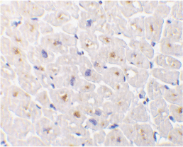
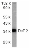
Left: Western blot analysis of DcR2 in HeLa whole cell lysate with DcR2 antibody at 1:1000 dilution.
Below: Immunocytochemistry staining of HeLa cells using DcR2 antibody at 10µg/ml.
Other Product Images
 Source : DcR2 antibody was raised against a peptide corresponding to amino acids 249 to 263 of human DcR2 precursor.
Purification : Affinity chromatography purified via peptide column
Clonality and Clone : This is a polyclonal antibody.
Host : DcR2 antibody was raised in rabbit. Please use anti-rabbit secondary antibodies.
Immunogen : Human DcR2 (Intracellular Domain) Peptide (Cat. No. 2021P)
Application : DcR2 antibody can be used for detection of DcR2 expression by Western blot at 1:1000 to 1:2000 dilution.
Tested Application(s) : E, WB, ICC
Buffer : Antibody is supplied in PBS containing 0.02% sodium azide.
Blocking Peptide : Cat. No. 2021P - DcR2 Peptide
Long-Term Storage : DcR2 antibody can be stored at 4ºC, stable for one year. As with all antibodies care should be taken to avoid repeated freeze thaw cycles. Antibodies should not be exposed to prolonged high temperatures.
Positive Control
1.Cat. No. 1201 - HeLa Cell Lysate
Species Reactivity :H, M, R
GI Number : 18203495
Accession Number : Q9UBN6
Short Description : (ID) Decoy Receptor 2 for TRAIL
References
1.Pan G; O'Rourke K; Chinnaiyan AM; O'Rourke K; Gentz R; Ebner R; Ni J; Dixit VM. The receptor for the cytotoxic ligand TRAIL. Science; 1997;276:111-113
2.Pan G, Ni J, Wei YF, Yu G, Gentz R, Dixit VM. An antagonist decoy receptor and a death domain-containing receptor for TRAIL. Science 1997;277:815-8
3.Sheridan JP, Marsters SA, Pitti RM, Gurney A, Skubatch M, Baldwin D, Ramakrishnan L, Gray CL, Baker K, Wood WI, Goddard AD, Godowski P, Ashkenazi A. Control of TRAIL-induced apoptosis by a family of signaling and decoy receptors. Science 1997;277:818-21
4.Marsters SA, Sheridan JP, Pitti RM, Huang A, Skubatch M, Baldwin D, Yuan J, Gurney A, Goddard AD, Godowski P, Ashkenazi A. A novel receptor for Apo2L/TRAIL contains a truncated death domain. Curr Biol 1997;7:1003-6
Catalog# : 2021
Apoptosis is induced by certain cytokines including TNF and Fas ligand in the TNF family through their death domain containing receptors. TRAIL/Apo2L is a new member of the TNF family and induces apoptosis of a variety of tumor cell lines. DR4 and DR5 are the recently identified functional receptors for TRAIL, and DcR1/TRID is a decoy receptor (1-3). Another member of the TRAIL receptor family was more recently identified and designated DcR2, TRAIL-R4, or TRUNDD (4-6). DcR2 has an extracellular TRAIL-binding domain but lacks intracellular death domain and does not induce apoptosis. Like DR4 and DR5, DcR2 transcript is widely expressed in normal human tissues. Overexpression of DcR2 attenuated TRAIL-induced apoptosis.
Additional Names : DcR2 (ID), TRAIL-R4
DescriptionLeft: Western blot analysis of DcR2 in HeLa whole cell lysate with DcR2 antibody at 1:1000 dilution.
Below: Immunocytochemistry staining of HeLa cells using DcR2 antibody at 10µg/ml.
Other Product Images
Source : DcR2 antibody was raised against a peptide corresponding to amino acids 249 to 263 of human DcR2 precursor.Purification : Affinity chromatography purified via peptide column
Clonality and Clone : This is a polyclonal antibody.
Host : DcR2 antibody was raised in rabbit. Please use anti-rabbit secondary antibodies.
Immunogen : Human DcR2 (Intracellular Domain) Peptide (Cat. No. 2021P)
Application : DcR2 antibody can be used for detection of DcR2 expression by Western blot at 1:1000 to 1:2000 dilution.
Tested Application(s) : E, WB, ICC
Buffer : Antibody is supplied in PBS containing 0.02% sodium azide.
Blocking Peptide : Cat. No. 2021P - DcR2 Peptide
Long-Term Storage : DcR2 antibody can be stored at 4ºC, stable for one year. As with all antibodies care should be taken to avoid repeated freeze thaw cycles. Antibodies should not be exposed to prolonged high temperatures.
Positive Control
1.Cat. No. 1201 - HeLa Cell Lysate
Species Reactivity :H, M, R
GI Number : 18203495
Accession Number : Q9UBN6
Short Description : (ID) Decoy Receptor 2 for TRAIL
References
1.Pan G; O'Rourke K; Chinnaiyan AM; O'Rourke K; Gentz R; Ebner R; Ni J; Dixit VM. The receptor for the cytotoxic ligand TRAIL. Science; 1997;276:111-113
2.Pan G, Ni J, Wei YF, Yu G, Gentz R, Dixit VM. An antagonist decoy receptor and a death domain-containing receptor for TRAIL. Science 1997;277:815-8
3.Sheridan JP, Marsters SA, Pitti RM, Gurney A, Skubatch M, Baldwin D, Ramakrishnan L, Gray CL, Baker K, Wood WI, Goddard AD, Godowski P, Ashkenazi A. Control of TRAIL-induced apoptosis by a family of signaling and decoy receptors. Science 1997;277:818-21
4.Marsters SA, Sheridan JP, Pitti RM, Huang A, Skubatch M, Baldwin D, Yuan J, Gurney A, Goddard AD, Godowski P, Ashkenazi A. A novel receptor for Apo2L/TRAIL contains a truncated death domain. Curr Biol 1997;7:1003-6
Wednesday, June 23, 2010
Custom DNA Synthesis Services
Since 1984, Bio-Synthesis, the first company to provide commercial custom DNA synthesis services, has remained a major force in advancing biotechnology research both as leading supplier and developer of new technologies for unmodified DNA synthesis, custom DNA modifications, antisense oligos and DNA chimera. Biochemical researchers choose BSI as their major supplier for oligonucleotide synthesis because of our core values: QUALITY, SPEED and HIGH THROUGHPUT capabilities. Quality is not just a goal, it is a GUARANTEE. Every custom DNA synthesized must pass strict quality control guidelines before delivered to our customers. In addition to high quality production, speed of our custom DNA has improved to next day delivery in most cases. Through improvement from traditional chemistry to development of advanced state-of-the-art proprietary instrumentations, BSI has achieved the goal of providing the most economical, high throughput, quality custom DNA synthesis.
As always, every custom DNA synthesis is:
Unmodified DNA Synthesis
We offered several scales of custom unmodified DNA synthesis at very competitive price. From small research custom DNA synthesis scale to large production of ASR and GMP DNA oligomers. We offer our client with one-stop solution for their custom DNA synthesis needs. See more...
Custom DNA Arrays/Libraries Oligo Synthesis
High-throughput custom DNA can be provided in 96 or 384 well plates. You can even request the plate type required for your system. Options for oligos in solution include equimolar concentration, forward and reverse primer in the same well, dilution in buffer or water, and equal or variable volume. Options for lyophilized oligos in plates includes equal nmole amounts or µg amount. See more...
DNA Modifications
BIO-SYNTHESIS offers thousand of custom DNA modifications to meet our clients needs. Modification such as DNA methylation, DNA base modification, backbone modification, and many others. See more...
Labeled DNA Probes
BSI's labeled DNA probes are routinely manufactured in 200 nmole or 1 umole scale. These fluorophore can be labeled at 5', 3' and internal position of an oligonucleotide. See more...
Antisense Oligos
Antisense oligos have been powerful tools to halt biological events, such as transcription, translation or splicing. At BSI, we offer hybrid synthesis of RNA-DNA with mix backbone linkages. See more...
Bridged Nucleic Acid (BNA) Sythesis
Synthesis BNA offers researchers with a powerful tool for diagnostic as well as for pharmaceutical applications such as antisense therapy, cellular delivery, drug delivery. See more...
DNA Chimera
Synthesis of chimeric DNA, chimeric DNA/RNA, chimeric DNA and peptide provides companies and researchers with a powerful tool for pharmaceutical applications. BSI offers BNA/DNA chimera synthesis at competitive prices and quick delivery. See more...
Analyte Specific Reagent (ASR oligos)
Bio-Synthesis Inc. is registered with the U.S. Food and Drug Administration as a Medical Device Class 1 manufacturer for Analyte Specific Reagent (ASR) products, which includes custom nucleic acid sequences for: in vitro diagnostic manufacturers, clinical laboratories or organizations that use the reagent to make tests for purposes other than providing diagnostic information to patients and practitioners. Note: According to 21CFR809.30 (d), Class I ASRs must be labeled as: "Analyte Specific Reagent. Analytical and performance characteristics are not established."
Bio-Synthesis's ASR products (GMP Oligos) are manufactured under good manufacturing practices (cGMP) in compliance with FDA regulations 21CFR864.4020 and 21CFR820.
As always, every custom DNA synthesis is:
- Deprotected, desalted, lyophilized, Ready-2-Use
- Quantitated 2x by UV spectrophotometry to provide an accurate measure of yield
- Quality checked by MALDI-TOF mass spectrometry, purity check by HPLC and/or PAGE
- Custom preparative and analytical services (optional)
- Quality Assurance document including: yields, TM, MW, ug/OD and more...
- Fast delivery with high throughput capacity
Unmodified DNA Synthesis
We offered several scales of custom unmodified DNA synthesis at very competitive price. From small research custom DNA synthesis scale to large production of ASR and GMP DNA oligomers. We offer our client with one-stop solution for their custom DNA synthesis needs. See more...
Custom DNA Arrays/Libraries Oligo Synthesis
High-throughput custom DNA can be provided in 96 or 384 well plates. You can even request the plate type required for your system. Options for oligos in solution include equimolar concentration, forward and reverse primer in the same well, dilution in buffer or water, and equal or variable volume. Options for lyophilized oligos in plates includes equal nmole amounts or µg amount. See more...
DNA Modifications
BIO-SYNTHESIS offers thousand of custom DNA modifications to meet our clients needs. Modification such as DNA methylation, DNA base modification, backbone modification, and many others. See more...
Labeled DNA Probes
BSI's labeled DNA probes are routinely manufactured in 200 nmole or 1 umole scale. These fluorophore can be labeled at 5', 3' and internal position of an oligonucleotide. See more...
Antisense Oligos
Antisense oligos have been powerful tools to halt biological events, such as transcription, translation or splicing. At BSI, we offer hybrid synthesis of RNA-DNA with mix backbone linkages. See more...
Bridged Nucleic Acid (BNA) Sythesis
Synthesis BNA offers researchers with a powerful tool for diagnostic as well as for pharmaceutical applications such as antisense therapy, cellular delivery, drug delivery. See more...
DNA Chimera
Synthesis of chimeric DNA, chimeric DNA/RNA, chimeric DNA and peptide provides companies and researchers with a powerful tool for pharmaceutical applications. BSI offers BNA/DNA chimera synthesis at competitive prices and quick delivery. See more...
Analyte Specific Reagent (ASR oligos)
Bio-Synthesis Inc. is registered with the U.S. Food and Drug Administration as a Medical Device Class 1 manufacturer for Analyte Specific Reagent (ASR) products, which includes custom nucleic acid sequences for: in vitro diagnostic manufacturers, clinical laboratories or organizations that use the reagent to make tests for purposes other than providing diagnostic information to patients and practitioners. Note: According to 21CFR809.30 (d), Class I ASRs must be labeled as: "Analyte Specific Reagent. Analytical and performance characteristics are not established."
Bio-Synthesis's ASR products (GMP Oligos) are manufactured under good manufacturing practices (cGMP) in compliance with FDA regulations 21CFR864.4020 and 21CFR820.
Tuesday, June 22, 2010
Array Related Service
Array Related Service
In addition to array chip printing and processing, we also provide other microarray related services, which include luminex assay services and genomics DNA/total RNA extraction from frozen or RNA later-kept tissues or cell pellets. In addition, we provide array result validation services, such as real-time PCR and Western blotting.
Luminex Assay Services
The Luminex platform utilizes flow cytometry, microspheres, lasers, digital signal processing and traditional chemistry to provide a flexible platform capable of measuring a few to 100 analytes, simultaneously. The platform is ideally suited to a wide range of applications, from basic research to drug discovery and diagnostics.
We provide high-quality, low-cost, short turn-around time service (usually a week after receiving your samples and assay kit) for most commercial or custom assays, including, but not limited to, the following:
 Sample Submissions
Please submit at least double the amount of sample (serum, plasma, TCS, cell extracts, nuclear extracts, or DNA) as required by the manufacturer's kit specifications, just in case the first trial fails. Send the samples in dry ice via a commercial overnight shipper.
Pricing
We charge a setup fee of $200, and a flat rate of $20 per sample. Price does not include assay kits. For a price quote, click here!
Other Array Related Services

Quality Control
The DNA/RNA we extract are subjected to quality checks. OD ratios of 260/280 and 260/230 are measured by the NanoDrop and MW profiling is performed by the Agilent 2100 bioanalyzer..
Sunday, June 20, 2010
Bio-Synthesis provides array processing services
Array Processing
Bio-Synthesis provides array processing services which involve target sample labeling, hybridization/incubation, staining/washing, scanning, and data analysis by using various commercial microarray platforms such as Affymetric, Illumina, Agilent and our own spotted arrays. Looking for a reliable source with flexible project execution ability? Contact us for details.
Service
Spotted array post processing
Volume
>10 slides
Price
Service
Spotted array RNA amplification
Price
$150
Service
Spotted array RNA labeling
Price
$150
Service
Spotted array hybridization
Price
$150
Service
Spotted array scanning
Price
$150
Service
Target amplification & labeling
Platform
Affymetrix array
Price
$ 450
Service
Hybridization & Scanning
Platform
Affymetrix array
Price
$ 250
Service
Platform
Volume
Price
Service
Full array processing services
Platform
Agilent array
Price
$ 350
Quality Control
For commercial array processing, we restrictively follow the manufacturers' QC guidelines. For our own spotted arrays, we use a wide range of quality controls (monitoring each processing step) to reach desired sensitivity, specificity and reproducibility, while keeping maximum flexibility for different applications.
Thursday, June 17, 2010
Bio-Synthesis provides comprehensive custom antibody packages
Bio-Synthesis provides comprehensive custom antibody packages and immunological services, including epitope prediction, custom antibody synthesis
,carrier protein conjugation, immunization and purification, ELISA analysis, antibody labeling, and antibody microarrays. BSI offers peptide antigen design, using a proprietary Antigen Identifier TM design tool. Anti-peptide polyclonal antibody production is performed with the best titer guarantee in the industry, by implementing a proprietary SP-MaxTM adjuvant, a new family of adjuvants with improved properties, but minimal toxicity. BSI works with host animals under USDA license and holds an NIH Animal Welfare Assurance from the Office of Laboratory Animal Welfare for immunization. All procedures performed during immunization are described in our Standard Operation Procedures (SOPs) to ensure a high-quality product.
All custom polyclonal antibody projects are treated with strict confidentiality. Used animals, developed antigens remain the exclusive property of the customer.
From antigen selection to antibody purfication, BSI has optimized each step of the antibody production process to provided the best antibodies available.
We offer 8 standard custom antibody protocols (combinations of hosts and protocol timeframe) and 6 standard antibody packages (combinations of provisions, antigen and deliverables). For information about a particular protocol, see our Polyclonal Antibody Services below or request a quote today.
Advanced antigen design
Comprehensive antibody packages
,carrier protein conjugation, immunization and purification, ELISA analysis, antibody labeling, and antibody microarrays. BSI offers peptide antigen design, using a proprietary Antigen Identifier TM design tool. Anti-peptide polyclonal antibody production is performed with the best titer guarantee in the industry, by implementing a proprietary SP-MaxTM adjuvant, a new family of adjuvants with improved properties, but minimal toxicity. BSI works with host animals under USDA license and holds an NIH Animal Welfare Assurance from the Office of Laboratory Animal Welfare for immunization. All procedures performed during immunization are described in our Standard Operation Procedures (SOPs) to ensure a high-quality product.
All custom polyclonal antibody projects are treated with strict confidentiality. Used animals, developed antigens remain the exclusive property of the customer.
From antigen selection to antibody purfication, BSI has optimized each step of the antibody production process to provided the best antibodies available.
We offer 8 standard custom antibody protocols (combinations of hosts and protocol timeframe) and 6 standard antibody packages (combinations of provisions, antigen and deliverables). For information about a particular protocol, see our Polyclonal Antibody Services below or request a quote today.
- Single anti-peptide polyclonal antibody
- Peptide phospho-specific polyclonal antibody
- Multiple-peptide co-immunization
- Multiple antigen peptide (MAP) polyclonal antibody
- User-supplied antigen
- Create-your-own antibody packages
Advanced antigen design
- Proprietary Antigen Identifier design tool with team of expert scientist to assist client for best peptide antigen selection
- Supreme adjuvant SP-MAX technology
- A new family of adjuvants with improved properties, but minimal toxicity which ensures high specificity and titer of the produced antibodies.
- ELISA Guarantee
- Our polyclonal antibodies have an ELISA titer of 1:20,000 or better for any host if peptide antigens were designed, synthesized, and conjugated by Bio-Synthesis.
Comprehensive antibody packages
- Single anti-peptide polyclonal antibody
- Peptide phospho-specific polyclonal antibody
- Multiple-peptide co-immunization
- Multiple antigen peptide (MAP) polyclonal antibody
- User-supplied antigen
- Create-your-own antibody packages
Wednesday, June 16, 2010
Custom Spotted Oligonucleotide Array Full Services
Custom Spotted Oligonucleotide Array Full Services
As researchers approach the developing field of genomics with custom microarray productions, the need for microarray services or productions that maximize flexibility and precision, without compromising speed and cost, has become critical.
To download more specific instructions and an appropriate order form, please click here.
Quality Control
Replicated spots and slides are used to monitor technical variations. In addition, pre-scanning with fast dye staining is conducted to make sure the spot size and morphology meet expectations. Sometimes a test hybridization is conducted, if needed..
As researchers approach the developing field of genomics with custom microarray productions, the need for microarray services or productions that maximize flexibility and precision, without compromising speed and cost, has become critical.
- In order to meet the demands for custom microarraying, BSI offers a complete suite of microarray services, which include:
- Experimental design consultation to provide expert support with DNA sequence quality control and assist clients with optimal probe selection.
- Synthesis chemistry and modifications custom synthesis of DNA, RNA, BNA or hybrid oligos with backbone modifications such as phosphorothioate for antisense study, or nucleotide modifications.
- Flexible chip fabrication, which allow fine-tune client's probe sets for high-density custom microarrays.
- Target sample labeling and hybridization
- Other related services DNA/RNA Extraction, RNA quality check, Real-Time PCR, Western-Blot.
- BioInformatic data analysis
To download more specific instructions and an appropriate order form, please click here.
Quality Control
Replicated spots and slides are used to monitor technical variations. In addition, pre-scanning with fast dye staining is conducted to make sure the spot size and morphology meet expectations. Sometimes a test hybridization is conducted, if needed..
Sunday, June 13, 2010
DcR1 Antibody
DcR1 Antibody
Catalog# : 2299
Apoptosis is induced by certain cytokines including TNF and Fas ligand in the TNF family through their death domain containing receptors. TRAIL/Apo2L is a new member of the TNF family and induces apoptosis of a variety of tumor cell lines. DR4 and DR5 are the recently identified functional receptors for TRAIL (1-3). Two decoy receptors for TRAIL have been identified and designated DcR1/TRID/TRAIL-R3/LIT (2-7) and DcR2/TRAIL-R4/TRUNDD (8-10). DcR1 has extracellular TRAIL-binding domain but lacks intracellular signaling domain. It is a glycophospholipid-anchored cell surface protein. DcR1 transcripts are expressed in many normal human tissues but not in most cancer cell lines. Overexpression of DcR1 did not induce apoptosis, but attenuated TRAIL-induced apoptosis.
Additional Names : DcR1, DcR1 (ED2), TRAIL-R3, TRID, LIT
 Description
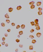
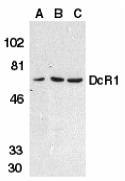
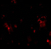
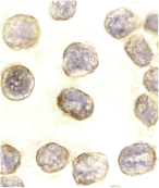
Left: Western blot analysis of DcR1 in HeLa cell (A), mouse (B) and rat (C) liver tissue lysates with DcR1 antibody at 1 µg/ml.
Below: Immunofluorescence of DcR1 in rat liver tissue with DcR1 antibody at 10 µg/ml.
Other Product Images
Source : DcR1 antibody was raised against a peptide corresponding to amino acids in the extracellular domain of human DcR1 precursor.
Purification : Affinity chromatography purified via peptide column
Clonality and Clone : This is a polyclonal antibody.
Host : DcR1 antibody was raised in rabbit. Please use anti-rabbit secondary antibodies.
Immunogen : Human DcR1 Peptide (Cat. No. 2299P)
Application : DcR1 antibody can be used for detection of DcR1 by Western blot at 0.5 to 1 µg/ml.An approximate 65 kDa band can be detected.
Tested Application(s) : E, WB, IF
Buffer : Antibody is supplied in PBS containing 0.02% sodium azide.
Blocking Peptide : Cat. No. 2299P - DcR1 Peptide
Long-Term Storage : DcR1 antibody can be stored at 4ºC, stable for one year. As with all antibodies care should be taken to avoid repeated freeze thaw cycles. Antibodies should not be exposed to prolonged high temperatures.
Positive Control
1.Cat. No. 1201 - HeLa Cell Lysate
2.Cat. No. 1404 - Mouse Liver Tissue Lysate
3.Cat. No. 1464 - Rat Liver Tissue Lysate
Species Reactivity :H, M, R
GI Number : 2338422
Accession Number : AAB67104
Short Description : (ED2) Decoy Receptor 1 for TRAIL
References
1.Pan G; O'Rourke K; Chinnaiyan et al.. The receptor for the cytotoxic ligand TRAIL. Science; 1997;276:111-113
2.Pan G, Ni J, Wei YF, et al. An antagonist decoy receptor and a death domain-containing receptor for TRAIL. Science 1997;277:815-8
3.Sheridan JP, Marsters SA, Pitti RM, et al. A. Control of TRAIL-induced apoptosis by a family of signaling and decoy receptors. Science 1997;277:818-21
4.Degli-Esposti MA, Smolak PJ, Walczak H, et al, Smith CA. Cloning and characterization of TRAIL-R3, a novel member of the emerging TRAIL receptor family. J Exp Med 1997;186(7):1165-70
Catalog# : 2299
Apoptosis is induced by certain cytokines including TNF and Fas ligand in the TNF family through their death domain containing receptors. TRAIL/Apo2L is a new member of the TNF family and induces apoptosis of a variety of tumor cell lines. DR4 and DR5 are the recently identified functional receptors for TRAIL (1-3). Two decoy receptors for TRAIL have been identified and designated DcR1/TRID/TRAIL-R3/LIT (2-7) and DcR2/TRAIL-R4/TRUNDD (8-10). DcR1 has extracellular TRAIL-binding domain but lacks intracellular signaling domain. It is a glycophospholipid-anchored cell surface protein. DcR1 transcripts are expressed in many normal human tissues but not in most cancer cell lines. Overexpression of DcR1 did not induce apoptosis, but attenuated TRAIL-induced apoptosis.
Additional Names : DcR1, DcR1 (ED2), TRAIL-R3, TRID, LIT
DescriptionLeft: Western blot analysis of DcR1 in HeLa cell (A), mouse (B) and rat (C) liver tissue lysates with DcR1 antibody at 1 µg/ml.
Below: Immunofluorescence of DcR1 in rat liver tissue with DcR1 antibody at 10 µg/ml.
Other Product Images
Source : DcR1 antibody was raised against a peptide corresponding to amino acids in the extracellular domain of human DcR1 precursor.
Purification : Affinity chromatography purified via peptide column
Clonality and Clone : This is a polyclonal antibody.
Host : DcR1 antibody was raised in rabbit. Please use anti-rabbit secondary antibodies.
Immunogen : Human DcR1 Peptide (Cat. No. 2299P)
Application : DcR1 antibody can be used for detection of DcR1 by Western blot at 0.5 to 1 µg/ml.An approximate 65 kDa band can be detected.
Tested Application(s) : E, WB, IF
Buffer : Antibody is supplied in PBS containing 0.02% sodium azide.
Blocking Peptide : Cat. No. 2299P - DcR1 Peptide
Long-Term Storage : DcR1 antibody can be stored at 4ºC, stable for one year. As with all antibodies care should be taken to avoid repeated freeze thaw cycles. Antibodies should not be exposed to prolonged high temperatures.
Positive Control
1.Cat. No. 1201 - HeLa Cell Lysate
2.Cat. No. 1404 - Mouse Liver Tissue Lysate
3.Cat. No. 1464 - Rat Liver Tissue Lysate
Species Reactivity :H, M, R
GI Number : 2338422
Accession Number : AAB67104
Short Description : (ED2) Decoy Receptor 1 for TRAIL
References
1.Pan G; O'Rourke K; Chinnaiyan et al.. The receptor for the cytotoxic ligand TRAIL. Science; 1997;276:111-113
2.Pan G, Ni J, Wei YF, et al. An antagonist decoy receptor and a death domain-containing receptor for TRAIL. Science 1997;277:815-8
3.Sheridan JP, Marsters SA, Pitti RM, et al. A. Control of TRAIL-induced apoptosis by a family of signaling and decoy receptors. Science 1997;277:818-21
4.Degli-Esposti MA, Smolak PJ, Walczak H, et al, Smith CA. Cloning and characterization of TRAIL-R3, a novel member of the emerging TRAIL receptor family. J Exp Med 1997;186(7):1165-70
DcR1 Antibody
DcR1 Antibody
Catalog# : 2179
Apoptosis is induced by certain cytokines including TNF and Fas ligand in the TNF family through their death domain containing receptors. TRAIL/Apo2L is a new member of the TNF family and induces apoptosis of a variety of tumor cell lines. DR4 and DR5 are the recently identified functional receptors for TRAIL. Two decoy receptors for TRAIL have been identified and designated DcR1/TRID/TRAIL-R3/LIT and DcR2/TRAIL-R4/TRUNDD. DcR1 has extracellular TRAIL-binding domain but lacks intracellular signaling domain. It is a glycophospholipid-anchored cell surface protein. DcR1 transcripts were expressed in many normal human tissues but not in most cancer cell lines. Overexpression of DcR1 did not induce apoptosis, but attenuated TRAIL-induced apoptosis.
Additional Names : DcR1 (ED), TRAIL-R3
 Description
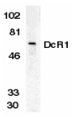
Left: Western blot analysis of DcR1 in HeLa whole cell lysate with DcR1 antibody (ED) at 1:500 dilution.
Below: Immunocytochemistry of DcR1 in HeLa cells with DcR1 antibody at 10 µg/ml.
Other Product Images
Source : DcR1 antibody was raised against a peptide corresponding to amino acids in a extracellular domain (ED) of human DcR1 precursor.
Purification : Affinity chromatography purified via peptide column
Clonality and Clone : This is a polyclonal antibody.
Host : DcR1 antibody was raised in rabbit. Please use anti-rabbit secondary antibodies.
Immunogen : Human DcR1 (Extracellular Domain) Peptide (Cat. No. 2179P)
Application : DcR1 antibody can be used for detection of DcR1 by Western blot at 1:500 to 1:1000 dilution. An approximate 65 kDa band can be detected (6). It is human, mouse, and rat reactive.
Tested Application(s) : E, WB, ICC
Buffer : Antibody is supplied in PBS containing 0.02% sodium azide.
Blocking Peptide : Cat. No. 2179P - DcR1 Peptide
Long-Term Storage : DcR1 antibody can be stored at 4ºC, stable for one year. As with all antibodies care should be taken to avoid repeated freeze thaw cycles. Antibodies should not be exposed to prolonged high temperatures.
Positive Control
1.Cat. No. 1201 - HeLa Cell Lysate
Species Reactivity :H, M, R
GI Number : 2338421
Accession Number : AF012536
Short Description : (ED) Decoy Receptor 1 for TRAIL
References
1.Pan G; O'Rourke K; Chinnaiyan et al.. The receptor for the cytotoxic ligand TRAIL. Science; 1997;276:111-113
2.Pan G, Ni J, Wei YF, et al. An antagonist decoy receptor and a death domain-containing receptor for TRAIL. Science 1997;277:815-8
3.Sheridan JP, Marsters SA, Pitti RM, et al. A. Control of TRAIL-induced apoptosis by a family of signaling and decoy receptors. Science 1997;277:818-21
4.Degli-Esposti MA, Smolak PJ, Walczak H, et al, Smith CA. Cloning and characterization of TRAIL-R3, a novel member of the emerging TRAIL receptor family. J Exp Med 1997;186(7):1165-70
Catalog# : 2179
Apoptosis is induced by certain cytokines including TNF and Fas ligand in the TNF family through their death domain containing receptors. TRAIL/Apo2L is a new member of the TNF family and induces apoptosis of a variety of tumor cell lines. DR4 and DR5 are the recently identified functional receptors for TRAIL. Two decoy receptors for TRAIL have been identified and designated DcR1/TRID/TRAIL-R3/LIT and DcR2/TRAIL-R4/TRUNDD. DcR1 has extracellular TRAIL-binding domain but lacks intracellular signaling domain. It is a glycophospholipid-anchored cell surface protein. DcR1 transcripts were expressed in many normal human tissues but not in most cancer cell lines. Overexpression of DcR1 did not induce apoptosis, but attenuated TRAIL-induced apoptosis.
Additional Names : DcR1 (ED), TRAIL-R3
DescriptionLeft: Western blot analysis of DcR1 in HeLa whole cell lysate with DcR1 antibody (ED) at 1:500 dilution.
Below: Immunocytochemistry of DcR1 in HeLa cells with DcR1 antibody at 10 µg/ml.
Other Product Images
Source : DcR1 antibody was raised against a peptide corresponding to amino acids in a extracellular domain (ED) of human DcR1 precursor.
Purification : Affinity chromatography purified via peptide column
Clonality and Clone : This is a polyclonal antibody.
Host : DcR1 antibody was raised in rabbit. Please use anti-rabbit secondary antibodies.
Immunogen : Human DcR1 (Extracellular Domain) Peptide (Cat. No. 2179P)
Application : DcR1 antibody can be used for detection of DcR1 by Western blot at 1:500 to 1:1000 dilution. An approximate 65 kDa band can be detected (6). It is human, mouse, and rat reactive.
Tested Application(s) : E, WB, ICC
Buffer : Antibody is supplied in PBS containing 0.02% sodium azide.
Blocking Peptide : Cat. No. 2179P - DcR1 Peptide
Long-Term Storage : DcR1 antibody can be stored at 4ºC, stable for one year. As with all antibodies care should be taken to avoid repeated freeze thaw cycles. Antibodies should not be exposed to prolonged high temperatures.
Positive Control
1.Cat. No. 1201 - HeLa Cell Lysate
Species Reactivity :H, M, R
GI Number : 2338421
Accession Number : AF012536
Short Description : (ED) Decoy Receptor 1 for TRAIL
References
1.Pan G; O'Rourke K; Chinnaiyan et al.. The receptor for the cytotoxic ligand TRAIL. Science; 1997;276:111-113
2.Pan G, Ni J, Wei YF, et al. An antagonist decoy receptor and a death domain-containing receptor for TRAIL. Science 1997;277:815-8
3.Sheridan JP, Marsters SA, Pitti RM, et al. A. Control of TRAIL-induced apoptosis by a family of signaling and decoy receptors. Science 1997;277:818-21
4.Degli-Esposti MA, Smolak PJ, Walczak H, et al, Smith CA. Cloning and characterization of TRAIL-R3, a novel member of the emerging TRAIL receptor family. J Exp Med 1997;186(7):1165-70
Daxx Antibody
Daxx Antibody
Catalog# : 1163
Apoptosis, or programmed cell death, occurs during normal cellular differentiation and development of multicellular organisms. Apoptosis is induced by certain cytokines including TNF and Fas ligand of the TNF family through their death domain containing receptors, TNFR1 and Fas. Cell death signals are transduced by death domain (DD)- containing adapter molecules and members of the ICE/CED-3 protease family. A novel DD-containing molecule was recently cloned from mouse, human and monkey and designated Daxx. Daxx binds specifically to the Fas death domain and enhances Fas induced apoptosis and activates the Jun N-terminal kinase (JNK) pathway. Daxx is widely expressed in fetal and adult human and mouse tissues indicating its important function in Fas signaling pathways.
Additional Names : Daxx (CT), Daxx
 Description
Left: Western blot analysis of Daxx in HeLa total cell lysate with Daxx antibody at 1 mg/ml.
Below: Immunocytochemistry of DAXX in HeLa cells with DAXX antibody at 10 µg/ml.
Other Product Imagesc
Source : Daxx antibody was raised against a peptide corresponding to amino acids near the carboxy-terminus of human Daxx.
Purification : Antibody is DEAE purified
Clonality and Clone : This is a polyclonal antibody.
Host : Daxx antibody was raised in rabbit. Please use anti-rabbit secondary antibodies.
Immunogen : Human Daxx (C-Terminus) Peptide (Cat. No. 1163P)
Application : Daxx antibody can be used for detection of Daxx by Western blot at 1 – 2 µg/ml.A 120 kDa major band can be detected.
Tested Application(s) : E, WB, ICC
Buffer : Antibody is supplied in PBS containing 0.02% sodium azide.
Blocking Peptide : Cat. No. 1163P - Daxx Peptide
Long-Term Storage : Daxx antibody can be stored at 4ºC, stable for one year. As with all antibodies care should be taken to avoid repeated freeze thaw cycles. Antibodies should not be exposed to prolonged high temperatures.
Positive Control
1.Cat. No. 1201 - HeLa Cell Lysate
Species Reactivity :H, M
GI Number : 48146287
Accession Number : CAG33366
Short Description : (CT) Adapter Molecule
References
1.Yang X, Khosravi-Far R, Chang HY, Baltimore D. Daxx, a novel Fas-binding protein that activates JNK and apoptosis. Cell 1997;89:1067-1076
2.Kiriakidou M, Driscoll DA, Lopez-Guisa JM, Strauss JF 3rd. Cloning and expression of primate Daxx cDNAs and mapping of the human gene to chromosome 6p21.3 in the MHC region. DNA Cell Biol 1997;16:1289-1298 (RD1299)
Catalog# : 1163
Apoptosis, or programmed cell death, occurs during normal cellular differentiation and development of multicellular organisms. Apoptosis is induced by certain cytokines including TNF and Fas ligand of the TNF family through their death domain containing receptors, TNFR1 and Fas. Cell death signals are transduced by death domain (DD)- containing adapter molecules and members of the ICE/CED-3 protease family. A novel DD-containing molecule was recently cloned from mouse, human and monkey and designated Daxx. Daxx binds specifically to the Fas death domain and enhances Fas induced apoptosis and activates the Jun N-terminal kinase (JNK) pathway. Daxx is widely expressed in fetal and adult human and mouse tissues indicating its important function in Fas signaling pathways.
Additional Names : Daxx (CT), Daxx
DescriptionLeft: Western blot analysis of Daxx in HeLa total cell lysate with Daxx antibody at 1 mg/ml.
Below: Immunocytochemistry of DAXX in HeLa cells with DAXX antibody at 10 µg/ml.
Other Product Imagesc
Source : Daxx antibody was raised against a peptide corresponding to amino acids near the carboxy-terminus of human Daxx.
Purification : Antibody is DEAE purified
Clonality and Clone : This is a polyclonal antibody.
Host : Daxx antibody was raised in rabbit. Please use anti-rabbit secondary antibodies.
Immunogen : Human Daxx (C-Terminus) Peptide (Cat. No. 1163P)
Application : Daxx antibody can be used for detection of Daxx by Western blot at 1 – 2 µg/ml.A 120 kDa major band can be detected.
Tested Application(s) : E, WB, ICC
Buffer : Antibody is supplied in PBS containing 0.02% sodium azide.
Blocking Peptide : Cat. No. 1163P - Daxx Peptide
Long-Term Storage : Daxx antibody can be stored at 4ºC, stable for one year. As with all antibodies care should be taken to avoid repeated freeze thaw cycles. Antibodies should not be exposed to prolonged high temperatures.
Positive Control
1.Cat. No. 1201 - HeLa Cell Lysate
Species Reactivity :H, M
GI Number : 48146287
Accession Number : CAG33366
Short Description : (CT) Adapter Molecule
References
1.Yang X, Khosravi-Far R, Chang HY, Baltimore D. Daxx, a novel Fas-binding protein that activates JNK and apoptosis. Cell 1997;89:1067-1076
2.Kiriakidou M, Driscoll DA, Lopez-Guisa JM, Strauss JF 3rd. Cloning and expression of primate Daxx cDNAs and mapping of the human gene to chromosome 6p21.3 in the MHC region. DNA Cell Biol 1997;16:1289-1298 (RD1299)
DAPK2 Antibody
DAPK2 Antibody
Catalog# : 2323
Apoptosis is mediated by death domain containing adapter molecules and a caspase family of proteases. Certain serine/threonine protein kinases, such as RIP and DAP kinase, are mediators of apoptosis. DAP kinase (DAPK) is pro-apoptotic calcium-regulated serine/threonine kinase containing death domain. Ectopic expression of DAPK induces cell death and suppresses oncogenic transformation. DAPK mediates IFNg induced apoptosis. A novel DAP kinase-related protein was recently identified and designated DAPK2 and DRP-1 (1, 2). Ectopicly expressed DAPK2 induced apoptosis in various types of cells (1,2). DAPK has high sequence homology to ZIP kinase and DRAK1/2, and they represent a novel family of serine/threonine kinases, which mediates apoptosis through their catalytic activities. The messenger RNA of DAPK2 is expressed in multiple human tissues (1).
Additional Names : DAPK2
 Description
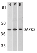
Left: Western blot analysis of DAPK2 in A431 (H), mouse spleen (M), and rat kidney (R) lysates with DAPK2 antibody at 1 µg/ml.
Below: Immunohistochemistry of DAPK2 in mouse spleen cells with DAPK2 antibody at 2 µg/ml.
Other Product Images

Source : DAPK2 antibody was raised against a peptide corresponding to amino acids near the carboxy terminus of human DAPK2. The sequence of this antigenic peptide is identical to the corresponding amino acids of mouse origin (1,2).
Purification : Affinity chromatography purified via peptide column
Clonality and Clone : This is a polyclonal antibody.
Host : DAPK2 antibody was raised in rabbit. Please use anti-rabbit secondary antibodies.
Immunogen : Human DAP kinase 2 Peptide (Cat. No. 2323P)
Application : DAPK2 antibody can be used for detection of DAPK2 by Western blot at 0.5 to 1 µg/ml.An approximately 42 kDa band can be detected. DAPK2 has no cross responses to DAPK1.
Tested Application(s) : E, WB, IHC
Buffer : Antibody is supplied in PBS containing 0.02% sodium azide.
Blocking Peptide : Cat. No. 2323P - DAPK2 Peptide
Long-Term Storage : DAPK2 antibody can be stored at 4ºC, stable for one year. As with all antibodies care should be taken to avoid repeated freeze thaw cycles. Antibodies should not be exposed to prolonged high temperatures.
Positive Control
1.Cat. No. 1202 - A431 Cell Lysate
2.Cat. No. 1406 - Mouse Spleen Tissue Lysate
3.Cat. No. 1465 - Rat Kidney Lysate
Species Reactivity :H, M, R
GI Number : 6521210
Accession Number : BAA88063
Short Description : Death-Associated Protein kinase 2
References
1.Kawai T, Nomura F, Hoshino K, Copeland NG, Gilbert DJ, Jenkins NA, Akira S. Death-associated protein kinase 2 is a new calcium/calmodulin-dependent protein kinase that signals apoptosis through its catalytic activity. Oncogene 1999;18(23):3471-80
2.Inbal B, Shani G, Cohen O, Kissil JL, Kimchi A. Death-associated protein kinase-related protein 1, a novel serine/threonine kinase involved in apoptosis. Mol Cell Biol 2000;20(3):1044-54 (WD0101)
Catalog# : 2323
Apoptosis is mediated by death domain containing adapter molecules and a caspase family of proteases. Certain serine/threonine protein kinases, such as RIP and DAP kinase, are mediators of apoptosis. DAP kinase (DAPK) is pro-apoptotic calcium-regulated serine/threonine kinase containing death domain. Ectopic expression of DAPK induces cell death and suppresses oncogenic transformation. DAPK mediates IFNg induced apoptosis. A novel DAP kinase-related protein was recently identified and designated DAPK2 and DRP-1 (1, 2). Ectopicly expressed DAPK2 induced apoptosis in various types of cells (1,2). DAPK has high sequence homology to ZIP kinase and DRAK1/2, and they represent a novel family of serine/threonine kinases, which mediates apoptosis through their catalytic activities. The messenger RNA of DAPK2 is expressed in multiple human tissues (1).
Additional Names : DAPK2
DescriptionLeft: Western blot analysis of DAPK2 in A431 (H), mouse spleen (M), and rat kidney (R) lysates with DAPK2 antibody at 1 µg/ml.
Below: Immunohistochemistry of DAPK2 in mouse spleen cells with DAPK2 antibody at 2 µg/ml.
Other Product Images
Source : DAPK2 antibody was raised against a peptide corresponding to amino acids near the carboxy terminus of human DAPK2. The sequence of this antigenic peptide is identical to the corresponding amino acids of mouse origin (1,2).
Purification : Affinity chromatography purified via peptide column
Clonality and Clone : This is a polyclonal antibody.
Host : DAPK2 antibody was raised in rabbit. Please use anti-rabbit secondary antibodies.
Immunogen : Human DAP kinase 2 Peptide (Cat. No. 2323P)
Application : DAPK2 antibody can be used for detection of DAPK2 by Western blot at 0.5 to 1 µg/ml.An approximately 42 kDa band can be detected. DAPK2 has no cross responses to DAPK1.
Tested Application(s) : E, WB, IHC
Buffer : Antibody is supplied in PBS containing 0.02% sodium azide.
Blocking Peptide : Cat. No. 2323P - DAPK2 Peptide
Long-Term Storage : DAPK2 antibody can be stored at 4ºC, stable for one year. As with all antibodies care should be taken to avoid repeated freeze thaw cycles. Antibodies should not be exposed to prolonged high temperatures.
Positive Control
1.Cat. No. 1202 - A431 Cell Lysate
2.Cat. No. 1406 - Mouse Spleen Tissue Lysate
3.Cat. No. 1465 - Rat Kidney Lysate
Species Reactivity :H, M, R
GI Number : 6521210
Accession Number : BAA88063
Short Description : Death-Associated Protein kinase 2
References
1.Kawai T, Nomura F, Hoshino K, Copeland NG, Gilbert DJ, Jenkins NA, Akira S. Death-associated protein kinase 2 is a new calcium/calmodulin-dependent protein kinase that signals apoptosis through its catalytic activity. Oncogene 1999;18(23):3471-80
2.Inbal B, Shani G, Cohen O, Kissil JL, Kimchi A. Death-associated protein kinase-related protein 1, a novel serine/threonine kinase involved in apoptosis. Mol Cell Biol 2000;20(3):1044-54 (WD0101)
DAD1 Antibody
DAD1 Antibody
Catalog# : 3313
Defender of cell death 1 (DAD1) was initially discovered in BHK21 cells as a negative regulator of programmed cell death (1), a process important for normal organism development and tissue homeostasis (2). DAD1 was later shown to be a subunit of the mammalian oligosaccharyltransferase complex and is required for its function and structural integrity (3,4). Mice lacking DAD1 express abnormal N-linked glycoproteins and undergo increased apoptotic-associated embryonic death (5,6). Furthermore, overexpression of DAD1 mRNA is seen in some human hepatocellular carcinomas (7), indicating it may also play a role in carcinogenesis. It should be noted that DAD1 is not related to the inhibitor of apoptosis proteins (IAP) family and does not contain any baculoviral IAP repeat (BIR) domains.
Additional Names : DAD1
 Description
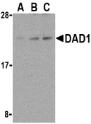
Left: Western blot analysis of DAD1 in HepG2 cell lysate with DAD1 antibody at (A) 0.5, (B) 1, and (C) 2 µg/ml.
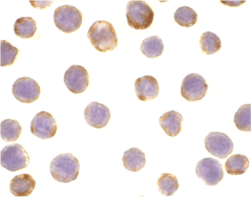
Below: Immunocytochemistry of DAD1 in HepG2 cells with DAD1 antibody at 10 µg/ml.
Other Product Images
Source : DAD1 antibody was raised against a peptide corresponding to 14 amino acids near the C-terminus of human DAD1.
Purification : Ion exchange chromatography purified
Clonality and Clone : This is a polyclonal antibody.
Host : DAD1 antibody was raised in rabbit. Please use anti-rabbit secondary antibodies.
Immunogen : Murine DAD1 Peptide (Cat. No. 3313P)
Application : DAD1 antibody can be used for detection of DAD1 by Western blot at 0.5 to 2 µg/ml.Despite its predicted molecular weight, DAD1 migrates at ~22 kDa in SDS-PAGE.
Tested Application(s) : E, WB, ICC
Buffer : Antibody is supplied in PBS containing 0.02% sodium azide.
Blocking Peptide : Cat. No. 3313P - DAD1 Peptide
Long-Term Storage : DAD1 antibody can be stored at 4ºC, stable for one year. As with all antibodies care should be taken to avoid repeated freeze thaw cycles. Antibodies should not be exposed to prolonged high temperatures.
Positive Control
1.Cat. No. 1211 - HepG2 Cell Lysate
Species Reactivity :H, M
GI Number : 14602573
Accession Number : AAH09798
Short Description : An inhibitor of apoptosis
References
1.Nakashima T, Sekiguchi T, Kuraoka A, et al. Molecular cloning of a human cDNA encoding a novel protein, DAD1, whose defect causes apoptotic cell death in hamster BHK21 cells. Mol. Cell Biol. 1993; 13:6367-74.
2.Stellar H. Mechanisms and genes of cellular suicide. Science 1995; 267:1445-9.
3.Gilmore R and Kelleher DJ. DAD1, the defender against apoptotic cell death, is a subunit of the mammalian oligosaccharyltransferase. Proc. Natl. Acad. Sci. USA 1997; 94:4994-9.
4.Sanjay A, Fu J, and Kreibich G. DAD1 is required for the function and the structural integrity of the oligosaccharyltransferase complex. J. Biol. Chem. 1998; 273:26094-9.
Catalog# : 3313
Defender of cell death 1 (DAD1) was initially discovered in BHK21 cells as a negative regulator of programmed cell death (1), a process important for normal organism development and tissue homeostasis (2). DAD1 was later shown to be a subunit of the mammalian oligosaccharyltransferase complex and is required for its function and structural integrity (3,4). Mice lacking DAD1 express abnormal N-linked glycoproteins and undergo increased apoptotic-associated embryonic death (5,6). Furthermore, overexpression of DAD1 mRNA is seen in some human hepatocellular carcinomas (7), indicating it may also play a role in carcinogenesis. It should be noted that DAD1 is not related to the inhibitor of apoptosis proteins (IAP) family and does not contain any baculoviral IAP repeat (BIR) domains.
Additional Names : DAD1
DescriptionLeft: Western blot analysis of DAD1 in HepG2 cell lysate with DAD1 antibody at (A) 0.5, (B) 1, and (C) 2 µg/ml.
Below: Immunocytochemistry of DAD1 in HepG2 cells with DAD1 antibody at 10 µg/ml.
Other Product Images
Source : DAD1 antibody was raised against a peptide corresponding to 14 amino acids near the C-terminus of human DAD1.
Purification : Ion exchange chromatography purified
Clonality and Clone : This is a polyclonal antibody.
Host : DAD1 antibody was raised in rabbit. Please use anti-rabbit secondary antibodies.
Immunogen : Murine DAD1 Peptide (Cat. No. 3313P)
Application : DAD1 antibody can be used for detection of DAD1 by Western blot at 0.5 to 2 µg/ml.Despite its predicted molecular weight, DAD1 migrates at ~22 kDa in SDS-PAGE.
Tested Application(s) : E, WB, ICC
Buffer : Antibody is supplied in PBS containing 0.02% sodium azide.
Blocking Peptide : Cat. No. 3313P - DAD1 Peptide
Long-Term Storage : DAD1 antibody can be stored at 4ºC, stable for one year. As with all antibodies care should be taken to avoid repeated freeze thaw cycles. Antibodies should not be exposed to prolonged high temperatures.
Positive Control
1.Cat. No. 1211 - HepG2 Cell Lysate
Species Reactivity :H, M
GI Number : 14602573
Accession Number : AAH09798
Short Description : An inhibitor of apoptosis
References
1.Nakashima T, Sekiguchi T, Kuraoka A, et al. Molecular cloning of a human cDNA encoding a novel protein, DAD1, whose defect causes apoptotic cell death in hamster BHK21 cells. Mol. Cell Biol. 1993; 13:6367-74.
2.Stellar H. Mechanisms and genes of cellular suicide. Science 1995; 267:1445-9.
3.Gilmore R and Kelleher DJ. DAD1, the defender against apoptotic cell death, is a subunit of the mammalian oligosaccharyltransferase. Proc. Natl. Acad. Sci. USA 1997; 94:4994-9.
4.Sanjay A, Fu J, and Kreibich G. DAD1 is required for the function and the structural integrity of the oligosaccharyltransferase complex. J. Biol. Chem. 1998; 273:26094-9.
Friday, June 11, 2010
CIDE-B Antibody
CIDE-B Antibody
Catalog# : 2321
Apoptosis is related to many diseases and induced by a family of cell death receptors and their ligands. Cell death signals are transduced by death domain containing adapter molecules and members of the caspase family of proteases. These death signals finally cause the degradation of chromosomal DNA by activated DNase. DFF45/ICAD has been identified as inhibitor of caspase activated DNase DFF40/CAD. DFF45 related proteins CIDE-A and CIDE-B (for cell death-inducing DFF-like effector A and B) were recently identified. CIDE contains a new type of domain termed CIDE-N, which has high homology with the regulatory domains of DFF45/ICAD and DFF40/CAD. Expression of CIDE-B induces apoptosis, which is inhibited by DFF45. CIDE-B is a DFF45-inhibitable effector that promotes cell death and DNA fragmentation. CIDE-B is expressed mainly in liver and at lower levels in spleen, kidney, peripheral blood lymphocytes and bone marrow. CIDE-B antibody has no cross activity to CIDE-A.
Additional Names : CIDE-B (IN), Cell death-inducing DFF-like effector B
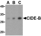 Description
Left: Western blot analysis of CIDE-B in mouse small intestine tissue lysate with CIDE-B antibody at (A) 0.5, (B) 1 and (C) 2 µg/ml.
Below: Immunohistochemistry of CIDE-B in human small intestine tissue with CIDE-B antibody at 5 µg/ml.
Other Product Images
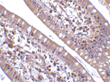 Source : CIDE-B antibody was raised against a peptide corresponding to 14 amino acids near the center of human CIDE-B.
Purification : Affinity chromatography purified via peptide column
Clonality and Clone : This is a polyclonal antibody.
Host : CIDE-B antibody was raised in rabbit. Please use anti-rabbit secondary antibodies.
Application : CIDE-B antibody can be used for detection of CIDE-B by Western blot at 0.5 – 1 µg/ml.
Tested Application(s) : E, WB, IHC
Buffer : Antibody is supplied in PBS containing 0.02% sodium azide.
Blocking Peptide : Cat.No. 2321P - CIDE-B Peptide
Long-Term Storage : CIDE-B antibody can be stored at 4ºC, stable for one year. As with all antibodies care should be taken to avoid repeated freeze thaw cycles. Antibodies should not be exposed to prolonged high temperatures.
Positive Control
1.Cat. No. 1408 - Mouse Small Intestine Tissue Lysate
2.Cat. No. 1308 - Human Small Intestine Tissue Lysate
Species Reactivity :H, M, R
GI Number : 6746627
Accession Number : AAF27658
Short Description : (IN) Cell death-inducing DFF-like effector B
References
1.Inohara N, Koseki T, Chen S,et al. CIDE, a novel family of cell death activators with homology to the 45 kDa subunit of the DNA fragmentation factor. EMBO J. 1998; 17:2526-33.
2.Inohara N, Koseki T, Chen S, et al. Identification of regulatory and catalytic domains in the apoptosis nuclease DFF40/CAD. J. Biol. Chem. 1999; 274:270-4.
Catalog# : 2321
Apoptosis is related to many diseases and induced by a family of cell death receptors and their ligands. Cell death signals are transduced by death domain containing adapter molecules and members of the caspase family of proteases. These death signals finally cause the degradation of chromosomal DNA by activated DNase. DFF45/ICAD has been identified as inhibitor of caspase activated DNase DFF40/CAD. DFF45 related proteins CIDE-A and CIDE-B (for cell death-inducing DFF-like effector A and B) were recently identified. CIDE contains a new type of domain termed CIDE-N, which has high homology with the regulatory domains of DFF45/ICAD and DFF40/CAD. Expression of CIDE-B induces apoptosis, which is inhibited by DFF45. CIDE-B is a DFF45-inhibitable effector that promotes cell death and DNA fragmentation. CIDE-B is expressed mainly in liver and at lower levels in spleen, kidney, peripheral blood lymphocytes and bone marrow. CIDE-B antibody has no cross activity to CIDE-A.
Additional Names : CIDE-B (IN), Cell death-inducing DFF-like effector B
DescriptionLeft: Western blot analysis of CIDE-B in mouse small intestine tissue lysate with CIDE-B antibody at (A) 0.5, (B) 1 and (C) 2 µg/ml.
Below: Immunohistochemistry of CIDE-B in human small intestine tissue with CIDE-B antibody at 5 µg/ml.
Other Product Images
Source : CIDE-B antibody was raised against a peptide corresponding to 14 amino acids near the center of human CIDE-B.Purification : Affinity chromatography purified via peptide column
Clonality and Clone : This is a polyclonal antibody.
Host : CIDE-B antibody was raised in rabbit. Please use anti-rabbit secondary antibodies.
Application : CIDE-B antibody can be used for detection of CIDE-B by Western blot at 0.5 – 1 µg/ml.
Tested Application(s) : E, WB, IHC
Buffer : Antibody is supplied in PBS containing 0.02% sodium azide.
Blocking Peptide : Cat.No. 2321P - CIDE-B Peptide
Long-Term Storage : CIDE-B antibody can be stored at 4ºC, stable for one year. As with all antibodies care should be taken to avoid repeated freeze thaw cycles. Antibodies should not be exposed to prolonged high temperatures.
Positive Control
1.Cat. No. 1408 - Mouse Small Intestine Tissue Lysate
2.Cat. No. 1308 - Human Small Intestine Tissue Lysate
Species Reactivity :H, M, R
GI Number : 6746627
Accession Number : AAF27658
Short Description : (IN) Cell death-inducing DFF-like effector B
References
1.Inohara N, Koseki T, Chen S,et al. CIDE, a novel family of cell death activators with homology to the 45 kDa subunit of the DNA fragmentation factor. EMBO J. 1998; 17:2526-33.
2.Inohara N, Koseki T, Chen S, et al. Identification of regulatory and catalytic domains in the apoptosis nuclease DFF40/CAD. J. Biol. Chem. 1999; 274:270-4.
Thursday, June 10, 2010
CIDE-B Antibody
CIDE-B Antibody
Catalog# : 2319
Apoptosis is related to many diseases and induced by a family of cell death receptors and their ligands. Cell death signals are transduced by death domain containing adapter molecules and members of the caspase family of proteases. These death signals finally cause the degradation of chromosomal DNA by activated DNase. DFF45/ICAD has been identified as inhibitor of caspase activated DNase DFF40/CAD. DFF45 related proteins CIDE-A and CIDE-B (for cell death-inducing DFF-like effector A and B) were recently identified. CIDE contains a new type of domain termed CIDE-N, which has high homology with the regulatory domains of DFF45/ICAD and DFF40/CAD (1,2). Expression of CIDE-B induces apoptosis, which is inhibited by DFF45. CIDE-B is a DFF45-inhibitable effector that promotes cell death and DNA fragmentation. CIDE-B is expressed mainly in liver and at lower levels in spleen, kidney, peripheral blood lymphocytes and bone marrow. CIDE-B antibody has no cross activity to CIDE-A.
Additional Names : CIDE-B (C2), Cell death-inducing DFF-like effector B
CIDE-B Antibody
CIDE-B Antibody
Catalog# : 2319
Apoptosis is related to many diseases and induced by a family of cell death receptors and their ligands. Cell death signals are transduced by death domain containing adapter molecules and members of the caspase family of proteases. These death signals finally cause the degradation of chromosomal DNA by activated DNase. DFF45/ICAD has been identified as inhibitor of caspase activated DNase DFF40/CAD. DFF45 related proteins CIDE-A and CIDE-B (for cell death-inducing DFF-like effector A and B) were recently identified. CIDE contains a new type of domain termed CIDE-N, which has high homology with the regulatory domains of DFF45/ICAD and DFF40/CAD (1,2). Expression of CIDE-B induces apoptosis, which is inhibited by DFF45. CIDE-B is a DFF45-inhibitable effector that promotes cell death and DNA fragmentation. CIDE-B is expressed mainly in liver and at lower levels in spleen, kidney, peripheral blood lymphocytes and bone marrow. CIDE-B antibody has no cross activity to CIDE-A.
Additional Names : CIDE-B (C2), Cell death-inducing DFF-like effector B
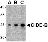 Description
Left: Western blot analysis of CIDE-B in mouse small intestine tissue lysate with CIDE-B antibody at (A) 0.5, (B) 1 and (C) 2 µg/ml.
Below: Immunohistochemistry of CIDE-B in human small intestine tissue with CIDE-B antibody at 5 µg/ml.
Other Product Images
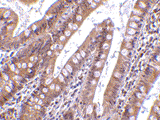 Source : CIDE-B antibody was raised against a peptide corresponding to 15 amino acids near the carboxy terminus of human CIDE-B.
Purification : Affinity chromatography purified via peptide column
Clonality and Clone : This is a polyclonal antibody.
Host : CIDE-B antibody was raised in rabbit. Please use anti-rabbit secondary antibodies.
Application : CIDE-B antibody can be used for detection of CIDE-B by Western blot at 0.5 – 1 µg/ml.
Tested Application(s) : E, WB, IHC
Buffer : Antibody is supplied in PBS containing 0.02% sodium azide.
Blocking Peptide : Cat.No. 2319P - CIDE-B Peptide
Long-Term Storage : CIDE-B antibody can be stored at 4ºC, stable for one year. As with all antibodies care should be taken to avoid repeated freeze thaw cycles. Antibodies should not be exposed to prolonged high temperatures.
Positive Control
1.Cat. No. 1408 - Mouse Small Intestine Tissue Lysate
2.Cat. No. 1308 - Human Small Intestine Tissue Lysate
Species Reactivity :H, M, R
GI Number : 6746627
Accession Number : AAF27658
Short Description : (C2) Cell death-inducing DFF-like effector B
References
1.Inohara N, Koseki T, Chen S,et al. CIDE, a novel family of cell death activators with homology to the 45 kDa subunit of the DNA fragmentation factor. EMBO J. 1998; 17:2526-33
2.Inohara N, Koseki T, Chen S, et al. Identification of regulatory and catalytic domains in the apoptosis nuclease DFF40/CAD. J. Biol. Chem. 1999; 274:270-4.
Catalog# : 2319
Apoptosis is related to many diseases and induced by a family of cell death receptors and their ligands. Cell death signals are transduced by death domain containing adapter molecules and members of the caspase family of proteases. These death signals finally cause the degradation of chromosomal DNA by activated DNase. DFF45/ICAD has been identified as inhibitor of caspase activated DNase DFF40/CAD. DFF45 related proteins CIDE-A and CIDE-B (for cell death-inducing DFF-like effector A and B) were recently identified. CIDE contains a new type of domain termed CIDE-N, which has high homology with the regulatory domains of DFF45/ICAD and DFF40/CAD (1,2). Expression of CIDE-B induces apoptosis, which is inhibited by DFF45. CIDE-B is a DFF45-inhibitable effector that promotes cell death and DNA fragmentation. CIDE-B is expressed mainly in liver and at lower levels in spleen, kidney, peripheral blood lymphocytes and bone marrow. CIDE-B antibody has no cross activity to CIDE-A.
Additional Names : CIDE-B (C2), Cell death-inducing DFF-like effector B
DescriptionLeft: Western blot analysis of CIDE-B in mouse small intestine tissue lysate with CIDE-B antibody at (A) 0.5, (B) 1 and (C) 2 µg/ml.
Below: Immunohistochemistry of CIDE-B in human small intestine tissue with CIDE-B antibody at 5 µg/ml.
Other Product Images
Source : CIDE-B antibody was raised against a peptide corresponding to 15 amino acids near the carboxy terminus of human CIDE-B.Purification : Affinity chromatography purified via peptide column
Clonality and Clone : This is a polyclonal antibody.
Host : CIDE-B antibody was raised in rabbit. Please use anti-rabbit secondary antibodies.
Application : CIDE-B antibody can be used for detection of CIDE-B by Western blot at 0.5 – 1 µg/ml.
Tested Application(s) : E, WB, IHC
Buffer : Antibody is supplied in PBS containing 0.02% sodium azide.
Blocking Peptide : Cat.No. 2319P - CIDE-B Peptide
Long-Term Storage : CIDE-B antibody can be stored at 4ºC, stable for one year. As with all antibodies care should be taken to avoid repeated freeze thaw cycles. Antibodies should not be exposed to prolonged high temperatures.
Positive Control
1.Cat. No. 1408 - Mouse Small Intestine Tissue Lysate
2.Cat. No. 1308 - Human Small Intestine Tissue Lysate
Species Reactivity :H, M, R
GI Number : 6746627
Accession Number : AAF27658
Short Description : (C2) Cell death-inducing DFF-like effector B
References
1.Inohara N, Koseki T, Chen S,et al. CIDE, a novel family of cell death activators with homology to the 45 kDa subunit of the DNA fragmentation factor. EMBO J. 1998; 17:2526-33
2.Inohara N, Koseki T, Chen S, et al. Identification of regulatory and catalytic domains in the apoptosis nuclease DFF40/CAD. J. Biol. Chem. 1999; 274:270-4.
CIDE-B Antibody
CIDE-B Antibody
Catalog# : 2091
Apoptosis is related to many diseases and induced by a family of cell death receptors and their ligands. Cell death signals are transduced by death domain containing adapter molecules and members of the caspase family of proteases. These death signals finally cause the degradation of chromosomal DNA by activated DNase. DFF45/ICAD has been identified as inhibitor of caspase activated DNase DFF40/CAD. DFF45 related proteins CIDE-A and CIDE-B (for cell death-inducing DFF-like effector A and B) were recently identified (1).?CIDE contains a new type of domain termed CIDE-N, which has high homology with the regulatory domains of DFF45/ICAD and DFF40/CAD (1,2). Expression of CIDE-B induces apoptosis, which is inhibited by DFF45. CIDE-B is a DFF45-inhibitable effector that promotes cell death and DNA fragmentation. CIDE-B is expressed mainly in liver and at lower levels in spleen, kidney, peripheral blood lymphocytes, and bone marrow (1).
Additional Names : CIDE-B (CT), CIDE-B
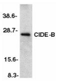 Description
Left: Western blot analysis of CIDE-B in mouse liver tissue lysate with CIDE-B antibody at 1:500 dilution.
Source : CIDE-B antibody was raised against a peptide corresponding to amino acids 204 to 219 of murine CIDE-B .
Purification : Affinity chromatography purified via peptide column
Clonality and Clone : This is a polyclonal antibody.
Host : CIDE-B antibody was raised in rabbit. Please use anti-rabbit secondary antibodies.
Immunogen : Mouse mCIDE-B (C-Terminus) Peptide (Cat. No. 2091P)
Application : CIDE-B antibody can be used for detection of CIDE-B by Western blot at 1:500 to 1:1000 dilution. An approximately 25 kDa band can be detected. It has no cross activity to CIDE-A.
Tested Application(s) : E, WB
Buffer : Antibody is supplied in PBS containing 0.02% sodium azide.
Blocking Peptide : Cat. No. 2091P - CIDE-B Peptide
Long-Term Storage : CIDE-B antibody can be stored at 4ºC, stable for one year. As with all antibodies care should be taken to avoid repeated freeze thaw cycles. Antibodies should not be exposed to prolonged high temperatures.
Positive Control
1.Cat. No. 1404 - Mouse Liver Tissue Lysate
Species Reactivity :M
GI Number : 3114593
Accession Number : AF041377
Short Description : (CT) Cell death-Inducing DFF-like Effector
References
1.Inohara N, Koseki T, Chen S, Wu X, Nunez G. CIDE, a novel family of cell death activators with homology to the 45 kDa subunit of the DNA fragmentation factor. EMBO J 1998;17:2526-33
2.Inohara N, Koseki T, Chen S, Benedict MA, Nunez G. Identification of regulatory and catalytic domains in the apoptosis nuclease DFF40/CAD. J Biol Chem 1999 ;274:270-4 (RD1299)
Catalog# : 2091
Apoptosis is related to many diseases and induced by a family of cell death receptors and their ligands. Cell death signals are transduced by death domain containing adapter molecules and members of the caspase family of proteases. These death signals finally cause the degradation of chromosomal DNA by activated DNase. DFF45/ICAD has been identified as inhibitor of caspase activated DNase DFF40/CAD. DFF45 related proteins CIDE-A and CIDE-B (for cell death-inducing DFF-like effector A and B) were recently identified (1).?CIDE contains a new type of domain termed CIDE-N, which has high homology with the regulatory domains of DFF45/ICAD and DFF40/CAD (1,2). Expression of CIDE-B induces apoptosis, which is inhibited by DFF45. CIDE-B is a DFF45-inhibitable effector that promotes cell death and DNA fragmentation. CIDE-B is expressed mainly in liver and at lower levels in spleen, kidney, peripheral blood lymphocytes, and bone marrow (1).
Additional Names : CIDE-B (CT), CIDE-B
DescriptionLeft: Western blot analysis of CIDE-B in mouse liver tissue lysate with CIDE-B antibody at 1:500 dilution.
Source : CIDE-B antibody was raised against a peptide corresponding to amino acids 204 to 219 of murine CIDE-B .
Purification : Affinity chromatography purified via peptide column
Clonality and Clone : This is a polyclonal antibody.
Host : CIDE-B antibody was raised in rabbit. Please use anti-rabbit secondary antibodies.
Immunogen : Mouse mCIDE-B (C-Terminus) Peptide (Cat. No. 2091P)
Application : CIDE-B antibody can be used for detection of CIDE-B by Western blot at 1:500 to 1:1000 dilution. An approximately 25 kDa band can be detected. It has no cross activity to CIDE-A.
Tested Application(s) : E, WB
Buffer : Antibody is supplied in PBS containing 0.02% sodium azide.
Blocking Peptide : Cat. No. 2091P - CIDE-B Peptide
Long-Term Storage : CIDE-B antibody can be stored at 4ºC, stable for one year. As with all antibodies care should be taken to avoid repeated freeze thaw cycles. Antibodies should not be exposed to prolonged high temperatures.
Positive Control
1.Cat. No. 1404 - Mouse Liver Tissue Lysate
Species Reactivity :M
GI Number : 3114593
Accession Number : AF041377
Short Description : (CT) Cell death-Inducing DFF-like Effector
References
1.Inohara N, Koseki T, Chen S, Wu X, Nunez G. CIDE, a novel family of cell death activators with homology to the 45 kDa subunit of the DNA fragmentation factor. EMBO J 1998;17:2526-33
2.Inohara N, Koseki T, Chen S, Benedict MA, Nunez G. Identification of regulatory and catalytic domains in the apoptosis nuclease DFF40/CAD. J Biol Chem 1999 ;274:270-4 (RD1299)
CIDE-A Antibody
CIDE-A Antibody
Catalog# : 2089
Apoptosis is related to many diseases and induced by a family of cell death receptors and their ligands. Cell death signals are transduced by death domain containing adapter molecules and members of the caspase family of proteases. These death signals finally cause the degradation of chromosomal DNA by activated DNase. DFF45/ICAD has been identified as inhibitor of caspase activated DNase DFF40/CAD. DFF45 related proteins CIDE-A and CIDE-B (for cell death-inducing DFF-like effector A and B) were recently identified (1). CIDE contains a new type of domain termed CIDE-N, which has high homology with the regulatory domains of DFF45/ICAD and DFF40/CAD (1,2). Expression of CIDE-A induces DNA fragmentation and activates apoptosis, which is inhibited by DFF45. CIDE-A is a DFF45-inhibitable effector that promotes cell death and DNA fragmentation. CIDE-A is expressed in many tissues.
Additional Names : CIDE-A (CT), CIDE-A
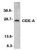 Description
Left: Western blot analysis of CIDE-A in mouse heart tissue lysate with CIDE-A antibody at 1:500 dilution.
Below: Immunohistochemistry of CIDE-A in mouse heart tissue with CIDE-A antibody at 5 µg/ml.
Other Product Images
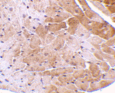 Source : CIDE-A antibody was raised against a peptide corresponding to amino acids 200 to 214 of mouse CIDE-A.
Purification : Affinity chromatography purified via peptide column
Clonality and Clone : This is a polyclonal antibody.
Host : CIDE-A antibody was raised in rabbit. Please use anti-rabbit secondary antibodies.
Immunogen : Mouse mCIDE-A (C-Terminus) Peptide (Cat. No. 2089P)
Application : CIDE-A antibody can be used for detection of CIDE-A by Western blot at 1:500 to 1:1000 dilution. An approximately 25 kDa band can be detected. It has no cross activity to CIDE-B.
Tested Application(s) : E, WB, IHC
Buffer : Antibody is supplied in PBS containing 0.02% sodium azide.
Blocking Peptide : Cat. No. 2089P - CIDE-A Peptide
Long-Term Storage : CIDE-A antibody can be stored at 4ºC, stable for one year. As with all antibodies care should be taken to avoid repeated freeze thaw cycles. Antibodies should not be exposed to prolonged high temperatures.
Positive Control
1.Cat. No. 1401 - Mouse Heart Tissue Lysate
Species Reactivity :M
GI Number : 3114592
Accession Number : AAC34985
Short Description : (CT) Cell death-Inducing DFF-like Effector
References
1.Inohara N, Koseki T, Chen S, Wu X, Nunez G. CIDE, a novel family of cell death activators with homology to the 45 kDa subunit of the DNA fragmentation factor. EMBO J 1998;17:2526-33
2.Inohara N, Koseki T, Chen S, Benedict MA, Nunez G. Identification of regulatory and catalytic domains in the apoptosis nuclease DFF40/CAD. J Biol Chem 1999 ;274:270-4 (RD1299)
Catalog# : 2089
Apoptosis is related to many diseases and induced by a family of cell death receptors and their ligands. Cell death signals are transduced by death domain containing adapter molecules and members of the caspase family of proteases. These death signals finally cause the degradation of chromosomal DNA by activated DNase. DFF45/ICAD has been identified as inhibitor of caspase activated DNase DFF40/CAD. DFF45 related proteins CIDE-A and CIDE-B (for cell death-inducing DFF-like effector A and B) were recently identified (1). CIDE contains a new type of domain termed CIDE-N, which has high homology with the regulatory domains of DFF45/ICAD and DFF40/CAD (1,2). Expression of CIDE-A induces DNA fragmentation and activates apoptosis, which is inhibited by DFF45. CIDE-A is a DFF45-inhibitable effector that promotes cell death and DNA fragmentation. CIDE-A is expressed in many tissues.
Additional Names : CIDE-A (CT), CIDE-A
DescriptionLeft: Western blot analysis of CIDE-A in mouse heart tissue lysate with CIDE-A antibody at 1:500 dilution.
Below: Immunohistochemistry of CIDE-A in mouse heart tissue with CIDE-A antibody at 5 µg/ml.
Other Product Images
Source : CIDE-A antibody was raised against a peptide corresponding to amino acids 200 to 214 of mouse CIDE-A.Purification : Affinity chromatography purified via peptide column
Clonality and Clone : This is a polyclonal antibody.
Host : CIDE-A antibody was raised in rabbit. Please use anti-rabbit secondary antibodies.
Immunogen : Mouse mCIDE-A (C-Terminus) Peptide (Cat. No. 2089P)
Application : CIDE-A antibody can be used for detection of CIDE-A by Western blot at 1:500 to 1:1000 dilution. An approximately 25 kDa band can be detected. It has no cross activity to CIDE-B.
Tested Application(s) : E, WB, IHC
Buffer : Antibody is supplied in PBS containing 0.02% sodium azide.
Blocking Peptide : Cat. No. 2089P - CIDE-A Peptide
Long-Term Storage : CIDE-A antibody can be stored at 4ºC, stable for one year. As with all antibodies care should be taken to avoid repeated freeze thaw cycles. Antibodies should not be exposed to prolonged high temperatures.
Positive Control
1.Cat. No. 1401 - Mouse Heart Tissue Lysate
Species Reactivity :M
GI Number : 3114592
Accession Number : AAC34985
Short Description : (CT) Cell death-Inducing DFF-like Effector
References
1.Inohara N, Koseki T, Chen S, Wu X, Nunez G. CIDE, a novel family of cell death activators with homology to the 45 kDa subunit of the DNA fragmentation factor. EMBO J 1998;17:2526-33
2.Inohara N, Koseki T, Chen S, Benedict MA, Nunez G. Identification of regulatory and catalytic domains in the apoptosis nuclease DFF40/CAD. J Biol Chem 1999 ;274:270-4 (RD1299)
CIDE-A Antibody
CIDE-A Antibody
Catalog# : 2089
Apoptosis is related to many diseases and induced by a family of cell death receptors and their ligands. Cell death signals are transduced by death domain containing adapter molecules and members of the caspase family of proteases. These death signals finally cause the degradation of chromosomal DNA by activated DNase. DFF45/ICAD has been identified as inhibitor of caspase activated DNase DFF40/CAD. DFF45 related proteins CIDE-A and CIDE-B (for cell death-inducing DFF-like effector A and B) were recently identified (1). CIDE contains a new type of domain termed CIDE-N, which has high homology with the regulatory domains of DFF45/ICAD and DFF40/CAD (1,2). Expression of CIDE-A induces DNA fragmentation and activates apoptosis, which is inhibited by DFF45. CIDE-A is a DFF45-inhibitable effector that promotes cell death and DNA fragmentation. CIDE-A is expressed in many tissues.
Additional Names : CIDE-A (CT), CIDE-A
Description
Left: Western blot analysis of CIDE-A in mouse heart tissue lysate with CIDE-A antibody at 1:500 dilution.
Below: Immunohistochemistry of CIDE-A in mouse heart tissue with CIDE-A antibody at 5 µg/ml.
Other Product Images
Source : CIDE-A antibody was raised against a peptide corresponding to amino acids 200 to 214 of mouse CIDE-A.
Purification : Affinity chromatography purified via peptide column
Clonality and Clone : This is a polyclonal antibody.
Host : CIDE-A antibody was raised in rabbit. Please use anti-rabbit secondary antibodies.
Immunogen : Mouse mCIDE-A (C-Terminus) Peptide (Cat. No. 2089P)
Application : CIDE-A antibody can be used for detection of CIDE-A by Western blot at 1:500 to 1:1000 dilution. An approximately 25 kDa band can be detected. It has no cross activity to CIDE-B.
Tested Application(s) : E, WB, IHC
Buffer : Antibody is supplied in PBS containing 0.02% sodium azide.
Blocking Peptide : Cat. No. 2089P - CIDE-A Peptide
Long-Term Storage : CIDE-A antibody can be stored at 4ºC, stable for one year. As with all antibodies care should be taken to avoid repeated freeze thaw cycles. Antibodies should not be exposed to prolonged high temperatures.
Positive Control
1.Cat. No. 1401 - Mouse Heart Tissue Lysate
Species Reactivity :M
GI Number : 3114592
Accession Number : AAC34985
Short Description : (CT) Cell death-Inducing DFF-like Effector
References
1.Inohara N, Koseki T, Chen S, Wu X, Nunez G. CIDE, a novel family of cell death activators with homology to the 45 kDa subunit of the DNA fragmentation factor. EMBO J 1998;17:2526-33
2.Inohara N, Koseki T, Chen S, Benedict MA, Nunez G. Identification of regulatory and catalytic domains in the apoptosis nuclease DFF40/CAD. J Biol Chem 1999 ;274:270-4 (RD1299)
Catalog# : 2089
Apoptosis is related to many diseases and induced by a family of cell death receptors and their ligands. Cell death signals are transduced by death domain containing adapter molecules and members of the caspase family of proteases. These death signals finally cause the degradation of chromosomal DNA by activated DNase. DFF45/ICAD has been identified as inhibitor of caspase activated DNase DFF40/CAD. DFF45 related proteins CIDE-A and CIDE-B (for cell death-inducing DFF-like effector A and B) were recently identified (1). CIDE contains a new type of domain termed CIDE-N, which has high homology with the regulatory domains of DFF45/ICAD and DFF40/CAD (1,2). Expression of CIDE-A induces DNA fragmentation and activates apoptosis, which is inhibited by DFF45. CIDE-A is a DFF45-inhibitable effector that promotes cell death and DNA fragmentation. CIDE-A is expressed in many tissues.
Additional Names : CIDE-A (CT), CIDE-A
DescriptionLeft: Western blot analysis of CIDE-A in mouse heart tissue lysate with CIDE-A antibody at 1:500 dilution.
Below: Immunohistochemistry of CIDE-A in mouse heart tissue with CIDE-A antibody at 5 µg/ml.
Other Product Images
Source : CIDE-A antibody was raised against a peptide corresponding to amino acids 200 to 214 of mouse CIDE-A.
Purification : Affinity chromatography purified via peptide column
Clonality and Clone : This is a polyclonal antibody.
Host : CIDE-A antibody was raised in rabbit. Please use anti-rabbit secondary antibodies.
Immunogen : Mouse mCIDE-A (C-Terminus) Peptide (Cat. No. 2089P)
Application : CIDE-A antibody can be used for detection of CIDE-A by Western blot at 1:500 to 1:1000 dilution. An approximately 25 kDa band can be detected. It has no cross activity to CIDE-B.
Tested Application(s) : E, WB, IHC
Buffer : Antibody is supplied in PBS containing 0.02% sodium azide.
Blocking Peptide : Cat. No. 2089P - CIDE-A Peptide
Long-Term Storage : CIDE-A antibody can be stored at 4ºC, stable for one year. As with all antibodies care should be taken to avoid repeated freeze thaw cycles. Antibodies should not be exposed to prolonged high temperatures.
Positive Control
1.Cat. No. 1401 - Mouse Heart Tissue Lysate
Species Reactivity :M
GI Number : 3114592
Accession Number : AAC34985
Short Description : (CT) Cell death-Inducing DFF-like Effector
References
1.Inohara N, Koseki T, Chen S, Wu X, Nunez G. CIDE, a novel family of cell death activators with homology to the 45 kDa subunit of the DNA fragmentation factor. EMBO J 1998;17:2526-33
2.Inohara N, Koseki T, Chen S, Benedict MA, Nunez G. Identification of regulatory and catalytic domains in the apoptosis nuclease DFF40/CAD. J Biol Chem 1999 ;274:270-4 (RD1299)
Subscribe to:
Posts (Atom)