Thursday, October 28, 2010
DNA Paternity Testing Labs
Biosyn is a leading Private Investigation company. Biosyn has team of professional scientist. Biosyn is work for lot of Legal attorneys, Civil Cases and Insurance companies. Biosyn also helps in immigration process. Biosyn is also work for searching evidence of adultery or other illegal conduct within marriage to establish grounds for a divorce. Biosyn is an active member of the AABB Parentage Testing Accreditation Program and also participates in proficiency testing by the College of American Pathologists.
DNA Testing
Deoxyribonucleic acid (DNA) is a nucleic acid that contains the genetic instructions used in the development and functioning of all known living organisms. The main role of DNA molecules is the long-term storage of information. DNA is often compared to a set of blueprints, since it contains the instructions needed to construct other components of cells, such as proteins and RNA molecules. The DNA segments that carry this genetic information are called genes, but other DNA sequences have structural purposes, or are involved in regulating the use of this genetic information.
Paternity Testing
A maternity or paternity identification test is conducted to establish whether a person is the biological parent of another person. A test to prove paternity (whether a man is someone's father) is known as a paternity test; a test to prove whether a woman is someone's mother) is called a maternity test.
Maternity Testing
Maternity DNA testing determines whether a woman could be the biological mother of a child. Like a DNA paternity test, it compares a child’s DNA pattern with that of the alleged mother to determine how likely it is that the child has inherited the DNA from the alleged mother.
Wednesday, October 27, 2010
EVER1 Antibody
EVER1 Antibody
Catalog# : 4549
Epidermodysplasia verruciformis (EV) is an autosomal recessive dermatosis characterized by abnormal susceptibility to human papillomaviruses (HPVs) and a high rate of progression to squamous cell carcinoma on sun-exposed skin. EV is caused by mutations in either of two adjacent genes, EVER1 and EVER2, located on chromosome 17q25.3. Both of these genes encode integral membrane proteins that localize to the endoplasmic reticulum and are predicted to form transmembrane channels. Both EVER1 and EVER2 are members of the transmembrane channel-like (TMC) protein family. EVER1 possesses eight trans-membrane domains and two leucine zipper motifs. EVER1 and EVER2 form a complex and interact with the zinc transporter 1 (ZnT-1), suggesting that EVER1 and EVER2 act to regulate cellular zinc balance. At least four isoforms of EVER1 are known to exist. This EVER1 antibody does not cross-react with EVER2.
Additional Names : EVER1 (IN), EV1, EVIN1, Transmembrane protein channel-like protein 6, TMC6
 Description
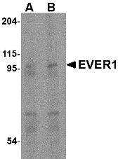
Left: Western blot analysis of EVER1 in human spleen tissue lysate with EVER1 antibody at (A) 1 and (B) 2 µg/ml.
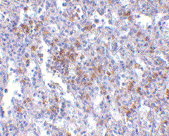
Below: Immunohistochemistry of EVER1 in human spleen with EVER1 antibody at 2.5 µg/ml.
Other Product Images.
 Source : EVER1 antibody was raised against a 15 amino acid peptide from near the center of human EVER1.
Purification : Affinity chromatography purified via peptide column
Clonality and Clone : This is a polyclonal antibody.
Host : EVER1 antibody was raised in rabbit. Please use anti-rabbit secondary antibodies.
Application : EVER1 antibody can be used for the detection of EVER1 by Western blot at 1 – 2 µg/ml.
Tested Application(s) : E, WB, IHC
Buffer : Antibody is supplied in PBS containing 0.02% sodium azide.
Blocking Peptide : Cat.No. 4549P - EVER1 Peptide
Long-Term Storage : EVER1 antibody can be stored at 4ºC, stable for one year. As with all antibodies care should be taken to avoid repeated freeze thaw cycles. Antibodies should not be exposed to prolonged high temperatures.
Positive Control
1. Cat. No. 1306 - Human Spleen Tissue Lysate
Species Reactivity :H, M
GI Number : 25527208
Accession Number : AAM44452
Short Description : (IN) Transmembrane protein channel-like protein 6
References
1. Majewski S, Jablonska J and Orth G. Epidermodysplasia verruciformis. Immunological and nonimmunological surveillance mechanisms: role in tumor progression. Clin. Dermatol. 1997; 15:321-34.
2. Ramoz N, Rueda L-A, Bouadjar B, et al. Mutations in two adjacent novel genes are associated with epidermodysplasia verruciformis. Nat. Genetics 2002; 32:579-81.
3. Keresztes G, Mutai H and Heller S. TMC and EVER genes belong to a larger novel family, the TMC gene family encoding transmembrane proteins. BMC Genomics 2003; 4:24-34.
4. Lazarczyk L, Pons C, Mendoza JA, et al. Regulation of cellular zinc balance as a potential mechanism of EVER-mediated protection against pathogenesis by cutaneous oncogenic human papillomaviruses. J. Exp. Med. 2008; 205:35-42.
Catalog# : 4549
Epidermodysplasia verruciformis (EV) is an autosomal recessive dermatosis characterized by abnormal susceptibility to human papillomaviruses (HPVs) and a high rate of progression to squamous cell carcinoma on sun-exposed skin. EV is caused by mutations in either of two adjacent genes, EVER1 and EVER2, located on chromosome 17q25.3. Both of these genes encode integral membrane proteins that localize to the endoplasmic reticulum and are predicted to form transmembrane channels. Both EVER1 and EVER2 are members of the transmembrane channel-like (TMC) protein family. EVER1 possesses eight trans-membrane domains and two leucine zipper motifs. EVER1 and EVER2 form a complex and interact with the zinc transporter 1 (ZnT-1), suggesting that EVER1 and EVER2 act to regulate cellular zinc balance. At least four isoforms of EVER1 are known to exist. This EVER1 antibody does not cross-react with EVER2.
Additional Names : EVER1 (IN), EV1, EVIN1, Transmembrane protein channel-like protein 6, TMC6
DescriptionLeft: Western blot analysis of EVER1 in human spleen tissue lysate with EVER1 antibody at (A) 1 and (B) 2 µg/ml.
Below: Immunohistochemistry of EVER1 in human spleen with EVER1 antibody at 2.5 µg/ml.
Other Product Images.
Source : EVER1 antibody was raised against a 15 amino acid peptide from near the center of human EVER1.Purification : Affinity chromatography purified via peptide column
Clonality and Clone : This is a polyclonal antibody.
Host : EVER1 antibody was raised in rabbit. Please use anti-rabbit secondary antibodies.
Application : EVER1 antibody can be used for the detection of EVER1 by Western blot at 1 – 2 µg/ml.
Tested Application(s) : E, WB, IHC
Buffer : Antibody is supplied in PBS containing 0.02% sodium azide.
Blocking Peptide : Cat.No. 4549P - EVER1 Peptide
Long-Term Storage : EVER1 antibody can be stored at 4ºC, stable for one year. As with all antibodies care should be taken to avoid repeated freeze thaw cycles. Antibodies should not be exposed to prolonged high temperatures.
Positive Control
1. Cat. No. 1306 - Human Spleen Tissue Lysate
Species Reactivity :H, M
GI Number : 25527208
Accession Number : AAM44452
Short Description : (IN) Transmembrane protein channel-like protein 6
References
1. Majewski S, Jablonska J and Orth G. Epidermodysplasia verruciformis. Immunological and nonimmunological surveillance mechanisms: role in tumor progression. Clin. Dermatol. 1997; 15:321-34.
2. Ramoz N, Rueda L-A, Bouadjar B, et al. Mutations in two adjacent novel genes are associated with epidermodysplasia verruciformis. Nat. Genetics 2002; 32:579-81.
3. Keresztes G, Mutai H and Heller S. TMC and EVER genes belong to a larger novel family, the TMC gene family encoding transmembrane proteins. BMC Genomics 2003; 4:24-34.
4. Lazarczyk L, Pons C, Mendoza JA, et al. Regulation of cellular zinc balance as a potential mechanism of EVER-mediated protection against pathogenesis by cutaneous oncogenic human papillomaviruses. J. Exp. Med. 2008; 205:35-42.
EVER1 Antibody
EVER1 Antibody
Catalog# : 4547
Epidermodysplasia verruciformis (EV) is an autosomal recessive dermatosis characterized by abnormal susceptibility to human papillomaviruses (HPVs) and a high rate of progression to squamous cell carcinoma on sun-exposed skin. EV is caused by mutations in either of two adjacent genes, EVER1 and EVER2, located on chromosome 17q25.3. Both of these genes encode integral membrane proteins that localize to the endoplasmic reticulum and are predicted to form transmembrane channels. Both EVER1 and EVER2 are members of the transmembrane channel-like (TMC) protein family. EVER1 possesses eight trans-membrane domains and two leucine zipper motifs. EVER1 and EVER2 form a complex and interact with the zinc transporter 1 (ZnT-1), suggesting that EVER1 and EVER2 act to regulate cellular zinc balance. At least four isoforms of EVER1 are known to exist. This EVER1 antibody does not cross-react with EVER2.
Additional Names : EVER1 (NT), EV1, EVIN1, Transmembrane protein channel-like protein 6, TMC6
 Description
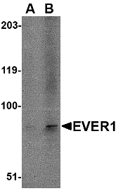
Left: Western blot analysis of EVER1 in A-20 cell lysate with EVER1 antibody at (A) 1 and (B) 2 µg/ml.
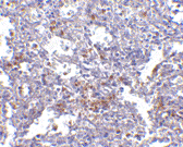
Below: Immunohistochemistry of EVER1 in human spleen with EVER1 antibody at 2.5 µg/ml.
Other Product Images
Source : EVER1 antibody was raised against a 14 amino acid peptide from near the amino terminus of human EVER1.
Purification : Affinity chromatography purified via peptide column
Clonality and Clone : This is a polyclonal antibody.
Host : EVER1 antibody was raised in rabbit. Please use anti-rabbit secondary antibodies.
Application : EVER1 antibody can be used for the detection of EVER1 by Western blot at 1 – 2 µg/ml.
Tested Application(s) : E, WB, IHC
Buffer : Antibody is supplied in PBS containing 0.02% sodium azide.
Blocking Peptide : Cat.No. 4547P - EVER1 Peptide
Long-Term Storage : EVER1 antibody can be stored at 4ºC, stable for one year. As with all antibodies care should be taken to avoid repeated freeze thaw cycles. Antibodies should not be exposed to prolonged high temperatures.
Positive Control
1. Cat. No. 1288 - A20 Cell Lysate
Species Reactivity :H, M, R
GI Number : 25527208
Accession Number : AAM44452
Short Description : (NT) Transmembrane protein channel-like protein 6
References
1. Majewski S, Jablonska J and Orth G. Epidermodysplasia verruciformis. Immunological and nonimmunological surveillance mechanisms: role in tumor progression. Clin. Dermatol. 1997; 15:321-34.
2. Ramoz N, Rueda L-A, Bouadjar B, et al. Mutations in two adjacent novel genes are associated with epidermodysplasia verruciformis. Nat. Genetics 2002; 32:579-81.
3. Keresztes G, Mutai H and Heller S. TMC and EVER genes belong to a larger novel family, the TMC gene family encoding transmembrane proteins. BMC Genomics 2003; 4:24-34.
4. Lazarczyk L, Pons C, Mendoza JA, et al. Regulation of cellular zinc balance as a potential mechanism of EVER-mediated protection against pathogenesis by cutaneous oncogenic human papillomaviruses. J. Exp. Med. 2008; 205:35-42.
Catalog# : 4547
Epidermodysplasia verruciformis (EV) is an autosomal recessive dermatosis characterized by abnormal susceptibility to human papillomaviruses (HPVs) and a high rate of progression to squamous cell carcinoma on sun-exposed skin. EV is caused by mutations in either of two adjacent genes, EVER1 and EVER2, located on chromosome 17q25.3. Both of these genes encode integral membrane proteins that localize to the endoplasmic reticulum and are predicted to form transmembrane channels. Both EVER1 and EVER2 are members of the transmembrane channel-like (TMC) protein family. EVER1 possesses eight trans-membrane domains and two leucine zipper motifs. EVER1 and EVER2 form a complex and interact with the zinc transporter 1 (ZnT-1), suggesting that EVER1 and EVER2 act to regulate cellular zinc balance. At least four isoforms of EVER1 are known to exist. This EVER1 antibody does not cross-react with EVER2.
Additional Names : EVER1 (NT), EV1, EVIN1, Transmembrane protein channel-like protein 6, TMC6
DescriptionLeft: Western blot analysis of EVER1 in A-20 cell lysate with EVER1 antibody at (A) 1 and (B) 2 µg/ml.
Below: Immunohistochemistry of EVER1 in human spleen with EVER1 antibody at 2.5 µg/ml.
Other Product Images
Source : EVER1 antibody was raised against a 14 amino acid peptide from near the amino terminus of human EVER1.
Purification : Affinity chromatography purified via peptide column
Clonality and Clone : This is a polyclonal antibody.
Host : EVER1 antibody was raised in rabbit. Please use anti-rabbit secondary antibodies.
Application : EVER1 antibody can be used for the detection of EVER1 by Western blot at 1 – 2 µg/ml.
Tested Application(s) : E, WB, IHC
Buffer : Antibody is supplied in PBS containing 0.02% sodium azide.
Blocking Peptide : Cat.No. 4547P - EVER1 Peptide
Long-Term Storage : EVER1 antibody can be stored at 4ºC, stable for one year. As with all antibodies care should be taken to avoid repeated freeze thaw cycles. Antibodies should not be exposed to prolonged high temperatures.
Positive Control
1. Cat. No. 1288 - A20 Cell Lysate
Species Reactivity :H, M, R
GI Number : 25527208
Accession Number : AAM44452
Short Description : (NT) Transmembrane protein channel-like protein 6
References
1. Majewski S, Jablonska J and Orth G. Epidermodysplasia verruciformis. Immunological and nonimmunological surveillance mechanisms: role in tumor progression. Clin. Dermatol. 1997; 15:321-34.
2. Ramoz N, Rueda L-A, Bouadjar B, et al. Mutations in two adjacent novel genes are associated with epidermodysplasia verruciformis. Nat. Genetics 2002; 32:579-81.
3. Keresztes G, Mutai H and Heller S. TMC and EVER genes belong to a larger novel family, the TMC gene family encoding transmembrane proteins. BMC Genomics 2003; 4:24-34.
4. Lazarczyk L, Pons C, Mendoza JA, et al. Regulation of cellular zinc balance as a potential mechanism of EVER-mediated protection against pathogenesis by cutaneous oncogenic human papillomaviruses. J. Exp. Med. 2008; 205:35-42.
CSN8 Antibody
CSN8 Antibody
Catalog# : 4601
The COP9 signalosome (CSN) is an evolutionarily conserved protein complex of the eight subunits that interacts with deubiquitinating enzymes and protein kinases and is highly homologous to the lid sub-complex of 26S proteasome. The CSN complex is an essential regulator of the ubiquitin conjugation pathway by mediating the deneddylation of the SCF-type E3 ligase complexes, which leads to a decrease in ubiquitin ligase activity of SCF-comlpexes such as SCF, CSA or DDB2. It is also involved in phosphorylation of p53, c-jun/JUN, ITPK1 and IRF8/ICSBP, possibly via its association with CK2 and PKD kinases. CSN8 encodes the smallest and the least conserved but first identified subunit of CSN. Recent studies show CSN8 is essential for Drosophila development and is essential for peripheral T cell homeostasis and antigen receptor-induced entry into the cell cycle from quiescence.
Additional Names : CSN8 (CT), COP9 signalosome complex subunit 8, SGN8, JAB1-containing signalosome subunit 8
 Description
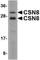
Left: Western blot analysis of CSN8 in Human liver lysate with CSN8 antibody at 2 µg/ml.
Source : CSN8 antibody was raised against a 15 amino acid peptide near the carboxy terminus of the human CSN8.
Purification : Affinity chromatography purified via peptide column
Clonality and Clone : This is a polyclonal antibody.
Host : CSN8 antibody was raised in rabbit. Please use anti-rabbit secondary antibodies.
Application : CSN8 antibody can be used for detection of CSN8 by Western blot at 1 - 2 µg/ml.
Tested Application(s) : E, WB
Buffer : Antibody is supplied in PBS containing 0.02% sodium azide.
Blocking Peptide : Cat.No. 4601P - CSN8 Peptide
Long-Term Storage : CSN8 antibody can be stored at 4ºC, stable for one year. As with all antibodies care should be taken to avoid repeated freeze thaw cycles. Antibodies should not be exposed to prolonged high temperatures.
Positive Control
1. Cat. No. 1304 - Human Liver Tissue Lysate
Species Reactivity :H, M, R
GI Number : 5729779
Accession Number : NP_006701
Short Description : (CT) COP9 signalosome complex subunit 8
References
1. Wei N and Deng XW. The COP9 signalosome. Annu. Rev. Cell Dev. Biol. 2003; 19:261-86.
2. Bech-Otschir D, Seeger M, and Dubiel W. The COP9 signalosome: at the interface between signal transduction and ubiquitin-dependent proteolysis. J. Cell Sci. 2002; 115:467-73.
3. Groisman R, Polanowska J, Kuraoka I, et al. The ubiquitin ligase activity in the DDB2 and regulated by the COP9 signalosome in response to DNA damage. Cell 2003; 113:357-67.
4. Uhle S, Medalia O, Waldron R, et al. Protein kinase CK2 and protein kinase D are associated with the COP9 signalosome. EMBO J. 2003; 22:1302-12.
Catalog# : 4601
The COP9 signalosome (CSN) is an evolutionarily conserved protein complex of the eight subunits that interacts with deubiquitinating enzymes and protein kinases and is highly homologous to the lid sub-complex of 26S proteasome. The CSN complex is an essential regulator of the ubiquitin conjugation pathway by mediating the deneddylation of the SCF-type E3 ligase complexes, which leads to a decrease in ubiquitin ligase activity of SCF-comlpexes such as SCF, CSA or DDB2. It is also involved in phosphorylation of p53, c-jun/JUN, ITPK1 and IRF8/ICSBP, possibly via its association with CK2 and PKD kinases. CSN8 encodes the smallest and the least conserved but first identified subunit of CSN. Recent studies show CSN8 is essential for Drosophila development and is essential for peripheral T cell homeostasis and antigen receptor-induced entry into the cell cycle from quiescence.
Additional Names : CSN8 (CT), COP9 signalosome complex subunit 8, SGN8, JAB1-containing signalosome subunit 8
DescriptionLeft: Western blot analysis of CSN8 in Human liver lysate with CSN8 antibody at 2 µg/ml.
Source : CSN8 antibody was raised against a 15 amino acid peptide near the carboxy terminus of the human CSN8.
Purification : Affinity chromatography purified via peptide column
Clonality and Clone : This is a polyclonal antibody.
Host : CSN8 antibody was raised in rabbit. Please use anti-rabbit secondary antibodies.
Application : CSN8 antibody can be used for detection of CSN8 by Western blot at 1 - 2 µg/ml.
Tested Application(s) : E, WB
Buffer : Antibody is supplied in PBS containing 0.02% sodium azide.
Blocking Peptide : Cat.No. 4601P - CSN8 Peptide
Long-Term Storage : CSN8 antibody can be stored at 4ºC, stable for one year. As with all antibodies care should be taken to avoid repeated freeze thaw cycles. Antibodies should not be exposed to prolonged high temperatures.
Positive Control
1. Cat. No. 1304 - Human Liver Tissue Lysate
Species Reactivity :H, M, R
GI Number : 5729779
Accession Number : NP_006701
Short Description : (CT) COP9 signalosome complex subunit 8
References
1. Wei N and Deng XW. The COP9 signalosome. Annu. Rev. Cell Dev. Biol. 2003; 19:261-86.
2. Bech-Otschir D, Seeger M, and Dubiel W. The COP9 signalosome: at the interface between signal transduction and ubiquitin-dependent proteolysis. J. Cell Sci. 2002; 115:467-73.
3. Groisman R, Polanowska J, Kuraoka I, et al. The ubiquitin ligase activity in the DDB2 and regulated by the COP9 signalosome in response to DNA damage. Cell 2003; 113:357-67.
4. Uhle S, Medalia O, Waldron R, et al. Protein kinase CK2 and protein kinase D are associated with the COP9 signalosome. EMBO J. 2003; 22:1302-12.
CPEB1 Antibody
CPEB1 Antibody
Catalog# : 4687
CPEB1 is an RNA binding protein that contains an RNA-recognition motif and a zinc finger-containing region found in a wide range of vertebrates and invertebrates. CPEB1 forms the nucleus of a complex of factors that regulate poly(A) elongation and promotes polyadenylation-induced translation. CPEB1 mediates many diverse biological processes such as germ cell development, cell division and senescence, and synaptic plasticity. Recently, it was discovered that CPEB1 is involved in beta-catenin mRNA translation and cell migration in astrocytes as well as regulating hypoxia-inducible factor (HIF)-1 expression, demonstrating the wide range of processes in which CPEB1 plays a role. At least four isoforms of CPEB1 are known to exist.
Additional Names : CPEB1 (NT), Cytoplasmic polyadenylation element-binding protein 1, CPE-B1, CEBP
 Description
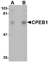
Left: Western blot analysis of CPEB1 in rat brain tissue lysate with CPEB1 antibody at (A) 1 and (B) 2 µg/ml.
Source : CPEB1 antibody was raised against a 14 amino acid peptide from near the amino terminus of human CPEB1.
Purification : Affinity chromatography purified via peptide column
Clonality and Clone : This is a polyclonal antibody.
Host : CPEB1 antibody was raised in rabbit. Please use anti-rabbit secondary antibodies.
Application : CPEB1 antibody can be used for detection of CPEB1 by Western blot at 1 – 2 µg/ml.
Tested Application(s) : E, WB
Buffer : Antibody is supplied in PBS containing 0.02% sodium azide.
Blocking Peptide : Cat.No. 4687P - CPEB1 Peptide
Long-Term Storage : CPEB1 antibody can be stored at 4ºC, stable for one year. As with all antibodies care should be taken to avoid repeated freeze thaw cycles. Antibodies should not be exposed to prolonged high temperatures.
Positive Control
1. Cat. No. 1463 - Rat Brain Tissue Lysate
Species Reactivity :H, M, R
GI Number : 74762720
Accession Number : Q9BZB8
Short Description : (NT) Cytoplasmic polyadenylation element-binding protein 1
References
1. Richter JD. CPEB: a life in translation. Trends in Biochem. Sci. 2007; 32:179-85.
2. Jones KJ, Korb E, Kundel MA, et al. CPEB1 regulates beta-catenin mRNA translation and cell migration in astrocytes. Glia 2008; 56:1401-13.
3. Hagele S, Kuhn U, Boning M, et al. Cytoplasmic polyadenylation element binding protein (CPEB)1 and 2 bind to the HIF-1alpha mRNA 3’UTR and modulate HIF-1 alpha protein expression. Biochem. J. 2008; epub.
Catalog# : 4687
CPEB1 is an RNA binding protein that contains an RNA-recognition motif and a zinc finger-containing region found in a wide range of vertebrates and invertebrates. CPEB1 forms the nucleus of a complex of factors that regulate poly(A) elongation and promotes polyadenylation-induced translation. CPEB1 mediates many diverse biological processes such as germ cell development, cell division and senescence, and synaptic plasticity. Recently, it was discovered that CPEB1 is involved in beta-catenin mRNA translation and cell migration in astrocytes as well as regulating hypoxia-inducible factor (HIF)-1 expression, demonstrating the wide range of processes in which CPEB1 plays a role. At least four isoforms of CPEB1 are known to exist.
Additional Names : CPEB1 (NT), Cytoplasmic polyadenylation element-binding protein 1, CPE-B1, CEBP
DescriptionLeft: Western blot analysis of CPEB1 in rat brain tissue lysate with CPEB1 antibody at (A) 1 and (B) 2 µg/ml.
Source : CPEB1 antibody was raised against a 14 amino acid peptide from near the amino terminus of human CPEB1.
Purification : Affinity chromatography purified via peptide column
Clonality and Clone : This is a polyclonal antibody.
Host : CPEB1 antibody was raised in rabbit. Please use anti-rabbit secondary antibodies.
Application : CPEB1 antibody can be used for detection of CPEB1 by Western blot at 1 – 2 µg/ml.
Tested Application(s) : E, WB
Buffer : Antibody is supplied in PBS containing 0.02% sodium azide.
Blocking Peptide : Cat.No. 4687P - CPEB1 Peptide
Long-Term Storage : CPEB1 antibody can be stored at 4ºC, stable for one year. As with all antibodies care should be taken to avoid repeated freeze thaw cycles. Antibodies should not be exposed to prolonged high temperatures.
Positive Control
1. Cat. No. 1463 - Rat Brain Tissue Lysate
Species Reactivity :H, M, R
GI Number : 74762720
Accession Number : Q9BZB8
Short Description : (NT) Cytoplasmic polyadenylation element-binding protein 1
References
1. Richter JD. CPEB: a life in translation. Trends in Biochem. Sci. 2007; 32:179-85.
2. Jones KJ, Korb E, Kundel MA, et al. CPEB1 regulates beta-catenin mRNA translation and cell migration in astrocytes. Glia 2008; 56:1401-13.
3. Hagele S, Kuhn U, Boning M, et al. Cytoplasmic polyadenylation element binding protein (CPEB)1 and 2 bind to the HIF-1alpha mRNA 3’UTR and modulate HIF-1 alpha protein expression. Biochem. J. 2008; epub.
DNA Double Helix
In geometry a double helix (plural helices) typically consists of two congruent helices with the same axis, differing by a translation along the axis, which may or may not be half-way.
Double Helix
In molecular biology, the double helix refers to the structure of DNA. The structure of DNA was first published in the journal Nature by James D. Watson and Francis Crick in 1953, based upon data from Maurice Wilkins and Rosalind Franklin. The DNA double helix is a right-handed spiral polymer of nucleic acids, held together by nucleotides which base pair together. A single turn of the helix constitutes ten nucleotides. The double helix structure of DNA contains a major groove and minor groove, the major groove being wider than the minor groove.
Nucleic Acid
A nucleic acid is a macromolecule composed of chains of monomeric nucleotides. In biochemistry these molecules carry genetic information or form structures within cells. The most common nucleic acids are deoxyribonucleic acid (DNA) and ribonucleic acid (RNA). The term "nucleic acid" is the generic name for a family of biopolymers, named for their role in the cell nucleus. The monomers from which nucleic acids are constructed are called nucleotides.Each nucleotide consists of three components: a nitrogenous heterocyclic base, which is either a purine or a pyrimidine; a pentose sugar; and a phosphate group.
Monday, October 25, 2010
CPEB1 Antibody
CPEB1 Antibody
Catalog# : 4687
CPEB1 is an RNA binding protein that contains an RNA-recognition motif and a zinc finger-containing region found in a wide range of vertebrates and invertebrates. CPEB1 forms the nucleus of a complex of factors that regulate poly(A) elongation and promotes polyadenylation-induced translation. CPEB1 mediates many diverse biological processes such as germ cell development, cell division and senescence, and synaptic plasticity. Recently, it was discovered that CPEB1 is involved in beta-catenin mRNA translation and cell migration in astrocytes as well as regulating hypoxia-inducible factor (HIF)-1 expression, demonstrating the wide range of processes in which CPEB1 plays a role. At least four isoforms of CPEB1 are known to exist.
Additional Names : CPEB1 (NT), Cytoplasmic polyadenylation element-binding protein 1, CPE-B1, CEBP
Description
Left: Western blot analysis of CPEB1 in rat brain tissue lysate with CPEB1 antibody at (A) 1 and (B) 2 µg/ml.
Source : CPEB1 antibody was raised against a 14 amino acid peptide from near the amino terminus of human CPEB1.
Purification : Affinity chromatography purified via peptide column
Clonality and Clone : This is a polyclonal antibody.
Host : CPEB1 antibody was raised in rabbit. Please use anti-rabbit secondary antibodies.
Application : CPEB1 antibody can be used for detection of CPEB1 by Western blot at 1 – 2 µg/ml.
Tested Application(s) : E, WB
Buffer : Antibody is supplied in PBS containing 0.02% sodium azide.
Blocking Peptide : Cat.No. 4687P - CPEB1 Peptide
Long-Term Storage : CPEB1 antibody can be stored at 4ºC, stable for one year. As with all antibodies care should be taken to avoid repeated freeze thaw cycles. Antibodies should not be exposed to prolonged high temperatures.
Positive Control
1. Cat. No. 1463 - Rat Brain Tissue Lysate
Species Reactivity :H, M, R
GI Number : 74762720
Accession Number : Q9BZB8
Short Description : (NT) Cytoplasmic polyadenylation element-binding protein 1
References
1. Richter JD. CPEB: a life in translation. Trends in Biochem. Sci. 2007; 32:179-85.
2. Jones KJ, Korb E, Kundel MA, et al. CPEB1 regulates beta-catenin mRNA translation and cell migration in astrocytes. Glia 2008; 56:1401-13.
3. Hagele S, Kuhn U, Boning M, et al. Cytoplasmic polyadenylation element binding protein (CPEB)1 and 2 bind to the HIF-1alpha mRNA 3’UTR and modulate HIF-1 alpha protein expression. Biochem. J. 2008; epub.
Catalog# : 4687
CPEB1 is an RNA binding protein that contains an RNA-recognition motif and a zinc finger-containing region found in a wide range of vertebrates and invertebrates. CPEB1 forms the nucleus of a complex of factors that regulate poly(A) elongation and promotes polyadenylation-induced translation. CPEB1 mediates many diverse biological processes such as germ cell development, cell division and senescence, and synaptic plasticity. Recently, it was discovered that CPEB1 is involved in beta-catenin mRNA translation and cell migration in astrocytes as well as regulating hypoxia-inducible factor (HIF)-1 expression, demonstrating the wide range of processes in which CPEB1 plays a role. At least four isoforms of CPEB1 are known to exist.
Additional Names : CPEB1 (NT), Cytoplasmic polyadenylation element-binding protein 1, CPE-B1, CEBP
DescriptionLeft: Western blot analysis of CPEB1 in rat brain tissue lysate with CPEB1 antibody at (A) 1 and (B) 2 µg/ml.
Source : CPEB1 antibody was raised against a 14 amino acid peptide from near the amino terminus of human CPEB1.
Purification : Affinity chromatography purified via peptide column
Clonality and Clone : This is a polyclonal antibody.
Host : CPEB1 antibody was raised in rabbit. Please use anti-rabbit secondary antibodies.
Application : CPEB1 antibody can be used for detection of CPEB1 by Western blot at 1 – 2 µg/ml.
Tested Application(s) : E, WB
Buffer : Antibody is supplied in PBS containing 0.02% sodium azide.
Blocking Peptide : Cat.No. 4687P - CPEB1 Peptide
Long-Term Storage : CPEB1 antibody can be stored at 4ºC, stable for one year. As with all antibodies care should be taken to avoid repeated freeze thaw cycles. Antibodies should not be exposed to prolonged high temperatures.
Positive Control
1. Cat. No. 1463 - Rat Brain Tissue Lysate
Species Reactivity :H, M, R
GI Number : 74762720
Accession Number : Q9BZB8
Short Description : (NT) Cytoplasmic polyadenylation element-binding protein 1
References
1. Richter JD. CPEB: a life in translation. Trends in Biochem. Sci. 2007; 32:179-85.
2. Jones KJ, Korb E, Kundel MA, et al. CPEB1 regulates beta-catenin mRNA translation and cell migration in astrocytes. Glia 2008; 56:1401-13.
3. Hagele S, Kuhn U, Boning M, et al. Cytoplasmic polyadenylation element binding protein (CPEB)1 and 2 bind to the HIF-1alpha mRNA 3’UTR and modulate HIF-1 alpha protein expression. Biochem. J. 2008; epub.
CLIP170 Antibody
CLIP170 Antibody
Catalog# : 4659
CLIP170 was initially identified as a new type of intermediate filament associated protein that is highly expressed in Reed-Sternberg cells, the tumoral cells diagnostic for Hodgkin’s disease. Later experiments showed that it is located at microtubule plus ends and is required for the binding of endocytic carrier vesicles. CLIP170 has also been suggested to act with LIS1, a protein implicated in brain development, to regulate dynein/dynactin binding microtubules. Other studies suggest that CLIP170 can influence the formation of lamellipodia and cell invasion by invasive breast cancer cells by regulating the release of kinesin and IQGAP1 from a complex of those proteins, CLIP170 and Rac1. At least two isoforms of CLIP170 are known to exist.
Additional Names : CLIP170, CAP-Gly domain-containing linker protein 1, CLIP, CYLN1, Restin, RSN
 Description
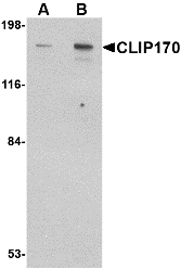
Left: Western blot analysis of CLIP170 in rat brain tissue lysate with CLIP170 antibody at (A) 0.5 and (B) 1 µg/ml.
Below: Immunofuorescence of CLIP170 in human brain tissue with CLIP170 antibody at 20 μg/ml.
Other Product Images
 Source : CLIP170 antibody was raised against a 17 amino acid peptide from near the carboxy terminus of human CLIP170.
Purification : Affinity chromatography purified via peptide column
Clonality and Clone : This is a polyclonal antibody.
Host : CLIP170 antibody was raised in rabbit. Please use anti-rabbit secondary antibodies.
Application : CLIP170 antibody can be used for detection of CLIP170 by Western blot at 0.5 – 1 µg/ml.
Tested Application(s) : E, WB
Buffer : Antibody is supplied in PBS containing 0.02% sodium azide.
Blocking Peptide : Cat.No. 4659P - CLIP170 Peptide
Long-Term Storage : CLIP170 antibody can be stored at 4ºC, stable for one year. As with all antibodies care should be taken to avoid repeated freeze thaw cycles. Antibodies should not be exposed to prolonged high temperatures.
Positive Control
1. Cat. No. 1463 - Rat Brain Tissue Lysate
Species Reactivity :H, M, R
GI Number : 4506751
Accession Number : NP_002947
Short Description : CAP-Gly domain-containing linker protein 1
References
1. Bilbe G, Delabie J, Bruggen J, et al. Restin: a novel intermediate filament-associated protein highly expressed in the Reed-Sternberg cells of Hodgkin’s disease. EMBO J. 1992; 11:2103-13.
2. Diamantopoulos GS, Perez F, Goodson HV, et al. Dynamic localization of CLIP-170 to microtubule plus ends is coupled to microtubule assembly. J. Cell Biol. 1999; 144:99-112.
3. Pierre P, Scheel J, Richard JE, et al. CLIP-170 links endocytic vesicles to microtubules. Cell 70:887-900.
4. Suzuki K and Takahashi K. Regulation of lamellipodia formation and cell invasion by CLIP-170 in invasive human breast cancer cells. Biochem. Biophys. Res. Commun. 2008; 368:199-204.
Catalog# : 4659
CLIP170 was initially identified as a new type of intermediate filament associated protein that is highly expressed in Reed-Sternberg cells, the tumoral cells diagnostic for Hodgkin’s disease. Later experiments showed that it is located at microtubule plus ends and is required for the binding of endocytic carrier vesicles. CLIP170 has also been suggested to act with LIS1, a protein implicated in brain development, to regulate dynein/dynactin binding microtubules. Other studies suggest that CLIP170 can influence the formation of lamellipodia and cell invasion by invasive breast cancer cells by regulating the release of kinesin and IQGAP1 from a complex of those proteins, CLIP170 and Rac1. At least two isoforms of CLIP170 are known to exist.
Additional Names : CLIP170, CAP-Gly domain-containing linker protein 1, CLIP, CYLN1, Restin, RSN
DescriptionLeft: Western blot analysis of CLIP170 in rat brain tissue lysate with CLIP170 antibody at (A) 0.5 and (B) 1 µg/ml.
Below: Immunofuorescence of CLIP170 in human brain tissue with CLIP170 antibody at 20 μg/ml.
Other Product Images
Source : CLIP170 antibody was raised against a 17 amino acid peptide from near the carboxy terminus of human CLIP170.Purification : Affinity chromatography purified via peptide column
Clonality and Clone : This is a polyclonal antibody.
Host : CLIP170 antibody was raised in rabbit. Please use anti-rabbit secondary antibodies.
Application : CLIP170 antibody can be used for detection of CLIP170 by Western blot at 0.5 – 1 µg/ml.
Tested Application(s) : E, WB
Buffer : Antibody is supplied in PBS containing 0.02% sodium azide.
Blocking Peptide : Cat.No. 4659P - CLIP170 Peptide
Long-Term Storage : CLIP170 antibody can be stored at 4ºC, stable for one year. As with all antibodies care should be taken to avoid repeated freeze thaw cycles. Antibodies should not be exposed to prolonged high temperatures.
Positive Control
1. Cat. No. 1463 - Rat Brain Tissue Lysate
Species Reactivity :H, M, R
GI Number : 4506751
Accession Number : NP_002947
Short Description : CAP-Gly domain-containing linker protein 1
References
1. Bilbe G, Delabie J, Bruggen J, et al. Restin: a novel intermediate filament-associated protein highly expressed in the Reed-Sternberg cells of Hodgkin’s disease. EMBO J. 1992; 11:2103-13.
2. Diamantopoulos GS, Perez F, Goodson HV, et al. Dynamic localization of CLIP-170 to microtubule plus ends is coupled to microtubule assembly. J. Cell Biol. 1999; 144:99-112.
3. Pierre P, Scheel J, Richard JE, et al. CLIP-170 links endocytic vesicles to microtubules. Cell 70:887-900.
4. Suzuki K and Takahashi K. Regulation of lamellipodia formation and cell invasion by CLIP-170 in invasive human breast cancer cells. Biochem. Biophys. Res. Commun. 2008; 368:199-204.
CIP75 Antibody
CIP75 Antibody
Catalog# : 5267
The ubiquitin-proteosome pathway of protein degradation is one of the mechanisms that ensure the proper level of cellular proteins. The ubiquitin-like (UbL) and ubiquitin-associated (UBA) domain containing protein family is thought to be involved in proteosomal degradation. One such protein is CIP75, also known as ubiquilin-4, interacts with a number of proteins such as Ataxin-1 and Connexin-43, resulting in an increased rate of turnover of these proteins. Overexpression CIP75 led to a significant reduction of Connexin-43 half-life, with the opposite being observed in siRNA knockdown experiments. CIP75 contains an N-terminal UbL domain which is thought to interact with proteins of the 26S proteosome complex and a C-terminal UBA domain which appears to mediate its interaction with Connexin-75. At least three isoforms of CIP75 are known to exist.
Additional Names : CIP75, Connexin-43-interatcting protein 75, ubiquilin-4, UBQLN4, Ataxin-1 ubiquitin-like-interacting protein, A1U, UBIN
 Description
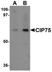
Left: Western blot analysis of CIP75 in 3T3 cell lysate with CIP75 antibody at (A) 1 and (B) 2 µg/ml.
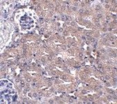
Below: Immunohistochemistry of CIP75 in mouse kidney tissue with CIP75 antibody at 5 μg/ml.
Other Product Images
 Source : CIP75 antibody was raised against an 18 amino acid peptide near the amino terminus of human CIP75.
Purification : Affinity chromatography purified via peptide column
Clonality and Clone : Polyclonal
Host : CIP75 antibody was raised in rabbit. Please use anti-rabbit secondary antibodies.
Application : CIP75 antibody can be used for detection of CIP75 by Western blot at 1 - 2 µg/ml.
Tested Application(s) : E, WB
Buffer : Antibody is supplied in PBS containing 0.02% sodium azide.
Blocking Peptide : Cat.No. 5267P - CIP75 Peptide
Long-Term Storage : CIP75 antibody can be stored at 4ºC, stable for one year. As with all antibodies care should be taken to avoid repeated freeze thaw cycles. Antibodies should not be exposed to prolonged high temperatures.
Positive Control
1. Cat. No. 1282 - 3T3 (NIH) Cell Lysate
Species Reactivity :H, M, R
GI Number : 45476982
Accession Number : Q9NRR5
Short Description : Connexin-43-interatcting protein 75
References
1. Su V and Lau AF. Ubiquitin-like and ubiquitin-associated domain proteins: significance in proteasomal degradation. Cell. Mol. Life Sci. 2009; 66:2819-33.
2. Davidson JD, Riley B, Burright EN, et al. Identification and characterization of an ataxin-1-interacting protein: A1Up, a ubiquitin-like protein. Hum. Mol. Genet. 2000; 9:2305-12.
3. Li X, Su V, Kurata WE, et al. A novel Connexin43-interacting protein, CIP75, which belongs to the UbL-UBA protein family, regulates the turnover of Connexin4 J. Biol. Chem. 2008; 283:5748-59.
Catalog# : 5267
The ubiquitin-proteosome pathway of protein degradation is one of the mechanisms that ensure the proper level of cellular proteins. The ubiquitin-like (UbL) and ubiquitin-associated (UBA) domain containing protein family is thought to be involved in proteosomal degradation. One such protein is CIP75, also known as ubiquilin-4, interacts with a number of proteins such as Ataxin-1 and Connexin-43, resulting in an increased rate of turnover of these proteins. Overexpression CIP75 led to a significant reduction of Connexin-43 half-life, with the opposite being observed in siRNA knockdown experiments. CIP75 contains an N-terminal UbL domain which is thought to interact with proteins of the 26S proteosome complex and a C-terminal UBA domain which appears to mediate its interaction with Connexin-75. At least three isoforms of CIP75 are known to exist.
Additional Names : CIP75, Connexin-43-interatcting protein 75, ubiquilin-4, UBQLN4, Ataxin-1 ubiquitin-like-interacting protein, A1U, UBIN
DescriptionLeft: Western blot analysis of CIP75 in 3T3 cell lysate with CIP75 antibody at (A) 1 and (B) 2 µg/ml.
Below: Immunohistochemistry of CIP75 in mouse kidney tissue with CIP75 antibody at 5 μg/ml.
Other Product Images
Source : CIP75 antibody was raised against an 18 amino acid peptide near the amino terminus of human CIP75.Purification : Affinity chromatography purified via peptide column
Clonality and Clone : Polyclonal
Host : CIP75 antibody was raised in rabbit. Please use anti-rabbit secondary antibodies.
Application : CIP75 antibody can be used for detection of CIP75 by Western blot at 1 - 2 µg/ml.
Tested Application(s) : E, WB
Buffer : Antibody is supplied in PBS containing 0.02% sodium azide.
Blocking Peptide : Cat.No. 5267P - CIP75 Peptide
Long-Term Storage : CIP75 antibody can be stored at 4ºC, stable for one year. As with all antibodies care should be taken to avoid repeated freeze thaw cycles. Antibodies should not be exposed to prolonged high temperatures.
Positive Control
1. Cat. No. 1282 - 3T3 (NIH) Cell Lysate
Species Reactivity :H, M, R
GI Number : 45476982
Accession Number : Q9NRR5
Short Description : Connexin-43-interatcting protein 75
References
1. Su V and Lau AF. Ubiquitin-like and ubiquitin-associated domain proteins: significance in proteasomal degradation. Cell. Mol. Life Sci. 2009; 66:2819-33.
2. Davidson JD, Riley B, Burright EN, et al. Identification and characterization of an ataxin-1-interacting protein: A1Up, a ubiquitin-like protein. Hum. Mol. Genet. 2000; 9:2305-12.
3. Li X, Su V, Kurata WE, et al. A novel Connexin43-interacting protein, CIP75, which belongs to the UbL-UBA protein family, regulates the turnover of Connexin4 J. Biol. Chem. 2008; 283:5748-59.
CDC42 Antibody
CDC42 Antibody
Catalog# : 5101
CDC42 is a small GTPase of the Rho-subfamily, which regulates signaling pathways that control diverse cellular functions including cell morphology, migration, endocytosis and cell cycle progression. This protein is highly similar to S. cerevisiae CDC42, and is able to complement the yeast cdc42-1 mutant. The product of oncogene Dbl was reported to specifically catalyze the dissociation of GDP from this protein. CDC42 is thought to regulate actin polymerization through its direct binding to Neural Wiskott-Aldrich syndrome protein (N-WASP), which subsequently activates Arp2/3 complex. At least two isoforms of CDC42 are known to exist.
Additional Names : CDC42, Cell division cycle 42, G25K
 Description
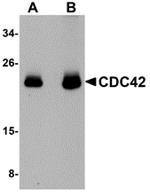
Left: Western blot analysis of CDC42 in human brain tissue lysate with CDC42 antibody at (A) 0.5 and (B) 1 µg/ml.
Source : CDC42 antibody was raised against a 17 amino acid peptide near the amino terminus of human CDC42.
Purification : Affinity chromatography purified via peptide column
Clonality and Clone : This is a polyclonal antibody.
Host : CDC42 antibody was raised in chicken. Please use anti-chicken secondary antibodies.
Application : CDC42 antibody can be used for detection of CDC42 by Western blot at 1 - 2 µg/ml.
Tested Application(s) : E, WB
Buffer : Antibody is supplied in PBS containing 0.02% sodium azide.
Blocking Peptide : Cat.No. 5101P - CDC42 Peptide
Long-Term Storage : CDC42 antibody can be stored at 4ºC, stable for one year. As with all antibodies care should be taken to avoid repeated freeze thaw cycles. Antibodies should not be exposed to prolonged high temperatures.
Positive Control
1. Cat. No. 1303 - Human Brain Tissue Lysate
Species Reactivity :H, M, R
GI Number : 4757952
Accession Number : NP_001782
Short Description : Cell division cycle 42
References
1. Erickson JW and Cerione RA. Multiple roles for Cdc42 in cell regulation. Curr. Opin. Cell Biol. 2001; 13:153-7.
2. Chen W, Lim HH, and Lim L. The CDC42 homologue from Caenorhabditis elegans. Complementation of yeast mutation. J. Biol. Chem. 1993; 268:13280-5.
3. Hart MJ, Eva A, Evans T, et al. Catalysis of guanine nucleotide exchange on the CDC42Hs protein by the dbl oncogene product. Nature 1991; 354:311-4.
4. Rohatgi R, Ma L, Miki H, et al. The interaction between N-WASP and the Arp2/3 complex links Cdc42-dependent signals to actin assembly. Cell 1999; 97:221-31.
Catalog# : 5101
CDC42 is a small GTPase of the Rho-subfamily, which regulates signaling pathways that control diverse cellular functions including cell morphology, migration, endocytosis and cell cycle progression. This protein is highly similar to S. cerevisiae CDC42, and is able to complement the yeast cdc42-1 mutant. The product of oncogene Dbl was reported to specifically catalyze the dissociation of GDP from this protein. CDC42 is thought to regulate actin polymerization through its direct binding to Neural Wiskott-Aldrich syndrome protein (N-WASP), which subsequently activates Arp2/3 complex. At least two isoforms of CDC42 are known to exist.
Additional Names : CDC42, Cell division cycle 42, G25K
DescriptionLeft: Western blot analysis of CDC42 in human brain tissue lysate with CDC42 antibody at (A) 0.5 and (B) 1 µg/ml.
Source : CDC42 antibody was raised against a 17 amino acid peptide near the amino terminus of human CDC42.
Purification : Affinity chromatography purified via peptide column
Clonality and Clone : This is a polyclonal antibody.
Host : CDC42 antibody was raised in chicken. Please use anti-chicken secondary antibodies.
Application : CDC42 antibody can be used for detection of CDC42 by Western blot at 1 - 2 µg/ml.
Tested Application(s) : E, WB
Buffer : Antibody is supplied in PBS containing 0.02% sodium azide.
Blocking Peptide : Cat.No. 5101P - CDC42 Peptide
Long-Term Storage : CDC42 antibody can be stored at 4ºC, stable for one year. As with all antibodies care should be taken to avoid repeated freeze thaw cycles. Antibodies should not be exposed to prolonged high temperatures.
Positive Control
1. Cat. No. 1303 - Human Brain Tissue Lysate
Species Reactivity :H, M, R
GI Number : 4757952
Accession Number : NP_001782
Short Description : Cell division cycle 42
References
1. Erickson JW and Cerione RA. Multiple roles for Cdc42 in cell regulation. Curr. Opin. Cell Biol. 2001; 13:153-7.
2. Chen W, Lim HH, and Lim L. The CDC42 homologue from Caenorhabditis elegans. Complementation of yeast mutation. J. Biol. Chem. 1993; 268:13280-5.
3. Hart MJ, Eva A, Evans T, et al. Catalysis of guanine nucleotide exchange on the CDC42Hs protein by the dbl oncogene product. Nature 1991; 354:311-4.
4. Rohatgi R, Ma L, Miki H, et al. The interaction between N-WASP and the Arp2/3 complex links Cdc42-dependent signals to actin assembly. Cell 1999; 97:221-31.
Peptide Interactions
Peptides are short polymers formed from the linking, in a defined order, of α-amino acids. The link between one amino acid residue and the next is known as an amide bond or a peptide bond. Proteins are polypeptide molecules (or consist of multiple polypeptide subunits). The distinction is that peptides are short and polypeptides/proteins are long. There are several different conventions to determine these, all of which have flaws.
Ribosomal Peptides
These are synthesized by translation of mRNA. They are often subjected to proteolysis to generate the mature form. These function, typically in higher organisms, as hormones and signaling molecules. Some lower organisms produce peptides as antibiotics, such as microcins.
Non-ribosomal Peptides
These peptides are assembled by enzymes that are specific to each peptide, rather than by the ribosome. The most common non-ribosomal peptide is glutathione, which is a component of the antioxidant defences of most aerobic organisms. Other nonribosomal peptides are most common in unicellular organisms, plants, and fungi and are synthesized by modular enzyme complexes called nonribosomal peptide synthesises.
Sunday, October 24, 2010
CCDC98 Antibody
CCDC98 Antibody
Catalog# : 4321
CCDC98, also known as Abraxas 1, was identified through protein binding studies using the breast and ovarian predisposition protein BRCA1 as the binding target. CCDC98 recruits RAP80, a ubiquitin-binding protein, to BRCA1, allowing the formation of BRCA1 foci in response to DNA damage caused by ionizing radiation. Both CCDC98 and RAP80 are required for DNA damage resistance, G2-M checkpoint control, and DNA repair. Cells depleted of either CCDC98 or RAP80 exhibited increased sensitivity to ionizing radiation, although not as much as in BRCA1-depleted cells, suggesting that CCDC98 and RAP80 control only part of the DNA damage response role of BRCA1. At least two isoforms of CCDC98 are known to exist.
Additional Names : CCDC98 (NT), Coiled-coil domain 98, Abraxas 1, ABRA1
 Description
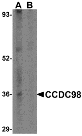
Left: Western blot analysis of CCDC98 in human breast tissue lysate in (A) the absence and (B) presence of blocking peptide with CCDC98 antibody at 1 µg/ml.
Below: Immunohistochemistry of CCDC98 in human breast tissue with CCDC98 antibody at 5 µg/ml.
Other Product Images
 Source : CCDC98 antibody was raised against a 15 amino acid peptide from near the amino terminus of human CCDC98.
Purification : Affinity chromatography purified via peptide column
Clonality and Clone : This is a polyclonal antibody.
Host : CCDC98 antibody was raised in rabbit. Please use anti-rabbit secondary antibodies.
Application : CCDC98 antibody can be used for detection of CCDC98 by Western blot at 1 – 2 µg/ml.
Tested Application(s) : E, WB, IHC
Buffer : Antibody is supplied in PBS containing 0.02% sodium azide.
Blocking Peptide : Cat.No. 4321P - CCDC98 Peptide
Long-Term Storage : CCDC98 antibody can be stored at 4ºC, stable for one year. As with all antibodies care should be taken to avoid repeated freeze thaw cycles. Antibodies should not be exposed to prolonged high temperatures.
Positive Control
1. Cat. No. 1311 - Human Breast Tissue Lysate
Species Reactivity :H, M
GI Number : 109148531
Accession Number : NP_620775
Short Description : (NT) coiled-coil domain 98
References
1. Wang B, Matsuoka S, Balliff BA, et al. Abraxas and RAP80 form a BRCA1 protein complex required for the DNA damage response. Science 2007; 316:1194-1198.
2. Kim H, Huang J, and Chen J. CCDC98 is a BRCA1-BRCT domain-binding protein involved in the DNA damage response. Nat. Struct. Mol. Biol. 2007; 14:710-5.
3. Liu Z, Wu J, and Yu X. CCDC98 targets BRCA1 to DNA damage sites. Nat. Struct. Mol. Biol. 2007; 14:716-20.
Catalog# : 4321
CCDC98, also known as Abraxas 1, was identified through protein binding studies using the breast and ovarian predisposition protein BRCA1 as the binding target. CCDC98 recruits RAP80, a ubiquitin-binding protein, to BRCA1, allowing the formation of BRCA1 foci in response to DNA damage caused by ionizing radiation. Both CCDC98 and RAP80 are required for DNA damage resistance, G2-M checkpoint control, and DNA repair. Cells depleted of either CCDC98 or RAP80 exhibited increased sensitivity to ionizing radiation, although not as much as in BRCA1-depleted cells, suggesting that CCDC98 and RAP80 control only part of the DNA damage response role of BRCA1. At least two isoforms of CCDC98 are known to exist.
Additional Names : CCDC98 (NT), Coiled-coil domain 98, Abraxas 1, ABRA1
DescriptionLeft: Western blot analysis of CCDC98 in human breast tissue lysate in (A) the absence and (B) presence of blocking peptide with CCDC98 antibody at 1 µg/ml.
Below: Immunohistochemistry of CCDC98 in human breast tissue with CCDC98 antibody at 5 µg/ml.
Other Product Images
Source : CCDC98 antibody was raised against a 15 amino acid peptide from near the amino terminus of human CCDC98.Purification : Affinity chromatography purified via peptide column
Clonality and Clone : This is a polyclonal antibody.
Host : CCDC98 antibody was raised in rabbit. Please use anti-rabbit secondary antibodies.
Application : CCDC98 antibody can be used for detection of CCDC98 by Western blot at 1 – 2 µg/ml.
Tested Application(s) : E, WB, IHC
Buffer : Antibody is supplied in PBS containing 0.02% sodium azide.
Blocking Peptide : Cat.No. 4321P - CCDC98 Peptide
Long-Term Storage : CCDC98 antibody can be stored at 4ºC, stable for one year. As with all antibodies care should be taken to avoid repeated freeze thaw cycles. Antibodies should not be exposed to prolonged high temperatures.
Positive Control
1. Cat. No. 1311 - Human Breast Tissue Lysate
Species Reactivity :H, M
GI Number : 109148531
Accession Number : NP_620775
Short Description : (NT) coiled-coil domain 98
References
1. Wang B, Matsuoka S, Balliff BA, et al. Abraxas and RAP80 form a BRCA1 protein complex required for the DNA damage response. Science 2007; 316:1194-1198.
2. Kim H, Huang J, and Chen J. CCDC98 is a BRCA1-BRCT domain-binding protein involved in the DNA damage response. Nat. Struct. Mol. Biol. 2007; 14:710-5.
3. Liu Z, Wu J, and Yu X. CCDC98 targets BRCA1 to DNA damage sites. Nat. Struct. Mol. Biol. 2007; 14:716-20.
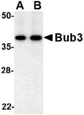
Bub3 Antibody
Bub3 Antibody
Catalog# : 4227
The mitotic checkpoint protein Bub3 is involved with the essential spindle checkpoint pathway which operates during early embryogenesis. Bub3 is important during G2 and early mitosis stages, permitting entry into mitosis depending upon the assembly state of microtubules, thus preventing premature sister chromatid separation, mis-segregation and aneuploidy. Bub3 contains four WD repeat domains and is required for the kinetochore localization of Bub1, a related kinase that is necessary for spindle assembly checkpoint function. Bub1 is able to autophosphorylate and can catalyze the phosphorylation of Bub3. Both Bub1 and Bub3 are mutually dependent for function. Altered Bub expression levels may significantly impair mitotic checkpoint function and is associated with tumor cell proliferation.
Additional Names : Bub3 (CT), Budding uninhibited by benzimidazoles 3, Bub3L
 Description
Left: Western blot analysis of bub3 in Jurkat cell lysate with bub3 antibody at (A) 0.5 and (B) 1 µg/ml.
Below: Immunocytochemistry of Bub3 in Jurkat cells with Bub3 antibody at 10 µg/ml.
Other Product Images
 Source : Bub3 antibody was raised against a 16 amino acid peptide from near the carboxy terminus of human bub3.
Purification : Affinity chromatography purified via peptide column
Clonality and Clone : This is a polyclonal antibody.
Host : Bub3 antibody was raised in rabbit. Please use anti-rabbit secondary antibodies.
Application : Bub3 antibody can be used for detection of bub3 by Western blot at 0.5 µg/ml.
Tested Application(s) : E, WB, ICC
Buffer : Antibody is supplied in PBS containing 0.02% sodium azide.
Blocking Peptide : Cat.No. 4227P - Bub3 Peptide
Long-Term Storage : Bub3 antibody can be stored at 4ºC, stable for one year. As with all antibodies care should be taken to avoid repeated freeze thaw cycles. Antibodies should not be exposed to prolonged high temperatures.
Positive Control
1. Cat. No. 1205 - Jurkat Cell Lysate
Species Reactivity :H, M
GI Number : 7387554
Accession Number : O43684
Short Description : (NT) a kinase involved in spindle formation
References
1. Kalitsis P, Earle E, Fowler KJ, et al. Bub3 gene disruption in mice reveals essential mitotic spindle checkpoint function during early embryogenesis. Genes Dev. 2000; 14:2277-82.
2. Taylor SS, Ha E and McKeon F. The human homologue of Bub3 is required for kinetochore localization of Bub1 and a Mad3/Bub1-related protein kinase. J. Cell Biol. 1998; 142:1-11.
3. Warren CD, Brady DM, Johnston RC, et al. Distinct chromosome segregation roles for spindle checkpoint proteins. Mol. Biol. Cell 2002; 13: 3029-41.
4. Roberts BT, Farr KA and Hoyt MA. The Saccharomyces cerevisiae checkpoint gene BUB1 encodes a novel protein kinase. Mol. Cell. Biol. 1994; 14:8282-91.
Catalog# : 4227
The mitotic checkpoint protein Bub3 is involved with the essential spindle checkpoint pathway which operates during early embryogenesis. Bub3 is important during G2 and early mitosis stages, permitting entry into mitosis depending upon the assembly state of microtubules, thus preventing premature sister chromatid separation, mis-segregation and aneuploidy. Bub3 contains four WD repeat domains and is required for the kinetochore localization of Bub1, a related kinase that is necessary for spindle assembly checkpoint function. Bub1 is able to autophosphorylate and can catalyze the phosphorylation of Bub3. Both Bub1 and Bub3 are mutually dependent for function. Altered Bub expression levels may significantly impair mitotic checkpoint function and is associated with tumor cell proliferation.
Additional Names : Bub3 (CT), Budding uninhibited by benzimidazoles 3, Bub3L
DescriptionLeft: Western blot analysis of bub3 in Jurkat cell lysate with bub3 antibody at (A) 0.5 and (B) 1 µg/ml.
Below: Immunocytochemistry of Bub3 in Jurkat cells with Bub3 antibody at 10 µg/ml.
Other Product Images
Source : Bub3 antibody was raised against a 16 amino acid peptide from near the carboxy terminus of human bub3.Purification : Affinity chromatography purified via peptide column
Clonality and Clone : This is a polyclonal antibody.
Host : Bub3 antibody was raised in rabbit. Please use anti-rabbit secondary antibodies.
Application : Bub3 antibody can be used for detection of bub3 by Western blot at 0.5 µg/ml.
Tested Application(s) : E, WB, ICC
Buffer : Antibody is supplied in PBS containing 0.02% sodium azide.
Blocking Peptide : Cat.No. 4227P - Bub3 Peptide
Long-Term Storage : Bub3 antibody can be stored at 4ºC, stable for one year. As with all antibodies care should be taken to avoid repeated freeze thaw cycles. Antibodies should not be exposed to prolonged high temperatures.
Positive Control
1. Cat. No. 1205 - Jurkat Cell Lysate
Species Reactivity :H, M
GI Number : 7387554
Accession Number : O43684
Short Description : (NT) a kinase involved in spindle formation
References
1. Kalitsis P, Earle E, Fowler KJ, et al. Bub3 gene disruption in mice reveals essential mitotic spindle checkpoint function during early embryogenesis. Genes Dev. 2000; 14:2277-82.
2. Taylor SS, Ha E and McKeon F. The human homologue of Bub3 is required for kinetochore localization of Bub1 and a Mad3/Bub1-related protein kinase. J. Cell Biol. 1998; 142:1-11.
3. Warren CD, Brady DM, Johnston RC, et al. Distinct chromosome segregation roles for spindle checkpoint proteins. Mol. Biol. Cell 2002; 13: 3029-41.
4. Roberts BT, Farr KA and Hoyt MA. The Saccharomyces cerevisiae checkpoint gene BUB1 encodes a novel protein kinase. Mol. Cell. Biol. 1994; 14:8282-91.
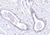
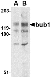
Bub1 Antibody
Bub1 Antibody
Catalog# : 4229
Bub1 was initially identified as the mammalian homolog to the yeast protein through a cDNA screen. Bub1 encodes a kinase involved in the formation of the spindle checkpoint, an important mechanism that ensures high fidelity mitotic chromosome segregation. It is thought to be required for assembly of a functional inner centromere, sister chromatid cohesion via targeting of the Shugoshim protein and metaphase congression. Bub1 functions by phosphorylating cdc20, a member of the mitotic checkpoint complex and activating the spindle checkpoint. A related protein kinase Bub3 interacts with Bub1 and targets it to kinetochores prior to chromosome alignment. Mutations in bub1 have been associated with aneuploidy and several forms of cancer.
Additional Names : Bub1 (NT), Budding uninhibited by benzimidazoles 1
 Description
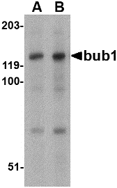
Left: Western blot analysis of bub1 in A-20 lysate with bub1 antibody at (A) 2 and (B) 4 µg/ml.
Below: Immunocytochemistry of Bub1 in A20 cells with Bub1 antibody at 10 µg/ml.
Other Product Images
 Source : Bub1 antibody was raised against a 19 amino acid peptide from near the amino terminus of human Bub1.
Purification : Affinity chromatography purified via peptide column
Clonality and Clone : This is a polyclonal antibody.
Host : Bub1 antibody was raised in rabbit. Please use anti-rabbit secondary antibodies.
Application : Bub1 antibody can be used for detection of bub1 by Western blot at 2 – 4 µg/ml.
Tested Application(s) : E, WB, ICC
Buffer : Antibody is supplied in PBS containing 0.02% sodium azide.
Blocking Peptide : Cat.No. 4229P - Bub1 Peptide
Long-Term Storage : Bub1 antibody can be stored at 4ºC, stable for one year. As with all antibodies care should be taken to avoid repeated freeze thaw cycles. Antibodies should not be exposed to prolonged high temperatures.
Positive Control
1. Cat. No. 1288 - A-20 Cell Lysate
Species Reactivity :H, M
GI Number : 8134347
Accession Number : O43683
Short Description : (NT) a kinase involved in spindle formation
References
1. Pangilinan F, Li Q, Weaver T, et al. Mammalian BUB1 protein kinases: map positions and in vivo expression. Genomics 1997; 46:379-88.
2. Williams GL, Roberts TM and Gjoerup OV. Bub1: Escapades in a cellular world. Cell Cycle 2007; 6:1699-704.
3. Meraldi P and Sorger PK. A dual role for Bub1 in the spindle checkpoint and chromosome congression. EMBO J. 2005; 24:1621-33.
4. Tang Z, Shu H, Oncel D, et al. Phosphorylation of cdc20 by bub1 provides a catalytic mechanism for APC/C inhibition by the spindle checkpoint. Mol. Cell 2004; 16:387-97.
Catalog# : 4229
Bub1 was initially identified as the mammalian homolog to the yeast protein through a cDNA screen. Bub1 encodes a kinase involved in the formation of the spindle checkpoint, an important mechanism that ensures high fidelity mitotic chromosome segregation. It is thought to be required for assembly of a functional inner centromere, sister chromatid cohesion via targeting of the Shugoshim protein and metaphase congression. Bub1 functions by phosphorylating cdc20, a member of the mitotic checkpoint complex and activating the spindle checkpoint. A related protein kinase Bub3 interacts with Bub1 and targets it to kinetochores prior to chromosome alignment. Mutations in bub1 have been associated with aneuploidy and several forms of cancer.
Additional Names : Bub1 (NT), Budding uninhibited by benzimidazoles 1
DescriptionLeft: Western blot analysis of bub1 in A-20 lysate with bub1 antibody at (A) 2 and (B) 4 µg/ml.
Below: Immunocytochemistry of Bub1 in A20 cells with Bub1 antibody at 10 µg/ml.
Other Product Images
Source : Bub1 antibody was raised against a 19 amino acid peptide from near the amino terminus of human Bub1.Purification : Affinity chromatography purified via peptide column
Clonality and Clone : This is a polyclonal antibody.
Host : Bub1 antibody was raised in rabbit. Please use anti-rabbit secondary antibodies.
Application : Bub1 antibody can be used for detection of bub1 by Western blot at 2 – 4 µg/ml.
Tested Application(s) : E, WB, ICC
Buffer : Antibody is supplied in PBS containing 0.02% sodium azide.
Blocking Peptide : Cat.No. 4229P - Bub1 Peptide
Long-Term Storage : Bub1 antibody can be stored at 4ºC, stable for one year. As with all antibodies care should be taken to avoid repeated freeze thaw cycles. Antibodies should not be exposed to prolonged high temperatures.
Positive Control
1. Cat. No. 1288 - A-20 Cell Lysate
Species Reactivity :H, M
GI Number : 8134347
Accession Number : O43683
Short Description : (NT) a kinase involved in spindle formation
References
1. Pangilinan F, Li Q, Weaver T, et al. Mammalian BUB1 protein kinases: map positions and in vivo expression. Genomics 1997; 46:379-88.
2. Williams GL, Roberts TM and Gjoerup OV. Bub1: Escapades in a cellular world. Cell Cycle 2007; 6:1699-704.
3. Meraldi P and Sorger PK. A dual role for Bub1 in the spindle checkpoint and chromosome congression. EMBO J. 2005; 24:1621-33.
4. Tang Z, Shu H, Oncel D, et al. Phosphorylation of cdc20 by bub1 provides a catalytic mechanism for APC/C inhibition by the spindle checkpoint. Mol. Cell 2004; 16:387-97.
Bub1 Antibody
Bub1 Antibody
Catalog# : 4239
Bub1 was initially identified as the mammalian homolog to the yeast protein through a cDNA screen. Bub1 encodes a kinase involved in the formation of the spindle checkpoint, an important mechanism that ensures high fidelity mitotic chromosome segregation. It is thought to be required for assembly of a functional inner centromere, sister chromatid cohesion via targeting of the Shugoshim protein and metaphase congression. Bub1 functions by phosphorylating cdc20, a member of the mitotic checkpoint complex and activating the spindle checkpoint. A related protein kinase Bub3 interacts with Bub1 and targets it to kinetochores prior to chromosome alignment. Mutations in bub1 have been associated with aneuploidy and several forms of cancer.
Additional Names : Bub1 (CT), Budding uninhibited by benzimidazoles 1
 Description
Left: Western blot analysis of bub1 in A-20 cell lysate with bub1 antibody at (A) 1 and (B) 2 µg/ml. Below: Immunocytochemistry of Bub1 in A20 cells with Bub1 antibody at 5 µg/ml.
Source : Bub1 antibody was raised against a 22 amino acid peptide from near the carboxy terminus of human Bub1.
Purification : Affinity chromatography purified via peptide column
Clonality and Clone : This is a polyclonal antibody.
Host : Bub1 antibody was raised in rabbit.
Application : Bub1 antibody can be used for detection of bub1 by Western blot at 1 – 2 µg/ml.
Tested Application(s) : E, WB, ICC
Buffer : Antibody is supplied in PBS containing 0.02% sodium azide.
Blocking Peptide : Cat.No. 4239P - Bub1 Peptide
Long-Term Storage : Bub1 antibody can be stored at 4ºC, stable for one year. As with all antibodies care should be taken to avoid repeated freeze thaw cycles. Antibodies should not be exposed to prolonged high temperatures.
Positive Control
1. Cat. No. 1288 - A20 Cell Lysate
Species Reactivity :H R
GI Number : 8134347
Accession Number : O43683
Short Description : (CT) a kinase involved in spindle formation
References
1. Pangilinan F, Li Q, Weaver T, et al. Mammalian BUB1 protein kinases: map positions and in vivo expression. Genomics 1997; 46:379-88.
2. Williams GL, Roberts TM and Gjoerup OV. Bub1: Escapades in a cellular world. Cell Cycle 2007; 6:1699-704.
3. Meraldi P and Sorger PK. A dual role for Bub1 in the spindle checkpoint and chromosome congression. EMBO J. 2005; 24:1621-33.
4. Tang Z, Shu H, Oncel D, et al. Phosphorylation of cdc20 by bub1 provides a catalytic mechanism for APC/C inhibition by the spindle checkpoint. Mol. Cell 2004; 16:387-97.
Catalog# : 4239
Bub1 was initially identified as the mammalian homolog to the yeast protein through a cDNA screen. Bub1 encodes a kinase involved in the formation of the spindle checkpoint, an important mechanism that ensures high fidelity mitotic chromosome segregation. It is thought to be required for assembly of a functional inner centromere, sister chromatid cohesion via targeting of the Shugoshim protein and metaphase congression. Bub1 functions by phosphorylating cdc20, a member of the mitotic checkpoint complex and activating the spindle checkpoint. A related protein kinase Bub3 interacts with Bub1 and targets it to kinetochores prior to chromosome alignment. Mutations in bub1 have been associated with aneuploidy and several forms of cancer.
Additional Names : Bub1 (CT), Budding uninhibited by benzimidazoles 1
DescriptionLeft: Western blot analysis of bub1 in A-20 cell lysate with bub1 antibody at (A) 1 and (B) 2 µg/ml. Below: Immunocytochemistry of Bub1 in A20 cells with Bub1 antibody at 5 µg/ml.
Source : Bub1 antibody was raised against a 22 amino acid peptide from near the carboxy terminus of human Bub1.
Purification : Affinity chromatography purified via peptide column
Clonality and Clone : This is a polyclonal antibody.
Host : Bub1 antibody was raised in rabbit.
Application : Bub1 antibody can be used for detection of bub1 by Western blot at 1 – 2 µg/ml.
Tested Application(s) : E, WB, ICC
Buffer : Antibody is supplied in PBS containing 0.02% sodium azide.
Blocking Peptide : Cat.No. 4239P - Bub1 Peptide
Long-Term Storage : Bub1 antibody can be stored at 4ºC, stable for one year. As with all antibodies care should be taken to avoid repeated freeze thaw cycles. Antibodies should not be exposed to prolonged high temperatures.
Positive Control
1. Cat. No. 1288 - A20 Cell Lysate
Species Reactivity :H R
GI Number : 8134347
Accession Number : O43683
Short Description : (CT) a kinase involved in spindle formation
References
1. Pangilinan F, Li Q, Weaver T, et al. Mammalian BUB1 protein kinases: map positions and in vivo expression. Genomics 1997; 46:379-88.
2. Williams GL, Roberts TM and Gjoerup OV. Bub1: Escapades in a cellular world. Cell Cycle 2007; 6:1699-704.
3. Meraldi P and Sorger PK. A dual role for Bub1 in the spindle checkpoint and chromosome congression. EMBO J. 2005; 24:1621-33.
4. Tang Z, Shu H, Oncel D, et al. Phosphorylation of cdc20 by bub1 provides a catalytic mechanism for APC/C inhibition by the spindle checkpoint. Mol. Cell 2004; 16:387-97.
Bub1 Antibody
Bub1 Antibody
Catalog# : 4239
Bub1 was initially identified as the mammalian homolog to the yeast protein through a cDNA screen. Bub1 encodes a kinase involved in the formation of the spindle checkpoint, an important mechanism that ensures high fidelity mitotic chromosome segregation. It is thought to be required for assembly of a functional inner centromere, sister chromatid cohesion via targeting of the Shugoshim protein and metaphase congression. Bub1 functions by phosphorylating cdc20, a member of the mitotic checkpoint complex and activating the spindle checkpoint. A related protein kinase Bub3 interacts with Bub1 and targets it to kinetochores prior to chromosome alignment. Mutations in bub1 have been associated with aneuploidy and several forms of cancer.
Additional Names : Bub1 (CT), Budding uninhibited by benzimidazoles 1
Description
Left: Western blot analysis of bub1 in A-20 cell lysate with bub1 antibody at (A) 1 and (B) 2 µg/ml. Below: Immunocytochemistry of Bub1 in A20 cells with Bub1 antibody at 5 µg/ml.
Source : Bub1 antibody was raised against a 22 amino acid peptide from near the carboxy terminus of human Bub1.
Purification : Affinity chromatography purified via peptide column
Clonality and Clone : This is a polyclonal antibody.
Host : Bub1 antibody was raised in rabbit.
Application : Bub1 antibody can be used for detection of bub1 by Western blot at 1 – 2 µg/ml.
Tested Application(s) : E, WB, ICC
Buffer : Antibody is supplied in PBS containing 0.02% sodium azide.
Blocking Peptide : Cat.No. 4239P - Bub1 Peptide
Long-Term Storage : Bub1 antibody can be stored at 4ºC, stable for one year. As with all antibodies care should be taken to avoid repeated freeze thaw cycles. Antibodies should not be exposed to prolonged high temperatures.
Positive Control
1. Cat. No. 1288 - A20 Cell Lysate
Species Reactivity :H R
GI Number : 8134347
Accession Number : O43683
Short Description : (CT) a kinase involved in spindle formation
References
1. Pangilinan F, Li Q, Weaver T, et al. Mammalian BUB1 protein kinases: map positions and in vivo expression. Genomics 1997; 46:379-88.
2. Williams GL, Roberts TM and Gjoerup OV. Bub1: Escapades in a cellular world. Cell Cycle 2007; 6:1699-704.
3. Meraldi P and Sorger PK. A dual role for Bub1 in the spindle checkpoint and chromosome congression. EMBO J. 2005; 24:1621-33.
4. Tang Z, Shu H, Oncel D, et al. Phosphorylation of cdc20 by bub1 provides a catalytic mechanism for APC/C inhibition by the spindle checkpoint. Mol. Cell 2004; 16:387-97.
Catalog# : 4239
Bub1 was initially identified as the mammalian homolog to the yeast protein through a cDNA screen. Bub1 encodes a kinase involved in the formation of the spindle checkpoint, an important mechanism that ensures high fidelity mitotic chromosome segregation. It is thought to be required for assembly of a functional inner centromere, sister chromatid cohesion via targeting of the Shugoshim protein and metaphase congression. Bub1 functions by phosphorylating cdc20, a member of the mitotic checkpoint complex and activating the spindle checkpoint. A related protein kinase Bub3 interacts with Bub1 and targets it to kinetochores prior to chromosome alignment. Mutations in bub1 have been associated with aneuploidy and several forms of cancer.
Additional Names : Bub1 (CT), Budding uninhibited by benzimidazoles 1
DescriptionLeft: Western blot analysis of bub1 in A-20 cell lysate with bub1 antibody at (A) 1 and (B) 2 µg/ml. Below: Immunocytochemistry of Bub1 in A20 cells with Bub1 antibody at 5 µg/ml.
Source : Bub1 antibody was raised against a 22 amino acid peptide from near the carboxy terminus of human Bub1.
Purification : Affinity chromatography purified via peptide column
Clonality and Clone : This is a polyclonal antibody.
Host : Bub1 antibody was raised in rabbit.
Application : Bub1 antibody can be used for detection of bub1 by Western blot at 1 – 2 µg/ml.
Tested Application(s) : E, WB, ICC
Buffer : Antibody is supplied in PBS containing 0.02% sodium azide.
Blocking Peptide : Cat.No. 4239P - Bub1 Peptide
Long-Term Storage : Bub1 antibody can be stored at 4ºC, stable for one year. As with all antibodies care should be taken to avoid repeated freeze thaw cycles. Antibodies should not be exposed to prolonged high temperatures.
Positive Control
1. Cat. No. 1288 - A20 Cell Lysate
Species Reactivity :H R
GI Number : 8134347
Accession Number : O43683
Short Description : (CT) a kinase involved in spindle formation
References
1. Pangilinan F, Li Q, Weaver T, et al. Mammalian BUB1 protein kinases: map positions and in vivo expression. Genomics 1997; 46:379-88.
2. Williams GL, Roberts TM and Gjoerup OV. Bub1: Escapades in a cellular world. Cell Cycle 2007; 6:1699-704.
3. Meraldi P and Sorger PK. A dual role for Bub1 in the spindle checkpoint and chromosome congression. EMBO J. 2005; 24:1621-33.
4. Tang Z, Shu H, Oncel D, et al. Phosphorylation of cdc20 by bub1 provides a catalytic mechanism for APC/C inhibition by the spindle checkpoint. Mol. Cell 2004; 16:387-97.
DNA Microarrays
A DNA microarray is a high-throughput technology used in molecular biology and in medicine. It consists of an arrayed series of thousands of microscopic spots of DNA oligonucleotides, called features, each containing picomoles of a specific DNA sequence.
DNA Microarrays
Dna Microarrays can be a short section of a gene or other DNA element that are used as probes to hybridize a cDNA or cRNA sample (called target) under high-stringency conditions. Probe-target hybridization is usually detected and quantified by fluorescence-based detection of fluorophore-labeled targets to determine relative abundance of nucleic acid sequences in the target.
Spotted Microarrays
In spotted microarrays, the probes are oligonucleotides, cDNA or small fragments of PCR products that correspond to mRNAs. The probes are synthesized prior to deposition on the array surface and are then "spotted" onto glass. A common approach utilizes an array of fine pins or needles controlled by a robotic arm that is dipped into wells containing DNA probes and then depositing each probe at designated locations on the array surface.
Oligonucleotide Microarrays
In oligonucleotide microarrays, the probes are short sequences designed to match parts of the sequence of known or predicted open reading frames. Although oligonucleotide probes are often used in "spotted" microarrays, the term "oligonucleotide array" most often refers to a specific technique of manufacturing. Oligonucleotide arrays are produced by printing short oligonucleotide sequences designed to represent a single gene or family of gene splice-variants by synthesizing this sequence directly onto the array surface instead of depositing intact sequences. Sequences may be longer (60-mer probes such as the Agilent design) or shorter (25-mer probes produced by Affymetrix) depending on the desired purpose; longer probes are more specific to individual target genes, shorter probes may be spotted in higher density across the array and are cheaper to manufacture
Friday, October 22, 2010
Large Scale Custom Peptide Synthesis
Bio-Synthesis has been producing synthetic peptides for over 25 years. Our expertise in custom synthetic polypeptide manufacturing allows us to produce the high-quality, large-scale, and GMP peptides with the highest success rate with long standing records. We have been delivered more than 100,000 peptides to customers worldwide, including very hydrophobic polypeptide, peptide with multiple disulfide bonds, multi-phosph0rylated peptides and extremely long peptides. Our large scale non-GMP ever delivered 5 kilograms of peptides on a single order and has the capacity of 10,000 peptides per month. Our capacity of GMP peptide is 10 kilograms.
Peptide Applications
Peptide applications play an important role in biochemistry, molecular biology, immunology and medicine. In the human body, most if not all biological/physiological processes are regulated by various forms of molecular recognition. Most of these processes involve initiation or inhibition trough protein-protein interaction. As we know peptides and proteins due to the vast number of conformational possibilities are ideal to carry out such complex control functions.
Use of Synthetic Peptides
Synthetic peptides have been widely used for many purposes like: structure-function studies of polypeptides, as peptide hormones and hormone analogues, in the preparation of cross-reacting antibodies, in the design of novel enzyme and in drug discovery.
Biologically Active Peptides
The last 40 years have seen an enormous growth in the methodologies available to obtain peptide and protein molecules. Through recombinant methods, most labs can now assemble genes, subcloned into expression vectors and obtain a wide range of endogenous proteins; likewise the pioneering work of Bruce Merrifield, makes it possible to obtain large scale with multikilo amounts of a number of biologically active peptides.
Thursday, October 21, 2010
Bub1 Antibody
Bub1 Antibody
Catalog# : 4239
Bub1 was initially identified as the mammalian homolog to the yeast protein through a cDNA screen. Bub1 encodes a kinase involved in the formation of the spindle checkpoint, an important mechanism that ensures high fidelity mitotic chromosome segregation. It is thought to be required for assembly of a functional inner centromere, sister chromatid cohesion via targeting of the Shugoshim protein and metaphase congression. Bub1 functions by phosphorylating cdc20, a member of the mitotic checkpoint complex and activating the spindle checkpoint. A related protein kinase Bub3 interacts with Bub1 and targets it to kinetochores prior to chromosome alignment. Mutations in bub1 have been associated with aneuploidy and several forms of cancer.
Additional Names : Bub1 (CT), Budding uninhibited by benzimidazoles 1
Description
Left: Western blot analysis of bub1 in A-20 cell lysate with bub1 antibody at (A) 1 and (B) 2 µg/ml. Below: Immunocytochemistry of Bub1 in A20 cells with Bub1 antibody at 5 µg/ml.
Source : Bub1 antibody was raised against a 22 amino acid peptide from near the carboxy terminus of human Bub1.
Purification : Affinity chromatography purified via peptide column
Clonality and Clone : This is a polyclonal antibody.
Host : Bub1 antibody was raised in rabbit.
Application : Bub1 antibody can be used for detection of bub1 by Western blot at 1 – 2 µg/ml.
Tested Application(s) : E, WB, ICC
Buffer : Antibody is supplied in PBS containing 0.02% sodium azide.
Blocking Peptide : Cat.No. 4239P - Bub1 Peptide
Long-Term Storage : Bub1 antibody can be stored at 4ºC, stable for one year. As with all antibodies care should be taken to avoid repeated freeze thaw cycles. Antibodies should not be exposed to prolonged high temperatures.
Positive Control
1. Cat. No. 1288 - A20 Cell Lysate
Species Reactivity :H R
GI Number : 8134347
Accession Number : O43683
Short Description : (CT) a kinase involved in spindle formation
References
1. Pangilinan F, Li Q, Weaver T, et al. Mammalian BUB1 protein kinases: map positions and in vivo expression. Genomics 1997; 46:379-88.
2. Williams GL, Roberts TM and Gjoerup OV. Bub1: Escapades in a cellular world. Cell Cycle 2007; 6:1699-704.
3. Meraldi P and Sorger PK. A dual role for Bub1 in the spindle checkpoint and chromosome congression. EMBO J. 2005; 24:1621-33.
4. Tang Z, Shu H, Oncel D, et al. Phosphorylation of cdc20 by bub1 provides a catalytic mechanism for APC/C inhibition by the spindle checkpoint. Mol. Cell 2004; 16:387-97.
Catalog# : 4239
Bub1 was initially identified as the mammalian homolog to the yeast protein through a cDNA screen. Bub1 encodes a kinase involved in the formation of the spindle checkpoint, an important mechanism that ensures high fidelity mitotic chromosome segregation. It is thought to be required for assembly of a functional inner centromere, sister chromatid cohesion via targeting of the Shugoshim protein and metaphase congression. Bub1 functions by phosphorylating cdc20, a member of the mitotic checkpoint complex and activating the spindle checkpoint. A related protein kinase Bub3 interacts with Bub1 and targets it to kinetochores prior to chromosome alignment. Mutations in bub1 have been associated with aneuploidy and several forms of cancer.
Additional Names : Bub1 (CT), Budding uninhibited by benzimidazoles 1
DescriptionLeft: Western blot analysis of bub1 in A-20 cell lysate with bub1 antibody at (A) 1 and (B) 2 µg/ml. Below: Immunocytochemistry of Bub1 in A20 cells with Bub1 antibody at 5 µg/ml.
Source : Bub1 antibody was raised against a 22 amino acid peptide from near the carboxy terminus of human Bub1.
Purification : Affinity chromatography purified via peptide column
Clonality and Clone : This is a polyclonal antibody.
Host : Bub1 antibody was raised in rabbit.
Application : Bub1 antibody can be used for detection of bub1 by Western blot at 1 – 2 µg/ml.
Tested Application(s) : E, WB, ICC
Buffer : Antibody is supplied in PBS containing 0.02% sodium azide.
Blocking Peptide : Cat.No. 4239P - Bub1 Peptide
Long-Term Storage : Bub1 antibody can be stored at 4ºC, stable for one year. As with all antibodies care should be taken to avoid repeated freeze thaw cycles. Antibodies should not be exposed to prolonged high temperatures.
Positive Control
1. Cat. No. 1288 - A20 Cell Lysate
Species Reactivity :H R
GI Number : 8134347
Accession Number : O43683
Short Description : (CT) a kinase involved in spindle formation
References
1. Pangilinan F, Li Q, Weaver T, et al. Mammalian BUB1 protein kinases: map positions and in vivo expression. Genomics 1997; 46:379-88.
2. Williams GL, Roberts TM and Gjoerup OV. Bub1: Escapades in a cellular world. Cell Cycle 2007; 6:1699-704.
3. Meraldi P and Sorger PK. A dual role for Bub1 in the spindle checkpoint and chromosome congression. EMBO J. 2005; 24:1621-33.
4. Tang Z, Shu H, Oncel D, et al. Phosphorylation of cdc20 by bub1 provides a catalytic mechanism for APC/C inhibition by the spindle checkpoint. Mol. Cell 2004; 16:387-97.
Bub1 Antibody
Bub1 Antibody
Catalog# : 4239
Bub1 was initially identified as the mammalian homolog to the yeast protein through a cDNA screen. Bub1 encodes a kinase involved in the formation of the spindle checkpoint, an important mechanism that ensures high fidelity mitotic chromosome segregation. It is thought to be required for assembly of a functional inner centromere, sister chromatid cohesion via targeting of the Shugoshim protein and metaphase congression. Bub1 functions by phosphorylating cdc20, a member of the mitotic checkpoint complex and activating the spindle checkpoint. A related protein kinase Bub3 interacts with Bub1 and targets it to kinetochores prior to chromosome alignment. Mutations in bub1 have been associated with aneuploidy and several forms of cancer.
Additional Names : Bub1 (CT), Budding uninhibited by benzimidazoles 1
Description
Left: Western blot analysis of bub1 in A-20 cell lysate with bub1 antibody at (A) 1 and (B) 2 µg/ml. Below: Immunocytochemistry of Bub1 in A20 cells with Bub1 antibody at 5 µg/ml.
Source : Bub1 antibody was raised against a 22 amino acid peptide from near the carboxy terminus of human Bub1.
Purification : Affinity chromatography purified via peptide column
Clonality and Clone : This is a polyclonal antibody.
Host : Bub1 antibody was raised in rabbit.
Application : Bub1 antibody can be used for detection of bub1 by Western blot at 1 – 2 µg/ml.
Tested Application(s) : E, WB, ICC
Buffer : Antibody is supplied in PBS containing 0.02% sodium azide.
Blocking Peptide : Cat.No. 4239P - Bub1 Peptide
Long-Term Storage : Bub1 antibody can be stored at 4ºC, stable for one year. As with all antibodies care should be taken to avoid repeated freeze thaw cycles. Antibodies should not be exposed to prolonged high temperatures.
Positive Control
1. Cat. No. 1288 - A20 Cell Lysate
Species Reactivity :H R
GI Number : 8134347
Accession Number : O43683
Short Description : (CT) a kinase involved in spindle formation
References
1. Pangilinan F, Li Q, Weaver T, et al. Mammalian BUB1 protein kinases: map positions and in vivo expression. Genomics 1997; 46:379-88.
2. Williams GL, Roberts TM and Gjoerup OV. Bub1: Escapades in a cellular world. Cell Cycle 2007; 6:1699-704.
3. Meraldi P and Sorger PK. A dual role for Bub1 in the spindle checkpoint and chromosome congression. EMBO J. 2005; 24:1621-33.
4. Tang Z, Shu H, Oncel D, et al. Phosphorylation of cdc20 by bub1 provides a catalytic mechanism for APC/C inhibition by the spindle checkpoint. Mol. Cell 2004; 16:387-97.
Catalog# : 4239
Bub1 was initially identified as the mammalian homolog to the yeast protein through a cDNA screen. Bub1 encodes a kinase involved in the formation of the spindle checkpoint, an important mechanism that ensures high fidelity mitotic chromosome segregation. It is thought to be required for assembly of a functional inner centromere, sister chromatid cohesion via targeting of the Shugoshim protein and metaphase congression. Bub1 functions by phosphorylating cdc20, a member of the mitotic checkpoint complex and activating the spindle checkpoint. A related protein kinase Bub3 interacts with Bub1 and targets it to kinetochores prior to chromosome alignment. Mutations in bub1 have been associated with aneuploidy and several forms of cancer.
Additional Names : Bub1 (CT), Budding uninhibited by benzimidazoles 1
DescriptionLeft: Western blot analysis of bub1 in A-20 cell lysate with bub1 antibody at (A) 1 and (B) 2 µg/ml. Below: Immunocytochemistry of Bub1 in A20 cells with Bub1 antibody at 5 µg/ml.
Source : Bub1 antibody was raised against a 22 amino acid peptide from near the carboxy terminus of human Bub1.
Purification : Affinity chromatography purified via peptide column
Clonality and Clone : This is a polyclonal antibody.
Host : Bub1 antibody was raised in rabbit.
Application : Bub1 antibody can be used for detection of bub1 by Western blot at 1 – 2 µg/ml.
Tested Application(s) : E, WB, ICC
Buffer : Antibody is supplied in PBS containing 0.02% sodium azide.
Blocking Peptide : Cat.No. 4239P - Bub1 Peptide
Long-Term Storage : Bub1 antibody can be stored at 4ºC, stable for one year. As with all antibodies care should be taken to avoid repeated freeze thaw cycles. Antibodies should not be exposed to prolonged high temperatures.
Positive Control
1. Cat. No. 1288 - A20 Cell Lysate
Species Reactivity :H R
GI Number : 8134347
Accession Number : O43683
Short Description : (CT) a kinase involved in spindle formation
References
1. Pangilinan F, Li Q, Weaver T, et al. Mammalian BUB1 protein kinases: map positions and in vivo expression. Genomics 1997; 46:379-88.
2. Williams GL, Roberts TM and Gjoerup OV. Bub1: Escapades in a cellular world. Cell Cycle 2007; 6:1699-704.
3. Meraldi P and Sorger PK. A dual role for Bub1 in the spindle checkpoint and chromosome congression. EMBO J. 2005; 24:1621-33.
4. Tang Z, Shu H, Oncel D, et al. Phosphorylation of cdc20 by bub1 provides a catalytic mechanism for APC/C inhibition by the spindle checkpoint. Mol. Cell 2004; 16:387-97.
BRCC45 Antibody
BRCC45 Antibody
Catalog# : 4329
BRCC45 was initially suggested to be a housekeeping protein that is highly expressed in brain and reproductive organs. Later experiments indicated BRCC45 forms a complex with the breast and ovarian predisposition proteins BRCA1 and BRCA2 as well as RAD51 and BRCC36. This complex has a ubiquitin E3 ligase activity and is thought to enhance cellular survival following DNA damage. BRCC45 has also been suggested to function as a death receptor-associated anti-apoptotic protein by inhibiting the BID-induced activation of the mitochondrial apoptotic pathway. Higher levels of BRCC45 were detected in the majority of hepatocellular carcinomas, suggesting that BRCC45 may promote tumorigenesis when overexpressed. At least three isoforms of BRCC45 are known to exist.
Additional Names : BRCC45, BRCA1/BRCA2-containing complex subunit 45, Brain and reproductive organ expressed, BRE, TNFRSF1A modulator
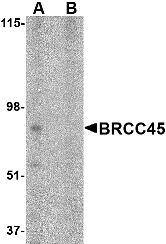 Description
Left: Western blot analysis of BRCC45 in HeLa cell lysate in (A) the absence and (B) presence of blocking peptide with BRCC45 antibody at 1 µg/ml.
Below: Immunohistochemistry of BRCC45 in human brain tissue with BRCC45 antibody at 2.5 µg/ml.
Other Product Images

Source : BRCC45 antibody was raised against a 18 amino acid peptide from near the amino terminus of human BRCC45.
Purification : Affinity chromatography purified via peptide column
Clonality and Clone : This is a polyclonal antibody.
Host : BRCC45 antibody was raised in rabbit. Please use anti-rabbit secondary antibodies.
Application : BRCC45 antibody can be used for detection of BRCC45 by Western blot at 1 – 2 µg/ml.
Tested Application(s) : E, WB, IHC
Buffer : Antibody is supplied in PBS containing 0.02% sodium azide.
Blocking Peptide : Cat.No. 4329P - BRCC45 Peptide
Long-Term Storage : BRCC45 antibody can be stored at 4ºC, stable for one year. As with all antibodies care should be taken to avoid repeated freeze thaw cycles. Antibodies should not be exposed to prolonged high temperatures.
Positive Control
1. Cat. No. 1201 - HeLa Cell Lysate
Species Reactivity :H, M, R
GI Number : 21361171
Accession Number : NP_004890
Short Description : BRCA1/BRCA2-containing complex subunit 45
References
1. Li L, Yoo H, Beckker FF, et al. Identification of a brain- and reproductive-organs-specific gene responsive to DNA damage and retinoic expression. Biochim. Biophys. Res. Commun. 1995; 206:764-74.
2. Dong Y, Hakimi MA, Chen X, et al. Regulation of BRCC, a holoenzyme complex containing BRCA1 and BRCA2, by a signalsome-like subunit and its role in DNA repair. Mol. Cell 2003; 12:1087-99.
3. Li Q, Ching AK, Chan BC, et al. A death receptor-associated anti-apoptotic protein, BRE, inhibits mitochondrial apoptotic pathway. J. Biol. Chem. 2004; 279:52106-16.
4. Chan BC, Ching AK, To KF, et al. BRE is an antiapoptotic protein in vivo and overexpressed in human hepatocellular carcinoma. Oncogene 2007; epub.
Catalog# : 4329
BRCC45 was initially suggested to be a housekeeping protein that is highly expressed in brain and reproductive organs. Later experiments indicated BRCC45 forms a complex with the breast and ovarian predisposition proteins BRCA1 and BRCA2 as well as RAD51 and BRCC36. This complex has a ubiquitin E3 ligase activity and is thought to enhance cellular survival following DNA damage. BRCC45 has also been suggested to function as a death receptor-associated anti-apoptotic protein by inhibiting the BID-induced activation of the mitochondrial apoptotic pathway. Higher levels of BRCC45 were detected in the majority of hepatocellular carcinomas, suggesting that BRCC45 may promote tumorigenesis when overexpressed. At least three isoforms of BRCC45 are known to exist.
Additional Names : BRCC45, BRCA1/BRCA2-containing complex subunit 45, Brain and reproductive organ expressed, BRE, TNFRSF1A modulator
DescriptionLeft: Western blot analysis of BRCC45 in HeLa cell lysate in (A) the absence and (B) presence of blocking peptide with BRCC45 antibody at 1 µg/ml.
Below: Immunohistochemistry of BRCC45 in human brain tissue with BRCC45 antibody at 2.5 µg/ml.
Other Product Images
Source : BRCC45 antibody was raised against a 18 amino acid peptide from near the amino terminus of human BRCC45.
Purification : Affinity chromatography purified via peptide column
Clonality and Clone : This is a polyclonal antibody.
Host : BRCC45 antibody was raised in rabbit. Please use anti-rabbit secondary antibodies.
Application : BRCC45 antibody can be used for detection of BRCC45 by Western blot at 1 – 2 µg/ml.
Tested Application(s) : E, WB, IHC
Buffer : Antibody is supplied in PBS containing 0.02% sodium azide.
Blocking Peptide : Cat.No. 4329P - BRCC45 Peptide
Long-Term Storage : BRCC45 antibody can be stored at 4ºC, stable for one year. As with all antibodies care should be taken to avoid repeated freeze thaw cycles. Antibodies should not be exposed to prolonged high temperatures.
Positive Control
1. Cat. No. 1201 - HeLa Cell Lysate
Species Reactivity :H, M, R
GI Number : 21361171
Accession Number : NP_004890
Short Description : BRCA1/BRCA2-containing complex subunit 45
References
1. Li L, Yoo H, Beckker FF, et al. Identification of a brain- and reproductive-organs-specific gene responsive to DNA damage and retinoic expression. Biochim. Biophys. Res. Commun. 1995; 206:764-74.
2. Dong Y, Hakimi MA, Chen X, et al. Regulation of BRCC, a holoenzyme complex containing BRCA1 and BRCA2, by a signalsome-like subunit and its role in DNA repair. Mol. Cell 2003; 12:1087-99.
3. Li Q, Ching AK, Chan BC, et al. A death receptor-associated anti-apoptotic protein, BRE, inhibits mitochondrial apoptotic pathway. J. Biol. Chem. 2004; 279:52106-16.
4. Chan BC, Ching AK, To KF, et al. BRE is an antiapoptotic protein in vivo and overexpressed in human hepatocellular carcinoma. Oncogene 2007; epub.
BRCC36 Antibody
BRCC36 Antibody
Catalog# : 4331
Together with the breast and ovarian predisposition proteins BRCA1 and BRCA2 and RAD51 and BRCC45, BRCC36 forms a holoenzyme complex that possesses a ubiquitin E3 ligase activity. Aberrant levels of BRCC36 were detected in sporadic breast tumors and depletion of either BRCC36 or BRCC45 by siRNA resulted in increased sensitivity to ionizing radiation and defects in the G2/M checkpoint, indicating that BRCC36 acts to enhance cellular survival following DNA damage. Recent experiments have shown that BRCC36 is essential for ionizing radiation-induced BRCA1 phosphorylation and nuclear foci formation, suggesting that BRCC36 may be an important target in the treatment of radiation-resistant breast tumors. At least two isoforms of BRCC36 are known to exist.
Additional Names : BRCC36, BRCA1/BRCA2-containing complex subunit 3, BRCC3
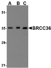 Description
Left: Western blot analysis of BRCC36 in MCF7 cell lysate with BRCC36 antibody at (A) 0.5, (B) 1 and (C) 2 µg/ml.
Below: Immunohistochemistry of BRCC36 in human brain tissue with BRCC36 antibody at 2.5 µg/ml.
Other Product Images
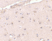 Source : BRCC36 antibody was raised against a 18 amino acid peptide from near the amino terminus of human BRCC36.
Purification : Affinity chromatography purified via peptide column
Clonality and Clone : This is a polyclonal antibody.
Host : BRCC36 antibody was raised in rabbit. Please use anti-rabbit secondary antibodies.
Application : BRCC36 antibody can be used for detection of BRCC36 by Western blot at 0.5 – 1 µg/ml.
Tested Application(s) : E, WB, IHC
Buffer : Antibody is supplied in PBS containing 0.02% sodium azide.
Blocking Peptide : Cat.No. 4331P - BRCC36 Peptide
Long-Term Storage : BRCC36 antibody can be stored at 4ºC, stable for one year. As with all antibodies care should be taken to avoid repeated freeze thaw cycles. Antibodies should not be exposed to prolonged high temperatures.
Positive Control
1. Cat. No. 1219 - MCF7 Cell Lysate
Species Reactivity :H, M, R
GI Number : 13236583
Accession Number : NP_077308
Short Description : BRCA1/BRCA2-containing complex subunit 3
References
1. Dong Y, Hakimi MA, Chen X, et al. Regulation of BRCC, a holoenzyme complex containing BRCA1 and BRCA2, by a signalsome-like subunit and its role in DNA repair. Mol. Cell 2003; 12:1087-99.
2. Chen X, Arciero CA, Wang C, et al. BRCC36 is essential for ionizing radiation-induced BRCA1 phosphorylation and nuclear foci formation. Cancer Res. 2006; 66:5039-46.
Catalog# : 4331
Together with the breast and ovarian predisposition proteins BRCA1 and BRCA2 and RAD51 and BRCC45, BRCC36 forms a holoenzyme complex that possesses a ubiquitin E3 ligase activity. Aberrant levels of BRCC36 were detected in sporadic breast tumors and depletion of either BRCC36 or BRCC45 by siRNA resulted in increased sensitivity to ionizing radiation and defects in the G2/M checkpoint, indicating that BRCC36 acts to enhance cellular survival following DNA damage. Recent experiments have shown that BRCC36 is essential for ionizing radiation-induced BRCA1 phosphorylation and nuclear foci formation, suggesting that BRCC36 may be an important target in the treatment of radiation-resistant breast tumors. At least two isoforms of BRCC36 are known to exist.
Additional Names : BRCC36, BRCA1/BRCA2-containing complex subunit 3, BRCC3
DescriptionLeft: Western blot analysis of BRCC36 in MCF7 cell lysate with BRCC36 antibody at (A) 0.5, (B) 1 and (C) 2 µg/ml.
Below: Immunohistochemistry of BRCC36 in human brain tissue with BRCC36 antibody at 2.5 µg/ml.
Other Product Images
Source : BRCC36 antibody was raised against a 18 amino acid peptide from near the amino terminus of human BRCC36.Purification : Affinity chromatography purified via peptide column
Clonality and Clone : This is a polyclonal antibody.
Host : BRCC36 antibody was raised in rabbit. Please use anti-rabbit secondary antibodies.
Application : BRCC36 antibody can be used for detection of BRCC36 by Western blot at 0.5 – 1 µg/ml.
Tested Application(s) : E, WB, IHC
Buffer : Antibody is supplied in PBS containing 0.02% sodium azide.
Blocking Peptide : Cat.No. 4331P - BRCC36 Peptide
Long-Term Storage : BRCC36 antibody can be stored at 4ºC, stable for one year. As with all antibodies care should be taken to avoid repeated freeze thaw cycles. Antibodies should not be exposed to prolonged high temperatures.
Positive Control
1. Cat. No. 1219 - MCF7 Cell Lysate
Species Reactivity :H, M, R
GI Number : 13236583
Accession Number : NP_077308
Short Description : BRCA1/BRCA2-containing complex subunit 3
References
1. Dong Y, Hakimi MA, Chen X, et al. Regulation of BRCC, a holoenzyme complex containing BRCA1 and BRCA2, by a signalsome-like subunit and its role in DNA repair. Mol. Cell 2003; 12:1087-99.
2. Chen X, Arciero CA, Wang C, et al. BRCC36 is essential for ionizing radiation-induced BRCA1 phosphorylation and nuclear foci formation. Cancer Res. 2006; 66:5039-46.
Bora Antibody
Bora Antibody
Catalog# : 5117
Bora (Protein aurora borealis) is a key activator of Aurora Related Protein Kinase A (ARK-1), which is a centrosome-associated serine/threonine kinase that regulates centrosome maturation, bipolar spindle assembly and chromosome segregation during mitosis. Bora is localized to the nucleus until mitosis is initiated, then translocates to the cytoplasm in a Cdc2 dependent manner. Activation of Cdc2 initiates the release of Bora into the cytoplasm where it can bind and activate ARK-1. PLK1 (polo-like kinase-1) interacts with Bora to control the accessibility of its activation loop for phosphorylation and activation by ARK-1. Bora and ARK-1 cooperatively activate PLK1 and control mitotic entry. Bora mutants result in multipolar spindles in mitosis identical to those observed when ARK-1 function is blocked. Thus, the ARK1-Bora-PLK1 regulatory circuit in mammalian cells elucidates a key mechanism in cell cycle regulation. At least three isoforms of Bora are known to exist.
Additional Names : Bora (NT), Protein aurora borealis, C13orf34, Chromosome 13 open reading frame 34
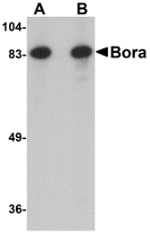 Description
Left: Western blot analysis of Bora in Jurkat cell lysate with Bora antibody at (A) 1 and (B) 2 µg/ml.
Below: Immunohistochemistry of Bora in mouse brain tissue with Bora antibody at 2.5 µg/ml.
Other Product Images
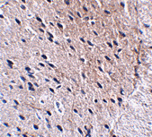 Source : Bora antibody was raised against a 15 amino acid peptide from near the amino terminus of human Bora.
Purification : Affinity chromatography purified via peptide column
Clonality and Clone : This is a polyclonal antibody.
Host : Bora antibody was raised in rabbit. Please use anti-rabbit secondary antibodies.
Application : Bora antibody can be used for detection of Bora by Western blot at 1-2 µg/ml.
Tested Application(s) : E, WB, IHC
Buffer : Antibody is supplied in PBS containing 0.02% sodium azide.
Blocking Peptide : Cat.No. 5117P - Bora Peptide
Long-Term Storage : Bora antibody can be stored at 4ºC, stable for one year. As with all antibodies care should be taken to avoid repeated freeze thaw cycles. Antibodies should not be exposed to prolonged high temperatures.
Positive Control
1. Cat. No. 1205 - Jurkat Cell Lysate
Species Reactivity :H, M, R
GI Number : 74737659
Accession Number : Q6PGQ7
Short Description : (NT) Protein aurora borealis
References
1. Berdnik D and Knoblich JA. Drosophila Aurora A is required for centrosome maturation and Actin-dependent asymmetric protein localization during mitosis. Curr. Biol. 2002; 12:640-647.
2. Wiese C and O’Brien LL. What's so Bor(a)ing about Aurora A activation? Dev. Cell 2006; 11:133-134.
3. Hutterer A, Berdnik D, Wirtz-Peitz F, et al. Mitotic activation of the kinase Aurora A requires its binding partner Bora. Dev. Cell 2006; 11:147-157.
4. Fu J, Bian M, Jiang Q, et al.. Roles of Aurora kinases in mitosis and tumorigenesis. Mol. Cancer Res. 2007; 5:1-10.
Catalog# : 5117
Bora (Protein aurora borealis) is a key activator of Aurora Related Protein Kinase A (ARK-1), which is a centrosome-associated serine/threonine kinase that regulates centrosome maturation, bipolar spindle assembly and chromosome segregation during mitosis. Bora is localized to the nucleus until mitosis is initiated, then translocates to the cytoplasm in a Cdc2 dependent manner. Activation of Cdc2 initiates the release of Bora into the cytoplasm where it can bind and activate ARK-1. PLK1 (polo-like kinase-1) interacts with Bora to control the accessibility of its activation loop for phosphorylation and activation by ARK-1. Bora and ARK-1 cooperatively activate PLK1 and control mitotic entry. Bora mutants result in multipolar spindles in mitosis identical to those observed when ARK-1 function is blocked. Thus, the ARK1-Bora-PLK1 regulatory circuit in mammalian cells elucidates a key mechanism in cell cycle regulation. At least three isoforms of Bora are known to exist.
Additional Names : Bora (NT), Protein aurora borealis, C13orf34, Chromosome 13 open reading frame 34
DescriptionLeft: Western blot analysis of Bora in Jurkat cell lysate with Bora antibody at (A) 1 and (B) 2 µg/ml.
Below: Immunohistochemistry of Bora in mouse brain tissue with Bora antibody at 2.5 µg/ml.
Other Product Images
Source : Bora antibody was raised against a 15 amino acid peptide from near the amino terminus of human Bora.Purification : Affinity chromatography purified via peptide column
Clonality and Clone : This is a polyclonal antibody.
Host : Bora antibody was raised in rabbit. Please use anti-rabbit secondary antibodies.
Application : Bora antibody can be used for detection of Bora by Western blot at 1-2 µg/ml.
Tested Application(s) : E, WB, IHC
Buffer : Antibody is supplied in PBS containing 0.02% sodium azide.
Blocking Peptide : Cat.No. 5117P - Bora Peptide
Long-Term Storage : Bora antibody can be stored at 4ºC, stable for one year. As with all antibodies care should be taken to avoid repeated freeze thaw cycles. Antibodies should not be exposed to prolonged high temperatures.
Positive Control
1. Cat. No. 1205 - Jurkat Cell Lysate
Species Reactivity :H, M, R
GI Number : 74737659
Accession Number : Q6PGQ7
Short Description : (NT) Protein aurora borealis
References
1. Berdnik D and Knoblich JA. Drosophila Aurora A is required for centrosome maturation and Actin-dependent asymmetric protein localization during mitosis. Curr. Biol. 2002; 12:640-647.
2. Wiese C and O’Brien LL. What's so Bor(a)ing about Aurora A activation? Dev. Cell 2006; 11:133-134.
3. Hutterer A, Berdnik D, Wirtz-Peitz F, et al. Mitotic activation of the kinase Aurora A requires its binding partner Bora. Dev. Cell 2006; 11:147-157.
4. Fu J, Bian M, Jiang Q, et al.. Roles of Aurora kinases in mitosis and tumorigenesis. Mol. Cancer Res. 2007; 5:1-10.
Bonzo Antibody
Bonzo Antibody
Catalog# : 1170
Human immunodeficiency virus (HIV) and simian immunodeficiency virus (SIV) require coreceptors, in addition to CD4, to infect target cells. Some G protein-coupled receptors including CCR5, CXCR4, CCR3, and CCR2b in the chemokine receptor family have been identified as HIV coreceptors. An orphan G protein-coupled receptor was recently cloned and designated Bonzo, STRL33 and TYMSTR, and identified as HIV and SIV coreceptor. Bonzo/STRL33 is used by SIV, HIV-2 and HIV-1. The messenger RNA of Bonzo/STRL33 is expressed in lymphoid tissues and activated peripheral blood lymphocytes.
Additional Names : Bonzo (CT), STRL33
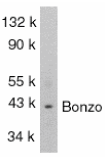 Description
Left: Western blot analysis of Bonzo in SW1353 total cells lysate with Bonzo antibody at 1:1000 dilution.
Source : Bonzo antibody was raised against a peptide corresponding to amino acids 319 to 338 of human origin. The sequence of this peptide is identical to those
Purification : Antibody is DEAE purified
Clonality and Clone : This is a polyclonal antibody.
Host : Bonzo antibody was raised in rabbit. Please use anti-rabbit secondary antibodies.
Immunogen : Human Bonzo (C-Terminus) Peptide (Cat. No. 1170P)
Application : Bonzo antibody can be used for detection of Bonzo by Western blot at 1:1000 dilution.
Tested Application(s) : E, WB
Buffer : Antibody is supplied in PBS containing 0.02% sodium azide.
Blocking Peptide : Cat. No. 1170P - Bonzo Peptide
Long-Term Storage : Bonzo antibody can be stored at 4ºC, stable for one year. As with all antibodies care should be taken to avoid repeated freeze thaw cycles. Antibodies should not be exposed to prolonged high temperatures.
Positive Control
1. Cat. No. 1214 - SW1353 Cell Lysate
Species Reactivity :H
GI Number : 2253422
Accession Number : AAB64221
Short Description : (CT) HIV coreceptor
References
1. Deng HK, Unutmaz D, KewalRamani VN, Littman DR. Expression cloning of new receptors used by simian and human immunodeficiency viruses. Nature 1997;388:296-300
2. Liao F, Alkhatib G, Peden KW, Sharma G, Berger EA, Farber JM. STRL33, A novel chemokine receptor-like protein, functions as a fusion cofactor for both macrophage-tropic and T cell line-tropic HIV-1. J Exp Med 1997;185:2015-23
3. Alkhatib G, Liao F, Berger EA, Farber JM, Peden KW. A new SIV co-receptor, STRL33. Nature 1997;388:238
4. Loetscher M, Amara A, Oberlin E, Brass N, Legler D, Loetscher P, D'Apuzzo M, Meese E, Rousset D, Virelizier JL, Baggiolini M, Arenzana-Seisdedos F, Moser B. TYMSTR, a putative chemokine receptor selectively expressed in activated T cells, exhibits HIV-1 coreceptor function. Curr Biol 1997;7:652-60 (RD1299)
Catalog# : 1170
Human immunodeficiency virus (HIV) and simian immunodeficiency virus (SIV) require coreceptors, in addition to CD4, to infect target cells. Some G protein-coupled receptors including CCR5, CXCR4, CCR3, and CCR2b in the chemokine receptor family have been identified as HIV coreceptors. An orphan G protein-coupled receptor was recently cloned and designated Bonzo, STRL33 and TYMSTR, and identified as HIV and SIV coreceptor. Bonzo/STRL33 is used by SIV, HIV-2 and HIV-1. The messenger RNA of Bonzo/STRL33 is expressed in lymphoid tissues and activated peripheral blood lymphocytes.
Additional Names : Bonzo (CT), STRL33
DescriptionLeft: Western blot analysis of Bonzo in SW1353 total cells lysate with Bonzo antibody at 1:1000 dilution.
Source : Bonzo antibody was raised against a peptide corresponding to amino acids 319 to 338 of human origin. The sequence of this peptide is identical to those
Purification : Antibody is DEAE purified
Clonality and Clone : This is a polyclonal antibody.
Host : Bonzo antibody was raised in rabbit. Please use anti-rabbit secondary antibodies.
Immunogen : Human Bonzo (C-Terminus) Peptide (Cat. No. 1170P)
Application : Bonzo antibody can be used for detection of Bonzo by Western blot at 1:1000 dilution.
Tested Application(s) : E, WB
Buffer : Antibody is supplied in PBS containing 0.02% sodium azide.
Blocking Peptide : Cat. No. 1170P - Bonzo Peptide
Long-Term Storage : Bonzo antibody can be stored at 4ºC, stable for one year. As with all antibodies care should be taken to avoid repeated freeze thaw cycles. Antibodies should not be exposed to prolonged high temperatures.
Positive Control
1. Cat. No. 1214 - SW1353 Cell Lysate
Species Reactivity :H
GI Number : 2253422
Accession Number : AAB64221
Short Description : (CT) HIV coreceptor
References
1. Deng HK, Unutmaz D, KewalRamani VN, Littman DR. Expression cloning of new receptors used by simian and human immunodeficiency viruses. Nature 1997;388:296-300
2. Liao F, Alkhatib G, Peden KW, Sharma G, Berger EA, Farber JM. STRL33, A novel chemokine receptor-like protein, functions as a fusion cofactor for both macrophage-tropic and T cell line-tropic HIV-1. J Exp Med 1997;185:2015-23
3. Alkhatib G, Liao F, Berger EA, Farber JM, Peden KW. A new SIV co-receptor, STRL33. Nature 1997;388:238
4. Loetscher M, Amara A, Oberlin E, Brass N, Legler D, Loetscher P, D'Apuzzo M, Meese E, Rousset D, Virelizier JL, Baggiolini M, Arenzana-Seisdedos F, Moser B. TYMSTR, a putative chemokine receptor selectively expressed in activated T cells, exhibits HIV-1 coreceptor function. Curr Biol 1997;7:652-60 (RD1299)
Subscribe to:
Posts (Atom)