Thursday, December 30, 2010
Gene Degradation
A gene is a locatable region of genomic sequence, corresponding to a unit of inheritance, which is associated with regulatory regions, transcribed regions and/or other functional sequence regions. The physical development and phenotype of organisms can be thought of as a product of genes interacting with each other and with the environment.
RNA Genes
In some cases, RNA is an intermediate product in the process of manufacturing proteins from genes. However, for other gene sequences, the RNA molecules are the actual functional products. For example, RNAs known as ribosome’s are capable of enzymatic function, and miRNAs have a regulatory role. The DNA sequences from which such RNAs are transcribed are known as RNA genes.
Gene Expression
In all organisms, there are two major steps separating a protein-coding gene from its protein: first, the DNA on which the gene resides must be transcribed from DNA to messenger RNA (mRNA), and second, it must be translated from mRNA to protein. RNA-coding genes must still go through the first step, but are not translated into protein. The process of producing a biologically functional molecule of either RNA or protein is called gene expression, and the resulting molecule itself is called a gene product.
Peptides play a crucial role in many cellular processes, however short peptides are devoid of well defined structures while in solution. In addition, the short half life in vivo due to rapid proteolytic degradation and poor cell permeability, remains a challenge to their therapeutic applications.[1] Thus common strategies aiming at improving receptor affinity, receptor efficacy, enzyme stability and pharmacokinetic properties are an active field of research.
Side chain to side chain cyclization of peptide is one common method applied whereby amino acid residues which have been identified as not involved in the receptor interaction are linked via lactam formation[2] or ring closing metathesis[3]. This method has been shown to mimic and stabilize a-helix structures which play an essential role in protein-protein interactions (PPI).
For instance, hydrocarbon stapled peptides mimicking BCL-2 domain (an important family of proteins that regulates apoptosis) showed increased protease resistance and cell permeability due to stabilized helical conformation[4]. This modification was further applied to NOTCH 1 transcription factor resulting to peptides with increased binding affinity towards NOTCH transactivation complex. NOTCH proteins have been shown to play a pivotal role in cellular differentiation, proliferation and apoptosis and their mutations have been linked with diseases like T-cell acute lymphoblastic leukemia.
These few examples show the potential of constrained synthetic peptides in PPI’s[5].
References:
1. A. Caporale, M. Sturlese, L. Gesiot, F. Zanta, A. Wittelsberger and C. Cabrele, J Med Chem 2010, 53, 8072-8079.
2. a) R. S. Harrison, G. Ruiz-Gomez, T. A. Hill, S. Y. Chow, N. E. Shepherd, R. J. Lohman, G. Abbenante, H. N. Hoang and D. P. Fairlie, J Med Chem 2010; b) E. N. Murage, J. C. Schroeder, M. Beinborn and J. M. Ahn, Bioorg Med Chem 2008, 16, 10106-10112.
3. a) H. E. Blackwell and R. H. Grubbs, Angewandte Chemie International Edition 1998, 37, 3281-3284; b) J. F. Reichwein, C. Versluis and R. M. Liskamp, J Org Chem 2000, 65, 6187-6195; c) J. Illesinghe, C. X. Guo, R. Garland, A. Ahmed, B. van Lierop, J. Elaridi, W. R. Jackson and A. J. Robinson, Chem Commun (Camb) 2009, 295-297.
4. L. D. Walensky, A. L. Kung, I. Escher, T. J. Malia, S. Barbuto, R. D. Wright, G. Wagner, G. L. Verdine and S. J. Korsmeyer, Science 2004, 305, 1466-1470.
5. R. E. Moellering, M. Cornejo, T. N. Davis, C. Del Bianco, J. C. Aster, S. C. Blacklow, A. L. Kung, D. G. Gilliland, G. L. Verdine and J. E. Bradner, Nature 2009, 462, 182-188.
Synthetic Conformationally Constrained Peptides
Most naturally occurring constrained peptides maintain the rigidity of the peptide framework by disulfide bond or N- to C-termini backbone cyclization. Commonly known antimicrobial peptides like Gramidicin S, Bacitracin and Polymyxin B are an example of backbone cyclic peptides that are used clinically (Table 1).[1]
Table 1: Natural cyclic peptides
 Parenthesis indicate amino acids that are cyclized, d the D-enantiomer; O, Ornithine; B, diaminobutyrate
Figure 1: Scheme showing reduction of disulfide bonds in cellular environment to acyclic thiol. In addition, availability of variety of protecting groups for amines and carboxylic acids which are cleavable under orthogonal conditions have made amide bond lactam bridges to be an alternative covalent linkage substituting disulfide bonds (Figure 2B).

Conotoxins are another class of small peptide ligands (typically 10-30 amino acids) highly crosslinked by disulfide bonds.[2] Despite their small size, these peptide ligands have very high affinities and selectivities to their cognate receptors and many of them have now become standard research tools in neuroscience.
Although cysteine bridges are quite common structural motifs in naturally occurring peptides like neurotoxins,[2] cyclotides,[3] somatostatin[4] and insulin superfamily[5}, disulfide bridges (Figure 1) are readily reduced to their acyclic thiol form in an intracellular environment. Thus scientists have derived new methods of inducing stable conformation constraint of many peptides. For instance, advancement of organometallic chemistry has led to use of phase transfer catalyst like Grubbs catalyst[6] in ring closing metathesis. This chemistry has been utilized by Gregory Verdine to synthesize stapled peptides which have been found with promising biological functions (Figure 2A, Table 2) [7].
Figure 2: (A) Hydrocarbon stapled peptides synthesized through ring closing metathesis, (B) amide bond lactam bridge.
As summarized in the table 2 below, several synthetic cyclic peptides have shown desirable biological properties over their linear counterparts.
 References
1. S. R. Woodward, L. J. Cruz, B. M. Olivera and D. R. Hillyard, EMBO J 1990, 9, 1015-1020.
2. B. M. Olivera, J. Rivier, C. Clark, C. A. Ramilo, G. P. Corpuz, F. C. Abogadie, E. E. Mena, S. R. Woodward, D. R. Hillyard and L. J. Cruz, Science 1990, 249, 257-263.
3. D. J. Craik, N. L. Daly, T. Bond and C. Waine, J Mol Biol 1999, 294, 1327-1336.
4. L. Pradayrol, H. Jornvall, V. Mutt and A. Ribet, FEBS Lett 1980, 109, 55-58.
5. S. J. Chan, Q. P. Cao and D. F. Steiner, Proc Natl Acad Sci U S A 1990, 87, 9319-9323.
6. G. C. Vougioukalakis and R. H. Grubbs, Chem Rev 2010, 110, 1746-1787.
7. L. D. Walensky, A. L. Kung, I. Escher, T. J. Malia, S. Barbuto, R. D. Wright, G. Wagner, G. L. Verdine and S. J. Korsmeyer, Science 2004, 305, 1466-1470.
Peptides as molecular imaging probes
Molecular imaging is a rapidly emerging biomedical research in diagnostics and therapeutic fields due to current indispensable tools in modern diagnostics[1]. Thus the need for highly sensitive and specific molecular imaging probes is still unmet. In order to design diagnostic imaging and radiotherapeutic agents, a high target/background ratio, target uptake and rapid removal of untargeted drug is desirable. Proteins, antibodies and their fragment are perfect candidates due to high specificity affinity and selectivity to the target organs. With the advancement of combinatorial chemistry and phage display methods, peptides are currently receiving considerable attention as molecular imaging probes. In addition, peptides have distinct advantages over proteins and antibodies due to many appealing characteristics:
1. Rapid clearance in the body ensures the radioisotopes are not retained in the body for a prolonged period of time.
2. Have low toxicity and immunogenicity
3. They can easily be synthesized, modified to optimize their binding affinity and improve their stability against proteolytic degradation and in vivo circulation providing a means to improve
4. With the well established peptide synthesis protocols they are easy to scale up and reproduce Phage display is a powerful technique that allows vast sequence space screening, peptide affinity and generate unique peptides that binds any given target [1]. Due to the diversified methods of peptide synthesis, peptides can be directly or indirectly labeled with imaging probes to provide or augment the imaging signals, depending on the modality. For example, several radioisotopes (99mTc, 18F, 64Cu, 111In, 123I, 68Ga) for PET and SPECT, organic near-infrared (NIR) fluorophores or quantum dots (QDs) for optical imaging and magnetic nanoparticles for MRI can be conjugated to peptides via organic linkers, macrocyclic or branched chelators (figure 1), polymers or nanoplatforms[2].
Several classic examples of peptides have been utilized as imaging agents for peptide-receptor interaction due to their high selectivity and affinity to their cognitive receptors as described below.
1.1 Somatostatin Analogs
Somatostatins (SST) are regulatory hormones with inhibitory effects on secretion of growth hormone, insulin, glucagon, gastrin, cholecystokinin, vasoactive intestinal peptide (VIP) and secretin[3]. These cyclopeptides are mainly produced by endocrine, gastrointestinal, immune and neuronal cells as well as by certain tumours[3]. Radionuclide labeled synthetic somatostatin analogs like octreotide have long been investigated for imaging neuroendocrine tumors due to their enhanced metabolic stability and biological activity compared to the native hormone. 111In-DTPA, 68GA-DOTA, 64Cu-DOTA and 18F-FP-Gluc-labeled SST analogues have been developed for imaging various tumors[2].
1.2 αvß3-Integrin Analogs
αvß3 Integrin Analogs are closely related with tumor progression and plays an important role during tumor angiogenesis and formation of new blood vessels. Integrin peptides contain an Arg-Gly-Asp (RGD) motif. αvß3 integrin has received considerable attention in diagnostic cancer imaging due to its overexpression in several forms of tumors including melanomas, ovarian and lung carcinoma, neuroblastomas, glioblastomas and breast cancer[2]. Of particular interest, 18F labeled cyclo(Arg-Gly-Asp-D-Tyr-Lys) analog has been used to identify αvß3 receptor expression in patients with melanoma, sarcoma, breast cancer and glioblastoma[4].
1.3 Bombesin analogs
Bombesin (BBN) is an amphibian homologue of mammalian gastrin-releasing peptide (GRP; pGlu1-Gln2-Arg3-Leu4-Gly5-Asn6-Gln7-Trp8-Ala9-Val10-Gly11-His12-Leu13-Met14-NH2) that exhibits high affinity and specificity to the GRP receptor (GRPr). Several BBN analogs have been screened and developed as imaging probes. 99mTc-EDDA/HYNIC, 111In-DOTA, 64Cu-DOTA and NOTA and 18F-Lys3 labeled BBN have been shown to have specific uptake in PC3-tumors in vivo[2].
 1.4 Cholecytoskinin analogs
Cholecytoskinin (CCK) and gastrin are structurally and functionally related peptide hormones that function in the gastrointestinal tract and central nervous system. They mediate their biological functions by interacting with CCK/gastrin receptors belonging to GPCR’s[5]. These receptors have been found to be overexpressed in certain human tumors like medullary thyroid cancer (MTC), astrocytomas, stromal ovarian tumors and gastroenteropancreatic cancers[6]. Several of 111In or 99mTc labeled CCK and minigastrin analogs have shown specificity in cells overexpressing CCK receptors and high tumor uptake. However the metabolic stability and in vivo bioavailability of these peptides needs to be improved for them to be clinically useful[2].
1.5 α-melanocyte stimulating hormone analogs
α-melanocyte stimulating hormone (α-MSH) is a 13 amino acid peptide produced by pituitary gland and is mainly responsible for the regulation of skin pigmentation. Studies have shown that a-MSH receptors are distributed in more than 80% human melanoma metastases[7]. Thus radiolabeled peptides that target these receptors are attractive probes for diagnosis of melanoma. 111In DOTA-conjugated, 18F-labeled a-MSH have been investigated in melanoma-bearing mice[8]. 99mTc labeled dual receptor targeting peptide ligands recognizing both integrin and α-MSH receptors have been designed and they demonstrate enhanced cellular intake in melanoma both in vitro and in vivo[9].
Other peptides like neurotensin (NT), a neurotransmitter is being investigated for ductal pancreatic adenocarcinomas which overexpressed NT receptors. Radiolabeled glucagon-like peptide-1 (GLP-1) and Exendin-4 have been developed to target insulinomas which overexpress the GLP-1 receptors.
All these peptides require custom design to improve their metabolic stability and half life in vivo. BIOSYNTHESIS, INC. a leading manufacturer of custom peptides in small scale research and bulk is now offering peptide conjugates containing metal chelators like DOTA, NOTA, TETA and DTPA.
References:
1. S. L. Deutscher, Chem Rev 2010, 110, 3196-3211.
2. S. Lee, J. Xie and X. Chen, Chem Rev 2010, 110, 3087-3111.
3. G. Weckbecker, I. Lewis, R. Albert, H. A. Schmid, D. Hoyer and C. Bruns, Nat Rev Drug Discov 2003, 2, 999-1017.
4. a) A. J. Beer, A. L. Grosu, J. Carlsen, A. Kolk, M. Sarbia, I. Stangier, P. Watzlowik, H. J. Wester, R. Haubner and M. Schwaiger, Clin Cancer Res 2007, 13, 6610-6616; b) A. J. Beer, M. Niemeyer, J. Carlsen, M. Sarbia, J. Nahrig, P. Watzlowik, H. J. Wester, N. Harbeck and M. Schwaiger, J Nucl Med 2008, 49, 255-259.
5. J. C. Reubi, J. C. Schaer and B. Waser, Cancer Res 1997, 57, 1377-1386.
6. J. C. Reubi and B. Waser, Int J Cancer 1996, 67, 644-647.
7. J. B. Tatro, Z. Wen, M. L. Entwistle, M. B. Atkins, T. J. Smith, S. Reichlin and J. R. Murphy, Cancer Res 1992, 52, 2545-2548.
8. Y. Miao, F. Gallazzi, H. Guo and T. P. Quinn, Bioconjug Chem 2008, 19, 539-547.
9. J. Yang, H. Guo, F. Gallazzi, M. Berwick, R. S. Padilla and Y. Miao, Bioconjug Chem 2009, 20, 1634-1642.
Wednesday, December 29, 2010
Deoxyoligonucleotides
An oligonucleotide (or oligo) is a short segment of RNA or DNA, typically with twenty or fewer bases. Although they can be formed by cleavage of longer segments, they are now more commonly synthesized by polymerizing individual nucleotide precursors. Bio-Synthesis provides oligonucleotide synthesis up to 250 bases.
DNA Micro Array
One subtype of DNA micro arrays can be described as substrates (nylon, glass, etc.) to which oligonucleotides have been bound at high density. Currently there exist three applications of DNA micro arrays: polymorphism studies, gene expression studies, and tracking down certain diseases.
Oligonucleotide Synthesis
Oligonucleotides are chemically synthesized using nucleotides, called phosphoramidites, normal nucleotides that have protection groups: preventing amine, hydroxyl groups and phosphate groups interacting incorrectly. One phosphoramidite is added at the time, the product's 5' phosphate is deprotected, and a new base is added, and so on (backwards); at the end, all the protection groups are removed.
Antisense Oligonucleotides
Antisense oligonucleotides are single strands of DNA or RNA that are complementary to a chosen sequence. In the case of antisense RNA they prevent protein translation of certain messenger RNA strands by binding to them. Antisense DNA can be used to target a specific, complementary (coding or non-coding) RNA. If binding takes places this DNA/RNA hybrid can be degraded by the enzyme RNase H.
LNA-DNA Chimeras in Cancer Treatments
Oncologists have been searching for a breakthrough in cancer research and medicine for a longtime now. Although a “cure” to cancer is still leaps and bounds out of reach, oncologists found the KRAS gene to which to turn their attention and target. It was found that a cancer with the wild type form of the KRAS gene responds better to EGFR inhibiting cancer drugs than cancers with a mutation in this gene. It is not a huge step towards ultimately preventing or reversing cancer, but it is important towards the practical treatment of cancer.
Oncologists had thus started using a technique involving LNA-DNA chimeras in order to detect and determine whether or not a particular person’s cancer cells contain any mutated form of the KRAS gene. Using LNA-DNA chimeras in order to block the amplification of wild type KRAS alleles, PCR is performed to amplify any possible trace amounts of existing mutated KRAS alleles. It was found that this simple and low-cost technique, called wild-type blocking PCR (WTB-PCR), lead to a higher success rate of detecting mutated alleles, which lead to more increased efficiency in cancer treatment.
LNA chimeras are becoming used much more widely and as possible uses are discovered for these nucleotides with high stability, research based around LNA will increase greatly.
Tuesday, December 28, 2010
Dengue Haemorrhagic Fever
Dengue fever cannot be prevented by vaccination or chemoprophylaxis - the only way to reduce the risk of being infected is by avoidance of mosquito bites. The most effective way of preventing dengue fever is to reduce the breeding sites of the Aedes mosquito.
Dengue Virus
The Dengue virus is a member of the virus family Flaviviridae and is transmitted to people through the bite of the mosquito’s Aedes aegypti and Aedes albopictus. Dengue virus is now believed to be the most common arthropod-borne disease in the world. Dengue is mainly found in the tropics because the mosquitoes require a warm climate.
Dengue Vaccination
Dengue fever cannot be prevented by vaccination or chemoprophylaxis - the only way to reduce the risk of being infected is by avoidance of mosquito bites. The most effective way of preventing dengue fever is to reduce the breeding sites of the Aedes mosquito.
Apoptosis Gene
Apoptosis is a form of programmed cell death in multicultural organisms. It is one of the main types of programmed cell deaths (PCD) and involves a series of biochemical events leading to a characteristic cell morphology and death, in more specific terms, a series of biochemical events that lead to a variety of morphological changes, including blebbing, changes to the cell membrane such as loss of membrane asymmetry and attachment, cell shrinkage, nuclear fragmentation, chromatin condensation, and chromosomal DNA fragmentation.
Cell termination
Apoptosis can occur when a cell is damaged beyond repair, infected with a virus, or undergoing stress conditions such as starvation. DNA damage from ionizing radiation or toxic chemicals can also induce apoptosis via the actions of the tumour-suppressing gene p53. The "decision" for apoptosis can come from the cell itself, from the surrounding tissue, or from a cell that is part of the immune system. In these cases apoptosis functions to remove the damaged cell, preventing it from sapping further nutrients from the organism, or to prevent the spread of viral infection.
Homeostasis
In the adult organism, the number of cells is kept relatively constant through cell death and division. Cells must be replaced when they become diseased or malfunctioning; but proliferation must be compensated by cell death. This balancing process is part of the homeostasis required by living organisms to maintain their internal states within certain limits.
Sunday, December 26, 2010
Chimeric RNA And Oligonucleotides
Ribonucleic acid (RNA) is a nucleic acid and consists of a long chain of nucleotide units. Each nucleotide consists of a nitrogenous base, a ribose sugar, and a phosphate. RNA is very similar to DNA, but differs in a few important structural details: in the cell RNA is usually single stranded, while DNA is usually double stranded. RNA nucleotides contain ribose while DNA contains deoxyribose (a type of ribose that lacks one oxygen atom), and RNA has the base uracil rather than thymine which is present in DNA.
Antisense RNA
Antisense RNA (aRNA) is single-stranded RNA that is complementary to a messenger RNA (mRNA) strand transcribed within a cell. Antisense RNA may be introduced into a cell to inhibit translation of a complementary mRNA by base pairing to it and physically obstructing the translation machinery. This effect is therefore stoichiometric. An example of naturally occurring mRNA antisense mechanism is the hok/sok system of the E.coli R1 plasmid. Antisense RNA has long been thought of as a promising technique for disease therapy; the only such case to have reached the market is the drug fomivirsen. Generally, antisense RNA still lack effective design, biological activity, and efficient route of administration.
Oligonucleotide
Oligonucleotide is a short segment of RNA or DNA, typically with twenty or fewer bases. Although they can be formed by cleavage of longer segments, they are now more commonly synthesized by polymerizing individual nucleotide precursors. Automated synthesizers allow the synthesis of oligonucleotides up to 160 to 200 bases. The length of the oligonucleotide is usually denoted by "mer" (from Greek meros, "part"). For example, a fragment of 25 bases would be called a 25-mer. Oligonucleotides are often used as probes for detecting DNA or RNA because they bind readily to their complements. Examples of procedures that use oligonucleotides include DNA microarrays, Southern blots, ASO analysis, fluorescent in situ hybridization (FISH), and the synthesis of artificial genes.
Wednesday, December 22, 2010
Custom Plasmid
A plasmid is an extra-chromosomal DNA molecule separate from the chromosomal DNA which is capable of replicating independently of the chromosomal DNA.In many cases; it is circular and double-stranded. Plasmids usually occur naturally in bacteria, but are sometimes found in eukaryotic organisms.
Gene Therapy
The success of gene therapy depends on the efficient insertion of therapeutic genes at the appropriate chromosomal target sites within the human genome, without causing cell injury, oncogenic mutations or an immune response. Plasmid vectors could be used for this purpose. Zinc finger nucleases (ZFNs) offer a way to cause a site-specific double strand break to the DNA genome and cause homologous recombination.
Ti Plasmid
Ti plasmid is a circular plasmid that often, but not always, is a part of the genetic equipment that Agrobacterium tumefactions and Agro bacterium rhizogenes use to transducer its genetic material to plants.
Custom Monoclonal Antibody
Monoclonal antibodies (mAb or moAb) are monospecific antibodies that are identical because they are produced by one type of immune cell that are all clones of a single parent cell. Given (almost) any substance, it is possible to create monoclonal antibodies that specifically bind to that substance; they can then serve to detect or purify that substance.
Hybridoma Cell Production
Monoclonal antibodies are typically made by fusing myeloma cells with the spleen cells from a mouse that has been immunized the desired antigen with. However, recent advances have allowed the use of rabbit B-cells. Polyethylene glycol is used to fuse adjacent plasma membranes, but the success rate is low so a selective medium is used in which only fused cells can grow. This is because myeloma cells have lost the ability to synthesize hypoxanthine-guanine-phosphoribosyl transferase.
Protein Micro Array
A protein microarray is a piece of glass on which different molecules of protein have been affixed at separate locations in an ordered manner thus forming a microscopic array. These are used to identify protein-protein interactions, to identify the substrates of protein kinases, or to identify the targets of biologically active small molecules.
Tuesday, December 21, 2010
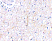
Plxdc2 Antibody
Plxdc2 Antibody
Catalog# :4419
Plxdc2, also known as Tumor endothelial marker 7-related (TEM7R) encodes a protein with 57% amino acid identity to TEM7, the most abundant tumor endothelial marker. Plxdc2 is strongly expressed in the endothelial cells of the tumor stroma, but not in the endothelial cells of normal colonic tissue. Plxdc2 is also expressed at high levels in vessels of some normal tissues, with highest expression in muscle and lung. Plxdc2 and TEM7 may be important for tumor angiogenesis in humans. Cortactin was identified as a protein capable of binding to the extracellular region of both TEM7 and Plxdc2, and may provide new opportunities for the delivery of therapeutic and imaging agents to the vessels of solid tumors.
Additional Names : Plxdc2 (CT), Plexin domain-containing protein 2, Tumor endothelial marker 7-related protein, TEM7R
 Description
Left: Western blot analysis of Plxdc2 in human colon tissue lysate with Plxdc2 antibody at (A) 0.5 (B) 1 µg/ml.
Below: Immunohistochemical staining of human brain tissue using Plxdc2 antibody at 2.5 µg/ml.
Other Product Images
 Source : Plxdc2 antibody was raised against a 17 amino acid peptide from near the carboxy terminus of human Plxdc2.
Purification : Affinity chromatography purified via peptide column
Clonality and Clone : This is a polyclonal antibody.
Host : Plxdc2 antibody was raised in rabbit. Please use anti-rabbit secondary antibodies.
Application : Plxdc2 antibody can be used for detection of Plxdc2 by Western blot at 0.5 – 1 µg/ml.
Tested Application(s) : E, WB, IHC
Buffer : Antibody is supplied in PBS containing 0.02% sodium azide.
Blocking Peptide :Cat.No. 4419P - Plxdc2 Peptide
Long-Term Storage : Plxdc2 antibody can be stored at 4ºC, stable for one year. As with all antibodies care should be taken to avoid repeated freeze thaw cycles. Antibodies should not be exposed to prolonged high temperatures.
Positive Control :
1. Cat. No. 1320 - Human Colon Tissue Lysate
Species Reactivity : H, M, R
GI Number : 74749416
Accession Number : Q6UX71
Short Description : (CT) TEM7 receptor
References
1. Carson-Weber EB, Watkins DN, Nanda A, et al. Cell surface tumor epithelial markers are conserved in mice and humans. Cancer Res. 2001; 61:6649-55.
2. Nabda A and St. Croix Bl. Tumor endothelial markers: new targets for cancer therapy. Curr Opin Oncol. 2004; 16:44-9.
3. St. Croix B, Rago C, Velculescu V, et al. Genes expressed in human tumor endothelium. Science 2000; 289:1197-202.
4. Nanda A, Buckhaults P, Seaman S, et al. Identification of a binding partner for the endothelial cell surface proteins TEM7 and TEM7R. Cancer Res. 2004; 64:8507-11.
Catalog# :4419
Plxdc2, also known as Tumor endothelial marker 7-related (TEM7R) encodes a protein with 57% amino acid identity to TEM7, the most abundant tumor endothelial marker. Plxdc2 is strongly expressed in the endothelial cells of the tumor stroma, but not in the endothelial cells of normal colonic tissue. Plxdc2 is also expressed at high levels in vessels of some normal tissues, with highest expression in muscle and lung. Plxdc2 and TEM7 may be important for tumor angiogenesis in humans. Cortactin was identified as a protein capable of binding to the extracellular region of both TEM7 and Plxdc2, and may provide new opportunities for the delivery of therapeutic and imaging agents to the vessels of solid tumors.
Additional Names : Plxdc2 (CT), Plexin domain-containing protein 2, Tumor endothelial marker 7-related protein, TEM7R
DescriptionLeft: Western blot analysis of Plxdc2 in human colon tissue lysate with Plxdc2 antibody at (A) 0.5 (B) 1 µg/ml.
Below: Immunohistochemical staining of human brain tissue using Plxdc2 antibody at 2.5 µg/ml.
Other Product Images
Source : Plxdc2 antibody was raised against a 17 amino acid peptide from near the carboxy terminus of human Plxdc2.Purification : Affinity chromatography purified via peptide column
Clonality and Clone : This is a polyclonal antibody.
Host : Plxdc2 antibody was raised in rabbit. Please use anti-rabbit secondary antibodies.
Application : Plxdc2 antibody can be used for detection of Plxdc2 by Western blot at 0.5 – 1 µg/ml.
Tested Application(s) : E, WB, IHC
Buffer : Antibody is supplied in PBS containing 0.02% sodium azide.
Blocking Peptide :Cat.No. 4419P - Plxdc2 Peptide
Long-Term Storage : Plxdc2 antibody can be stored at 4ºC, stable for one year. As with all antibodies care should be taken to avoid repeated freeze thaw cycles. Antibodies should not be exposed to prolonged high temperatures.
Positive Control :
1. Cat. No. 1320 - Human Colon Tissue Lysate
Species Reactivity : H, M, R
GI Number : 74749416
Accession Number : Q6UX71
Short Description : (CT) TEM7 receptor
References
1. Carson-Weber EB, Watkins DN, Nanda A, et al. Cell surface tumor epithelial markers are conserved in mice and humans. Cancer Res. 2001; 61:6649-55.
2. Nabda A and St. Croix Bl. Tumor endothelial markers: new targets for cancer therapy. Curr Opin Oncol. 2004; 16:44-9.
3. St. Croix B, Rago C, Velculescu V, et al. Genes expressed in human tumor endothelium. Science 2000; 289:1197-202.
4. Nanda A, Buckhaults P, Seaman S, et al. Identification of a binding partner for the endothelial cell surface proteins TEM7 and TEM7R. Cancer Res. 2004; 64:8507-11.
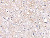
Plxdc2 Antibody
Plxdc2 Antibody
Catalog# :4417
Plxdc2, also known as Tumor endothelial marker 7-related (TEM7R) encodes a protein with 57% amino acid identity to TEM7, the most abundant tumor endothelial marker. Plxdc2 is strongly expressed in the endothelial cells of the tumor stroma, but not in the endothelial cells of normal colonic tissue. Plxdc2 is also expressed at high levels in vessels of some normal tissues, with highest expression in muscle and lung. Plxdc2 and TEM7 may be important for tumor angiogenesis in humans. Cortactin was identified as a protein capable of binding to the extracellular region of both TEM7 and Plxdc2, and may provide new opportunities for the delivery of therapeutic and imaging agents to the vessels of solid tumors.
Additional Names : Plxdc2 (NT), Plexin domain-containing protein 2, Tumor endothelial marker 7-related protein, TEM7R
 Description
Left: Western blot analysis of Plxdc2 in human brain tissue lysate with Plxdc2 antibody at (A) 0.5 (B) 1 µg/ml.
Below: Immunohistochemical staining of human brain tissue using Plxdc2 antibody at 2.5 µg/ml.
Other Product Images
Source :Plxdc2 antibody was raised against a 18 amino acid peptide from near the amino terminus of human Plxdc2.
Purification : Affinity chromatography purified via peptide column
Clonality and Clone : This is a polyclonal antibody.
Host : Plxdc2 antibody was raised in rabbit. Please use anti-rabbit secondary antibodies.
Application : Plxdc2 antibody can be used for detection of Plxdc2 by Western blot at 0.5 – 1 µg/ml.
Tested Application(s) : E, WB, IHC
Buffer : Antibody is supplied in PBS containing 0.02% sodium azide.
Blocking Peptide :Cat.No. 4417P - Plxdc2 Peptide
Long-Term Storage : Plxdc2 antibody can be stored at 4ºC, stable for one year. As with all antibodies care should be taken to avoid repeated freeze thaw cycles. Antibodies should not be exposed to prolonged high temperatures.
Positive Control :
1. Cat. No. 1303 - Human Brain Tissue Lysate
Species Reactivity : H, M, R
GI Number : 74749416
Accession Number : Q6UX71
Short Description : (NT) TEM7 receptor
References
1. Carson-Weber EB, Watkins DN, Nanda A, et al. Cell surface tumor epithelial markers are conserved in mice and humans. Cancer Res. 2001; 61:6649-55.
2. Nabda A and St. Croix Bl. Tumor endothelial markers: new targets for cancer therapy. Curr Opin Oncol. 2004; 16:44-9.
3. St. Croix B, Rago C, Velculescu V, et al. Genes expressed in human tumor endothelium. Science 2000; 289:1197-202.
4. Nanda A, Buckhaults P, Seaman S, et al. Identification of a binding partner for the endothelial cell surface proteins TEM7 and TEM7R. Cancer Res. 2004; 64:8507-11.
Catalog# :4417
Plxdc2, also known as Tumor endothelial marker 7-related (TEM7R) encodes a protein with 57% amino acid identity to TEM7, the most abundant tumor endothelial marker. Plxdc2 is strongly expressed in the endothelial cells of the tumor stroma, but not in the endothelial cells of normal colonic tissue. Plxdc2 is also expressed at high levels in vessels of some normal tissues, with highest expression in muscle and lung. Plxdc2 and TEM7 may be important for tumor angiogenesis in humans. Cortactin was identified as a protein capable of binding to the extracellular region of both TEM7 and Plxdc2, and may provide new opportunities for the delivery of therapeutic and imaging agents to the vessels of solid tumors.
Additional Names : Plxdc2 (NT), Plexin domain-containing protein 2, Tumor endothelial marker 7-related protein, TEM7R
DescriptionLeft: Western blot analysis of Plxdc2 in human brain tissue lysate with Plxdc2 antibody at (A) 0.5 (B) 1 µg/ml.
Below: Immunohistochemical staining of human brain tissue using Plxdc2 antibody at 2.5 µg/ml.
Other Product Images
Source :Plxdc2 antibody was raised against a 18 amino acid peptide from near the amino terminus of human Plxdc2.
Purification : Affinity chromatography purified via peptide column
Clonality and Clone : This is a polyclonal antibody.
Host : Plxdc2 antibody was raised in rabbit. Please use anti-rabbit secondary antibodies.
Application : Plxdc2 antibody can be used for detection of Plxdc2 by Western blot at 0.5 – 1 µg/ml.
Tested Application(s) : E, WB, IHC
Buffer : Antibody is supplied in PBS containing 0.02% sodium azide.
Blocking Peptide :Cat.No. 4417P - Plxdc2 Peptide
Long-Term Storage : Plxdc2 antibody can be stored at 4ºC, stable for one year. As with all antibodies care should be taken to avoid repeated freeze thaw cycles. Antibodies should not be exposed to prolonged high temperatures.
Positive Control :
1. Cat. No. 1303 - Human Brain Tissue Lysate
Species Reactivity : H, M, R
GI Number : 74749416
Accession Number : Q6UX71
Short Description : (NT) TEM7 receptor
References
1. Carson-Weber EB, Watkins DN, Nanda A, et al. Cell surface tumor epithelial markers are conserved in mice and humans. Cancer Res. 2001; 61:6649-55.
2. Nabda A and St. Croix Bl. Tumor endothelial markers: new targets for cancer therapy. Curr Opin Oncol. 2004; 16:44-9.
3. St. Croix B, Rago C, Velculescu V, et al. Genes expressed in human tumor endothelium. Science 2000; 289:1197-202.
4. Nanda A, Buckhaults P, Seaman S, et al. Identification of a binding partner for the endothelial cell surface proteins TEM7 and TEM7R. Cancer Res. 2004; 64:8507-11.
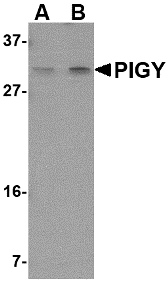
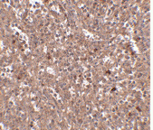
PIG-Y Antibody
PIG-Y Antibody
Catalog# :4955
Glycosylphosphatidylinositol (GPI) lipid anchoring is an important post-translational modification of proteins that takes place in the endoplasmic reticulum. The synthesis of GPI is initiated by GPI-N-acetylglucosaminyltransferase (GPI-GnT), a complex of proteins including PIG-A, PIG-H, PIG-C, GPI1, and DPM2. PIG-Y, the mammalian homolog to yeast Eri1p, is also thought to be involved in the biosynthesis of GPI. The PIG-Y gene encodes two proteins, one of which arises from leaky scanning of the mRNA. This antibody only detects isoform 1 of PIG-Y. Despite its predicted molecular weight, PIG-Y often migrates at 28-30 kDa in SDS-PAGE.
Additional Names : PIG-Y (IN), Phosphatidylinositol glycan anchor biosynthesis class Y, PreY, PIGY
 Description
Left: Western blot analysis of PIG-Y in human spleen tissue lysate with PIG-Y antibody at (A) 1 and (B) 2 µg/ml.
Below: Immunohistochemistry of PIG-Y in human spleen tissue with PIG-Y antibody at 2.5 µg/ml.
Other Product Images
 Source : PIG-Y antibody was raised against a 16 amino acid peptide near the center of human PIG-Y.
Purification : Affinity chromatography purified via peptide column
Clonality and Clone : This is a polyclonal antibody.
Host : PIG-Y antibody was raised in rabbit. Please use anti-rabbit secondary antibodies.
Application : PIG-Y antibody can be used for detection of PIG-Y by Western blot at 1 – 2 µg/ml.
Tested Application(s) : E, WB, IHC
Buffer : Antibody is supplied in PBS containing 0.02% sodium azide.
Blocking Peptide :Cat.No. 4955P - PIG-Y Peptide
Long-Term Storage : PIG-Y antibody can be stored at 4ºC, stable for one year. As with all antibodies care should be taken to avoid repeated freeze thaw cycles. Antibodies should not be exposed to prolonged high temperatures.
Positive Control :
1. Cat. No. 1306 - Human Spleen Tissue Lysate
Species Reactivity : H, M
GI Number : 14249680
Accession Number : NP_116295
Short Description : (IN) Phosphatidylinositol glycan anchor biosynthesis class Y
References
1. Eisenhaber B, Maurer-Stroh S, Novatchkova M, et al. Enzymes and auxiliary factors for GPI lipid anchor biosynthesis and post-translational transfer to proteins. Bioessays 2003; 25:367-85.
2. Watanabe R, Murakami Y, Marmor MD, et al. Initial enzyme for glycosylphosphatidylinositol biosynthesis requires PIG-P and is regulated by DPM2. EMBO J. 2000; 19:4402-11.
3. Murakami Y, Siripanyaphinoyo U, Hong Y, et al. The initial enzyme for glycosylphosphatidylinositol biosynthesis requires PIG-Y, a seventh component. Mol. Biol. Cell 2005; 16:5236-46.
Catalog# :4955
Glycosylphosphatidylinositol (GPI) lipid anchoring is an important post-translational modification of proteins that takes place in the endoplasmic reticulum. The synthesis of GPI is initiated by GPI-N-acetylglucosaminyltransferase (GPI-GnT), a complex of proteins including PIG-A, PIG-H, PIG-C, GPI1, and DPM2. PIG-Y, the mammalian homolog to yeast Eri1p, is also thought to be involved in the biosynthesis of GPI. The PIG-Y gene encodes two proteins, one of which arises from leaky scanning of the mRNA. This antibody only detects isoform 1 of PIG-Y. Despite its predicted molecular weight, PIG-Y often migrates at 28-30 kDa in SDS-PAGE.
Additional Names : PIG-Y (IN), Phosphatidylinositol glycan anchor biosynthesis class Y, PreY, PIGY
DescriptionLeft: Western blot analysis of PIG-Y in human spleen tissue lysate with PIG-Y antibody at (A) 1 and (B) 2 µg/ml.
Below: Immunohistochemistry of PIG-Y in human spleen tissue with PIG-Y antibody at 2.5 µg/ml.
Other Product Images
Source : PIG-Y antibody was raised against a 16 amino acid peptide near the center of human PIG-Y.Purification : Affinity chromatography purified via peptide column
Clonality and Clone : This is a polyclonal antibody.
Host : PIG-Y antibody was raised in rabbit. Please use anti-rabbit secondary antibodies.
Application : PIG-Y antibody can be used for detection of PIG-Y by Western blot at 1 – 2 µg/ml.
Tested Application(s) : E, WB, IHC
Buffer : Antibody is supplied in PBS containing 0.02% sodium azide.
Blocking Peptide :Cat.No. 4955P - PIG-Y Peptide
Long-Term Storage : PIG-Y antibody can be stored at 4ºC, stable for one year. As with all antibodies care should be taken to avoid repeated freeze thaw cycles. Antibodies should not be exposed to prolonged high temperatures.
Positive Control :
1. Cat. No. 1306 - Human Spleen Tissue Lysate
Species Reactivity : H, M
GI Number : 14249680
Accession Number : NP_116295
Short Description : (IN) Phosphatidylinositol glycan anchor biosynthesis class Y
References
1. Eisenhaber B, Maurer-Stroh S, Novatchkova M, et al. Enzymes and auxiliary factors for GPI lipid anchor biosynthesis and post-translational transfer to proteins. Bioessays 2003; 25:367-85.
2. Watanabe R, Murakami Y, Marmor MD, et al. Initial enzyme for glycosylphosphatidylinositol biosynthesis requires PIG-P and is regulated by DPM2. EMBO J. 2000; 19:4402-11.
3. Murakami Y, Siripanyaphinoyo U, Hong Y, et al. The initial enzyme for glycosylphosphatidylinositol biosynthesis requires PIG-Y, a seventh component. Mol. Biol. Cell 2005; 16:5236-46.
Phosphorylated Peptide
Peptides are short polymers formed from the linking, in a defined order, of α-amino acids. The link between one amino acid residue and the next is known as an amide bond or a peptide bond. Proteins are polypeptide molecules (or consist of multiple polypeptide subunits). The distinction is that peptides are short and polypeptides/proteins are long. There are several different conventions to determine these, all of which have caveats and nuances.
Protein Phosphorylation
The phosphorylation state of a protein is important to signal transduction pathways. The primary players are protein kinases that catalyze the transfer of the terminal phosphate group of ATP to acceptor protein target(s) at serine, threonine and tyrosine residues. Over 500 kinases are present in cells and roughly one fifth of these are tyrosine kinases. Kinases are involved in most aspects of cell regulation, including cell growth, differentiation and metabolism. Some kinases exert their effects through transmembrane receptors, such as the insulin receptor, while others act inside the cell.
Kinases
Kinases are important drug targets because of their involvement in so many pathways and regulatory events. Cancer, inflammation and other disruptions of normal cell regulation often can be attributed to kinase dis-regulation. Protein phosphatases play opposite roles to protein kinases by dephosphorylating the protein target.
Sunday, December 19, 2010
DNA RNA Hybrid
The helix is unwound, the coding strand consists of unpaired bases, whilst the template strand consists of an RNA:DNA hybrid, followed by a number of unpaired bases at the rear. This hybrid consists of the most-recently-added nucleotides of the RNA transcript, complementary base-paired to the template strand. The number of base-pairs in the hybrid is under investigation, but it has been suggested that the hybrid is formed from the last 10 nucleotides added. Bio-Synthesis provides custom DNA RNA hybrid synthesis with various base, ribose, and backbone modifications.
One-Hybrid
The one-hybrid variation of this technique is designed to investigate protein-DNA interactions and uses a single fusion protein in which the AD is linked directly to the binding domain. The binding domain in this case however is not necessarily of fixed sequence as in two-hybrid protein-protein analysis but may be constituted by a library. This library can be selected against the desired target sequence which is inserted in the promoter region of the reporter gene construct. In a positive-selection system, a binding domain which successfully binds the UAS and allows transcription will thus be selected.
Two-hybrid Screening
Two-hybrid screening or 2-hybrid screening is a molecular biology technique used to discover protein-protein interactions and protein-DNA interactions by testing for physical interactions (such as binding) between two proteins or a single protein and a DNA molecule, respectively.
Three-Hybrid
RNA-protein interactions have been investigated through a three-hybrid variation of the two-hybrid technique. In this case, a hybrid RNA molecule serves to adjoin together the two protein fusion domains which aren't intended to interact with each other but rather the intermediary RNA molecule.
Saturday, December 18, 2010
Nucleotide Polymorphisms
In molecular biology and bioinformatics, a SNP array is a type of DNA micro array which is used to detect polymorphisms within a population. A single nucleotide polymorphism (SNP), a variation at a single site in DNA, is the most frequent type of variation in the genome. For example, there are around 10 million SNPs that have been identified in the human genome. As SNPs are highly conserved throughout evolution and within a population, the map of SNPs serves as an excellent genotypic marker for research.
SNP Arrays Applications
An SNP array is a useful tool to study the whole genome. The most important application of SNP array is in determining disease susceptibility and consequently, in pharmacogenomics by measuring the efficacy of drug therapies specifically for the individual. As each individual has many single nucleotide polymorphisms that together create a unique DNA sequence, SNP-based genetic linkage analysis could be performed to map disease loci, and hence determine disease susceptibility genes for an individual. The combination of SNP maps and high density SNP array allows the use of SNPs as the markers for Mendelian diseases with complex traits efficiently.
Single Nucleotide Polymorphism
A single nucleotide polymorphism (SNP, pronounced snip), is a DNA sequence variation occurring when a single nucleotide - A, T, C, or G - in the genome (or other shared sequence) differs between members of a species (or between paired chromosomes in an individual). For example, two sequenced DNA fragments from different individuals, AAGCCTA to AAGCTTA, contain a difference in a single nucleotide. In this case we say that there are two alleles: C and T. Almost all common SNPs have only two alleles. For a variation to be considered a SNP, it must occur in at least 1% of the population.
Friday, December 17, 2010
DNA Membrane
Introduction DNA, or Deoxyribonucleic acid, is a chemical structure that forms chromosomes which differentiate people’s individual traits. The structural build of a DNA molecule is a double helix which consists of two strands of genetic material spiraling around one another. The strands of DNA vary by having different orders of base pairs. There are millions of base pairs in each individual strand of DNA, so using these sequences would be time consuming. Instead, scientists use the repeating pattern found on DNA to determine whether two DNA samples are from the same person and of if the donors are related.
Exogenous DNA
Exogenous DNA refers to any deoxyribonucleic acid that originates outside of the organism of concern or study. The introduction of exogenous DNA into a cell is called transfection. This can take place naturally, as occurs when a virus infects cells, or artificially. Commonly used transfection methods are: (a) chemical methods, including calcium phosphate precipitation, DEAE-dextran complexation and lipid-mediated DNA transfer; (b) physical methods, including electroporation, microinjection, and biolistic particle delivery (gene gun); and (c) using recombinant, lab manipulated viruses as vectors.
Southern Blot Result
Hybridization of the probe to a specific DNA fragment on the filter membrane indicates that this fragment contains DNA sequence that is complementary to the probe. The transfer step of the DNA from the electrophoresis gel to a membrane permits easy binding of the labeled hybridization probe to the size-fractionated DNA. It also allows for the fixation of the target-probe hybrids, required for analysis by autoradiography or other detection methods. Southern blots performed with restriction enzyme-digested genomic DNA may be used to determine the number of sequences (e.g., gene copies) in a genome.
Thursday, December 16, 2010
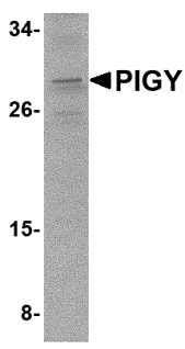
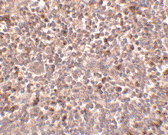
PIG-Y Antibody
PIG-Y Antibody
Catalog# :4945
Glycosylphosphatidylinositol (GPI) lipid anchoring is an important post-translational modification of proteins that takes place in the endoplasmic reticulum. The synthesis of GPI is initiated by GPI-N-acetylglucosaminyltransferase (GPI-GnT), a complex of proteins including PIG-A, PIG-H, PIG-C, GPI1, and DPM2. PIG-Y, the mammalian homolog to yeast Eri1p, is also thought to be involved in the biosynthesis of GPI. The PIG-Y gene encodes two proteins, one of which arises from leaky scanning of the mRNA. This antibody only detects isoform 1 of PIG-Y. Despite its predicted molecular weight, PIG-Y often migrates at 28-30 kDa in SDS-PAGE.
Additional Names : PIG-Y (CT), Phosphatidylinositol glycan anchor biosynthesis class Y, PreY, PIGY
 Description
Left: Western blot analysis of PIG-Y in A-20 cell lysate with PIG-Y antibody at 2 µg/ml.
Below: Immunohistochemistry of PIGY in human spleen tissue with PIGY antibody at 2.5 µg/ml.
Other Product Images
Source : PIG-Y antibody was raised against a 16 amino acid peptide near the carboxy terminus of human PIG-Y.
Purification : Affinity chromatography purified via peptide column
Clonality and Clone : This is a polyclonal antibody.
Host : PIG-Y antibody was raised in rabbit. Please use anti-rabbit secondary antibodies.
Application : PIG-Y antibody can be used for detection of PIG-Y by Western blot at 1 – 2 µg/ml.
Tested Application(s) : E, WB, IHC
Buffer : Antibody is supplied in PBS containing 0.02% sodium azide.
Blocking Peptide :Cat.No. 4945P - PIG-Y Peptide
Long-Term Storage : PIG-Y antibody can be stored at 4ºC, stable for one year. As with all antibodies care should be taken to avoid repeated freeze thaw cycles. Antibodies should not be exposed to prolonged high temperatures.
Positive Control :
1. Cat. No. 1288 - A20 Cell Lysate
Species Reactivity : H, M
GI Number : 14249680
Accession Number : NP_116295
Short Description : (CT) Phosphatidylinositol glycan anchor biosynthesis class Y
References
1. Eisenhaber B, Maurer-Stroh S, Novatchkova M, et al. Enzymes and auxiliary factors for GPI lipid anchor biosynthesis and post-translational transfer to proteins. Bioessays 2003; 25:367-85.
2. Watanabe R, Murakami Y, Marmor MD, et al. Initial enzyme for glycosylphosphatidylinositol biosynthesis requires PIG-P and is regulated by DPM2. EMBO J. 2000; 19:4402-11.
3. Murakami Y, Siripanyaphinoyo U, Hong Y, et al. The initial enzyme for glycosylphosphatidylinositol biosynthesis requires PIG-Y, a seventh component. Mol. Biol. Cell 2005; 16:5236-46.
Catalog# :4945
Glycosylphosphatidylinositol (GPI) lipid anchoring is an important post-translational modification of proteins that takes place in the endoplasmic reticulum. The synthesis of GPI is initiated by GPI-N-acetylglucosaminyltransferase (GPI-GnT), a complex of proteins including PIG-A, PIG-H, PIG-C, GPI1, and DPM2. PIG-Y, the mammalian homolog to yeast Eri1p, is also thought to be involved in the biosynthesis of GPI. The PIG-Y gene encodes two proteins, one of which arises from leaky scanning of the mRNA. This antibody only detects isoform 1 of PIG-Y. Despite its predicted molecular weight, PIG-Y often migrates at 28-30 kDa in SDS-PAGE.
Additional Names : PIG-Y (CT), Phosphatidylinositol glycan anchor biosynthesis class Y, PreY, PIGY
DescriptionLeft: Western blot analysis of PIG-Y in A-20 cell lysate with PIG-Y antibody at 2 µg/ml.
Below: Immunohistochemistry of PIGY in human spleen tissue with PIGY antibody at 2.5 µg/ml.
Other Product Images
Source : PIG-Y antibody was raised against a 16 amino acid peptide near the carboxy terminus of human PIG-Y.
Purification : Affinity chromatography purified via peptide column
Clonality and Clone : This is a polyclonal antibody.
Host : PIG-Y antibody was raised in rabbit. Please use anti-rabbit secondary antibodies.
Application : PIG-Y antibody can be used for detection of PIG-Y by Western blot at 1 – 2 µg/ml.
Tested Application(s) : E, WB, IHC
Buffer : Antibody is supplied in PBS containing 0.02% sodium azide.
Blocking Peptide :Cat.No. 4945P - PIG-Y Peptide
Long-Term Storage : PIG-Y antibody can be stored at 4ºC, stable for one year. As with all antibodies care should be taken to avoid repeated freeze thaw cycles. Antibodies should not be exposed to prolonged high temperatures.
Positive Control :
1. Cat. No. 1288 - A20 Cell Lysate
Species Reactivity : H, M
GI Number : 14249680
Accession Number : NP_116295
Short Description : (CT) Phosphatidylinositol glycan anchor biosynthesis class Y
References
1. Eisenhaber B, Maurer-Stroh S, Novatchkova M, et al. Enzymes and auxiliary factors for GPI lipid anchor biosynthesis and post-translational transfer to proteins. Bioessays 2003; 25:367-85.
2. Watanabe R, Murakami Y, Marmor MD, et al. Initial enzyme for glycosylphosphatidylinositol biosynthesis requires PIG-P and is regulated by DPM2. EMBO J. 2000; 19:4402-11.
3. Murakami Y, Siripanyaphinoyo U, Hong Y, et al. The initial enzyme for glycosylphosphatidylinositol biosynthesis requires PIG-Y, a seventh component. Mol. Biol. Cell 2005; 16:5236-46.
PCDH12 Antibody
PCDH12 Antibody
Catalog# :5071
Protocadherins comprise the largest group within the cadherin family of calcium-dependent cell-cell adhesion molecules. Protocadherin 12 (PCDH12) was initially identified through PCR screening of mouse heart microvascular endothelial cell RNA; further experiments revealed its mRNA to be strongly expressed in highly vascularized organs such as lung and kidney, in addition to glycogen-rich trophoblasts in the placenta. PCDH12-null mice are viable and fertile, but show reduced placental and embryonic sizes when compared to wild-type mice. Further studies showed significant expression changes in 2,289 genes, including those involved in tissue morphogenesis, angiogenesis, cell-matrix adhesion and migration, immune response and chromatin remodeling. This antibody is predicted to not cross-react with PCDH18.
Additional Names : PCDH12, Protocadherin 12, vascular endothelial cadherin 2, VE-cadherin 2, VECAD2
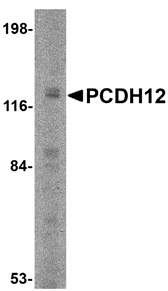 Description
Left: Western blot analysis of PCDH12 in K562 cell lysate with PCDH12 antibody at 2 µg/ml.
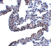
Below: Immunohistochemistry of PCDH12 in rat lung tissue with PCDH12 antibody at 5 µg/ml.
Other Product Images
Source :
PCDH12 antibody was raised against a 17 amino acid peptide near the amino terminus of human PCDH12.
Purification : Affinity chromatography purified via peptide column
Clonality and Clone : This is a polyclonal antibody.
Host : PCDH12 antibody was raised in rabbit. Please use anti-rabbit secondary antibodies.
Application : PCDH12 antibody can be used for detection of PCDH12 by Western blot at 1 – 2 µg/ml.
Tested Application(s) : E, WB, IHC
Buffer : Antibody is supplied in PBS containing 0.02% sodium azide.
Blocking Peptide :Cat.No. 5071P - PCDH12 Peptide
Long-Term Storage : PCDH12 antibody can be stored at 4ºC, stable for one year. As with all antibodies care should be taken to avoid repeated freeze thaw cycles. Antibodies should not be exposed to prolonged high temperatures.
Positive Control :
1. Cat. No. 1204 - K562 Cell Lysate
Species Reactivity : H, M, R
GI Number : 7706113
Accession Number : NP_057664
Short Description : Protocadherin 12
References
1. Frank M and Kemler R. Protocadherins. Curr. Opin. Cell Biol. 2002; 14:557-62.
2. Telo P, Breviaro F, Huber P, et al. Identification of a novel cadherin (vascular endothelial cadherin-2) located at intercellular junctions in endothelial cells. J. Biol. Chem. 1998; 28:17565-72.
3. Rampon C, Prandini MH, Bouillot S, et al. Protocadherin 12 (VE-cadherin 2) is expressed in endothelial, trophoblast, and mesangial cells. Exp. Cell Res. 2005; 302:48-60.
4. Rampon C, Bouillot S, Climescu-Haulica A, et al. Protocadherin 12 deficiency alters morphogenesis and transcriptional profile of the placenta. Physiol. Genomics 2008; 34:193-204.
Catalog# :5071
Protocadherins comprise the largest group within the cadherin family of calcium-dependent cell-cell adhesion molecules. Protocadherin 12 (PCDH12) was initially identified through PCR screening of mouse heart microvascular endothelial cell RNA; further experiments revealed its mRNA to be strongly expressed in highly vascularized organs such as lung and kidney, in addition to glycogen-rich trophoblasts in the placenta. PCDH12-null mice are viable and fertile, but show reduced placental and embryonic sizes when compared to wild-type mice. Further studies showed significant expression changes in 2,289 genes, including those involved in tissue morphogenesis, angiogenesis, cell-matrix adhesion and migration, immune response and chromatin remodeling. This antibody is predicted to not cross-react with PCDH18.
Additional Names : PCDH12, Protocadherin 12, vascular endothelial cadherin 2, VE-cadherin 2, VECAD2
DescriptionLeft: Western blot analysis of PCDH12 in K562 cell lysate with PCDH12 antibody at 2 µg/ml.
Below: Immunohistochemistry of PCDH12 in rat lung tissue with PCDH12 antibody at 5 µg/ml.
Other Product Images
Source :
PCDH12 antibody was raised against a 17 amino acid peptide near the amino terminus of human PCDH12.
Purification : Affinity chromatography purified via peptide column
Clonality and Clone : This is a polyclonal antibody.
Host : PCDH12 antibody was raised in rabbit. Please use anti-rabbit secondary antibodies.
Application : PCDH12 antibody can be used for detection of PCDH12 by Western blot at 1 – 2 µg/ml.
Tested Application(s) : E, WB, IHC
Buffer : Antibody is supplied in PBS containing 0.02% sodium azide.
Blocking Peptide :Cat.No. 5071P - PCDH12 Peptide
Long-Term Storage : PCDH12 antibody can be stored at 4ºC, stable for one year. As with all antibodies care should be taken to avoid repeated freeze thaw cycles. Antibodies should not be exposed to prolonged high temperatures.
Positive Control :
1. Cat. No. 1204 - K562 Cell Lysate
Species Reactivity : H, M, R
GI Number : 7706113
Accession Number : NP_057664
Short Description : Protocadherin 12
References
1. Frank M and Kemler R. Protocadherins. Curr. Opin. Cell Biol. 2002; 14:557-62.
2. Telo P, Breviaro F, Huber P, et al. Identification of a novel cadherin (vascular endothelial cadherin-2) located at intercellular junctions in endothelial cells. J. Biol. Chem. 1998; 28:17565-72.
3. Rampon C, Prandini MH, Bouillot S, et al. Protocadherin 12 (VE-cadherin 2) is expressed in endothelial, trophoblast, and mesangial cells. Exp. Cell Res. 2005; 302:48-60.
4. Rampon C, Bouillot S, Climescu-Haulica A, et al. Protocadherin 12 deficiency alters morphogenesis and transcriptional profile of the placenta. Physiol. Genomics 2008; 34:193-204.
OVGP1 Antibody
OVGP1 Antibody
Catalog# :4763
Oviductins belong to a family of glycoproteins that have been suggested to play several roles during the early processes of reproduction. OVGP1 is a large carbohydrate-rich, epithelial glycoprotein with numerous O-glycosylation sites within threonine, serine, and proline-rich tandem repeats. The protein is secreted from non-ciliated oviductal epithelial cells and associates with ovulated oocytes, blastomeres, and spermatozoan acrosomal regions. Despite its predicted molecular weight, OVGP1 will often run at higher molecular weight in SDS-PAGE. At least two isoforms of OVGP are known to exist.
Additional Names : OVGP1, OVGP, Oviductin, oviduct-specific glycoprotein, muc9
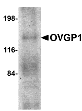 Description
Left: Western blot analysis of OVGP1 in human placenta tissue lysate with OVGP1 antibody at 1 µg/ml.
Below: Immunocytochemistry of OVGP1 in 293 cells with OVGP1L antibody at 2.5 μg/ml.
Other Product Images
 Source :
OVGP1 antibody was raised against a 18 amino acid peptide from near the amino terminus of human OVGP1.
Purification : Affinity chromatography purified via peptide column
Clonality and Clone : This is a polyclonal antibody.
Host : OVGP1 antibody was raised in rabbit. Please use anti-rabbit secondary antibodies.
Application : OVGP1 antibody can be used for the detection of OVGP1 by Western blot at 1 µg/ml.
Tested Application(s) : E, WB
Buffer : Antibody is supplied in PBS containing 0.02% sodium azide.
Blocking Peptide :Cat.No. 4763P - OVGP1 Peptide
Long-Term Storage : OVGP1 antibody can be stored at 4ºC, stable for one year. As with all antibodies care should be taken to avoid repeated freeze thaw cycles. Antibodies should not be exposed to prolonged high temperatures.
Positive Control :
1. Cat. No. 1309 - Human Placenta Lysate
Species Reactivity : H, M, R
GI Number : 58386720
Accession Number : NP_002548
Short Description : an oviduct-specific protein
References
1. Buhi WC. Characterization and biological roles of oviduct-specific, oestrogen-dependent glycoprotein. Reproduction 123:355-62.
2. Arias EB, Verhage HG and Jaffe RC. Complementary deoxyribonucleic acid cloning and molecular characterization of an estrogen-dependent human oviductal glycoprotein. Biol. Reprod. 1994; 51:685-94.
Catalog# :4763
Oviductins belong to a family of glycoproteins that have been suggested to play several roles during the early processes of reproduction. OVGP1 is a large carbohydrate-rich, epithelial glycoprotein with numerous O-glycosylation sites within threonine, serine, and proline-rich tandem repeats. The protein is secreted from non-ciliated oviductal epithelial cells and associates with ovulated oocytes, blastomeres, and spermatozoan acrosomal regions. Despite its predicted molecular weight, OVGP1 will often run at higher molecular weight in SDS-PAGE. At least two isoforms of OVGP are known to exist.
Additional Names : OVGP1, OVGP, Oviductin, oviduct-specific glycoprotein, muc9
DescriptionLeft: Western blot analysis of OVGP1 in human placenta tissue lysate with OVGP1 antibody at 1 µg/ml.
Below: Immunocytochemistry of OVGP1 in 293 cells with OVGP1L antibody at 2.5 μg/ml.
Other Product Images
Source :OVGP1 antibody was raised against a 18 amino acid peptide from near the amino terminus of human OVGP1.
Purification : Affinity chromatography purified via peptide column
Clonality and Clone : This is a polyclonal antibody.
Host : OVGP1 antibody was raised in rabbit. Please use anti-rabbit secondary antibodies.
Application : OVGP1 antibody can be used for the detection of OVGP1 by Western blot at 1 µg/ml.
Tested Application(s) : E, WB
Buffer : Antibody is supplied in PBS containing 0.02% sodium azide.
Blocking Peptide :Cat.No. 4763P - OVGP1 Peptide
Long-Term Storage : OVGP1 antibody can be stored at 4ºC, stable for one year. As with all antibodies care should be taken to avoid repeated freeze thaw cycles. Antibodies should not be exposed to prolonged high temperatures.
Positive Control :
1. Cat. No. 1309 - Human Placenta Lysate
Species Reactivity : H, M, R
GI Number : 58386720
Accession Number : NP_002548
Short Description : an oviduct-specific protein
References
1. Buhi WC. Characterization and biological roles of oviduct-specific, oestrogen-dependent glycoprotein. Reproduction 123:355-62.
2. Arias EB, Verhage HG and Jaffe RC. Complementary deoxyribonucleic acid cloning and molecular characterization of an estrogen-dependent human oviductal glycoprotein. Biol. Reprod. 1994; 51:685-94.
op18 Antibody
op18 Antibody
Catalog# :4237
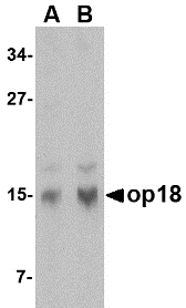
Op18 belongs to the stathmin family of genes and encodes a ubiquitous cytosolic phosphoprotein that may function as an intracellular relay integrating several signaling pathways such as those involved in cell proliferation and differentiation. Op18 has also been shown to be involved in the regulation of the microtubule filament system by destabilizing microtubules, thereby preventing assembly and promoting the disassembly of microtubules. More recently, op18 has been implicated as a potential target of the ASK1-p38 MAP kinase cascade, suggesting that the ASK1-p38 cascade may regulate microtubule dynamics through op18. Op18 is highly expressed in a wide variety of human malignancies, including leukemia, prostate cancer, ovarian carcinoma, and breast carcinoma, suggesting that op18 may be an ideal target for anti-cancer therapeutics.
Additional Names : op18 (NT), oncoprotein 18, STMN1, Stathmin 1, Lag, LAP18
 Description
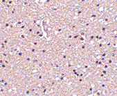
Left: Western blot analysis of op18 in human brain tissue lysate with op18 antibody at (A) 0.5 and (B) 1 µg/ml.
Below: Immunohistochemistry of op18 in human brain tissue with op18 antibody at 2.5 µg/ml.
Other Product Images
Source : Op18 antibody was raised against a 17 amino acid peptide from near the amino terminus of human op18.
Purification : Affinity chromatography purified via peptide column
Clonality and Clone : This is a polyclonal antibody.
Host : op18 antibody was raised in rabbit. Please use anti-rabbit secondary antibodies.
Application : Op18 antibody can be used for detection of op18 by Western blot at 0.5 – 1 µg/ml.
Tested Application(s) : E, WB, IHC
Buffer : Antibody is supplied in PBS containing 0.02% sodium azide.
Blocking Peptide :Cat.No. 4237P - op18 Peptide
Long-Term Storage : op18 antibody can be stored at 4ºC, stable for one year. As with all antibodies care should be taken to avoid repeated freeze thaw cycles. Antibodies should not be exposed to prolonged high temperatures.
Positive Control :
1. Cat. No. 1303 - Human Brain Tissue Lysate
Species Reactivity : H, M, R
GI Number : 51895905
Accession Number : AAH82228
Short Description : (NT) a member of the Stahmin family of proteins
References
1. Curmi PA, Gavet O, Charbaut E, et al. Stathmin and its phosphoprotein family: general properties, biochemical and functional interaction with tubulin. Cell Structure and Function 1999; 24:345-57.
2. Belmont LD and Mitchison TJ. Identification of a protein that interacts with tubulin dimers and increases catastrophe rate of microtubules. Cell 1996; 84:623-31.
3. Mizumura K, Takeda K, Hashimoto S, et al. Identification of op18/stathmin as a potential target of ASK1-p38 MAP kinase cascade. J. Cell Physiol. 2006; 206:363-70.
4. Mistry SJ and Atweh GF. Role of stathmin in the regulation of the mitotic spindle. Mount Sinai J. Med. 2002; 69:299-304
Catalog# :4237
Op18 belongs to the stathmin family of genes and encodes a ubiquitous cytosolic phosphoprotein that may function as an intracellular relay integrating several signaling pathways such as those involved in cell proliferation and differentiation. Op18 has also been shown to be involved in the regulation of the microtubule filament system by destabilizing microtubules, thereby preventing assembly and promoting the disassembly of microtubules. More recently, op18 has been implicated as a potential target of the ASK1-p38 MAP kinase cascade, suggesting that the ASK1-p38 cascade may regulate microtubule dynamics through op18. Op18 is highly expressed in a wide variety of human malignancies, including leukemia, prostate cancer, ovarian carcinoma, and breast carcinoma, suggesting that op18 may be an ideal target for anti-cancer therapeutics.
Additional Names : op18 (NT), oncoprotein 18, STMN1, Stathmin 1, Lag, LAP18
DescriptionLeft: Western blot analysis of op18 in human brain tissue lysate with op18 antibody at (A) 0.5 and (B) 1 µg/ml.
Below: Immunohistochemistry of op18 in human brain tissue with op18 antibody at 2.5 µg/ml.
Other Product Images
Source : Op18 antibody was raised against a 17 amino acid peptide from near the amino terminus of human op18.
Purification : Affinity chromatography purified via peptide column
Clonality and Clone : This is a polyclonal antibody.
Host : op18 antibody was raised in rabbit. Please use anti-rabbit secondary antibodies.
Application : Op18 antibody can be used for detection of op18 by Western blot at 0.5 – 1 µg/ml.
Tested Application(s) : E, WB, IHC
Buffer : Antibody is supplied in PBS containing 0.02% sodium azide.
Blocking Peptide :Cat.No. 4237P - op18 Peptide
Long-Term Storage : op18 antibody can be stored at 4ºC, stable for one year. As with all antibodies care should be taken to avoid repeated freeze thaw cycles. Antibodies should not be exposed to prolonged high temperatures.
Positive Control :
1. Cat. No. 1303 - Human Brain Tissue Lysate
Species Reactivity : H, M, R
GI Number : 51895905
Accession Number : AAH82228
Short Description : (NT) a member of the Stahmin family of proteins
References
1. Curmi PA, Gavet O, Charbaut E, et al. Stathmin and its phosphoprotein family: general properties, biochemical and functional interaction with tubulin. Cell Structure and Function 1999; 24:345-57.
2. Belmont LD and Mitchison TJ. Identification of a protein that interacts with tubulin dimers and increases catastrophe rate of microtubules. Cell 1996; 84:623-31.
3. Mizumura K, Takeda K, Hashimoto S, et al. Identification of op18/stathmin as a potential target of ASK1-p38 MAP kinase cascade. J. Cell Physiol. 2006; 206:363-70.
4. Mistry SJ and Atweh GF. Role of stathmin in the regulation of the mitotic spindle. Mount Sinai J. Med. 2002; 69:299-304
Wednesday, December 15, 2010
Primer Hybridization
Primers are short artificial DNA strands that are used in genetic methods such as DNA sequencing, hybridization probes, and Polymerase Chain Reaction (PCR). The actual construction of primers starts with 3'-hydroxyl nucleosides attached to a so-called Controlled-Pore Glass (CPG). The 5'-hydroxyl of the nucleosides is covered dimethoxytrityl (DMT), which prevents the building of a nucleotide chain. To add a nucleotide, DMT is chemically removed removed, and the nucleotide is added. The 5'-hydroxyl of the new nucleotide is blocked by DMT, preventing the addition of more than one nucleotide to each chain.
Polymerase Chain Reaction
Polymerase chain reaction (PCR) is a technique widely used in molecular biology. It derives its name from one of its key components, a DNA polymerase used to amplify a piece of DNA by in vitro enzymatic replication. As PCR progresses, the DNA thus generated is itself used as a template for replication. This sets in motion a chain reaction in which the DNA template is exponentially amplified. With PCR it is possible to amplify a single or few copies of a piece of DNA across several orders of magnitude, generating millions or more copies of the DNA piece. PCR can be extensively modified to perform a wide array of genetic manipulations.
PCR Optimization
In practice, PCR can fail for various reasons, in part due to its sensitivity to contamination causing amplification of spurious DNA products. Because of this, a number of techniques and procedures have been developed for optimizing PCR conditions. Contamination with extraneous DNA is addressed with lab protocols and procedures that separate pre-PCR mixtures from potential DNA contaminants. This usually involves spatial separation of PCR-setup areas from areas for analysis or purification of PCR products, and thoroughly cleaning the work surface between reaction setups.
Tuesday, December 14, 2010
DNA RNA Protein
Tanscription is the synthesis of RNA under the direction of DNA. RNA synthesis, or transcription, is the process of transcribing DNA nucleotide sequence information into RNA sequence information. Both nucleic acid sequences use complementary language, and the information is simply transcribed, or copied, from one molecule to the other. DNA sequence is enzymatic ally copied by RNA polymerase to produce a complementary nucleotide RNA strand, called messenger RNA (mRNA), because it carries a genetic message from the DNA to the protein-synthesizing machinery of the cell.
Protein Biosynthesis
Protein biosynthesis (synthesis) is the process in which cells build proteins. The term is sometimes used to refer only to protein translation but more often it refers to a multi-step process, beginning with amino acid synthesis and transcription which are then used for translation. Protein biosynthesis, although very similar, differs between prokaryotes and eukaryotes.
DNA Replication
As in DNA replication, RNA is synthesized in the 5' → 3' direction (from the point of view of the growing RNA transcript). Only one of the two DNA strands is transcribed. This strand is called the template strand, because it provides the template for ordering the sequence of nucleotides in an RNA transcript. The other strand is called the coding strand, because its sequence is the same as the newly created RNA transcript.
Promoter Clearance
After the first bond is synthesized the RNA polymerase must clear the promoter. During this time there is a tendency to release the RNA transcript and produce truncated transcripts. This is called abortive initiation and is common for both Eukaryotes and Prokaroytes. Once the transcript reaches approximately 23 nucleotides it no longer slips and Elongation can occur. This is an ATP dependent process.Promoter clearance also coincides with Phosphorylation of serine 5 on the carboxy terminal domain which is phosphorylated by TFIIH.
Monday, December 13, 2010
DNA Maternity Test
A maternity or paternity identification test is conducted to establish whether a person is the biological parent of another person. A test to prove paternity (whether a man is someone's father) is known as a paternity test; a test to prove maternity (whether a woman is someone's mother) is called a maternity test. Although paternity tests are more common than maternity tests, there may be circumstances in which the biological mother of the child is unclear. Examples include cases of an adopted child attempting to reunify with his or her biological mother, potential hospital mix-ups, and in vitro fertilization where the laboratory may have implanted an unrelated embryo inside the mother.
Maternity Test
Maternity DNA testing determines whether a woman could be the biological mother of a child. Like a DNA paternity test, it compares a child’s DNA pattern with that of the alleged mother to determine how likely it is that the child has inherited the DNA from the alleged mother.
DNA Sample Collection
Samples for nearly all our DNA tests are collected using the simple and pain-free buccal swab technique. Buccal swabs are similar to the cotton wool buds found in most people’s bathroom cupboards. During DNA sample collection, four buccal swabs are rubbed against the inside of the person’s cheeks (two swabs are used on each cheek). The rubbing motion gathers loose cheek cells, and these cells contain the DNA used in the genetic test.
op18 Antibody
op18 Antibody
Catalog# :4235
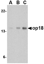
Op18 belongs to the stathmin family of genes and encodes a ubiquitous cytosolic phosphoprotein that may function as an intracellular relay integrating several signaling pathways such as those involved in cell proliferation and differentiation. Op18 has also been shown to be involved in the regulation of the microtubule filament system by destabilizing microtubules, thereby preventing assembly and promoting the disassembly of microtubules. More recently, op18 has been implicated as a potential target of the ASK1-p38 MAP kinase cascade, suggesting that the ASK1-p38 cascade may regulate microtubule dynamics through op18. Op18 is highly expressed in a wide variety of human malignancies, including leukemia, prostate cancer, ovarian carcinoma, and breast carcinoma, suggesting that op18 may be an ideal target for anti-cancer therapeutics.
Additional Names : op18 (CT), oncoprotein 18, STMN1, Stathmin 1, Lag, LAP18
 Description
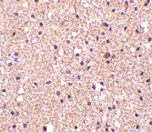
Left: Western blot analysis of op18 in EL4 cell lysate with op18 antibody at (A) 0.5, (B) 1 and (C) 2 µg/ml.
Below: Immunohistochemistry of op18 in human brain tissue with op18 antibody at 2.5 µg/ml.
Other Product Images
 Source : Op18 antibody was raised against a 20 amino acid peptide from near the carboxy terminus of human op18.
Purification : Affinity chromatography purified via peptide column
Clonality and Clone : This is a polyclonal antibody.
Host : op18 antibody was raised in rabbit. Please use anti-rabbit secondary antibodies.
Application : Op18 antibody can be used for detection of op18 by Western blot at 0.5 – 1 µg/ml.
Tested Application(s) : E, WB, IHC
Buffer : Antibody is supplied in PBS containing 0.02% sodium azide.
Blocking Peptide :Cat.No. 4235P - op18 Peptide
Long-Term Storage : op18 antibody can be stored at 4ºC, stable for one year. As with all antibodies care should be taken to avoid repeated freeze thaw cycles. Antibodies should not be exposed to prolonged high temperatures.
Positive Control :
1.Cat. No. 1287 - EL4 Lysate
2.Cat. No. 1303 - Human Brain Tissue Lysate
Species Reactivity : H, M
GI Number : 51895905
Accession Number : AAH82228
Short Description : (CT) a member of the Stahmin family of proteins
References
1. Curmi PA, Gavet O, Charbaut E, et al. Stathmin and its phosphoprotein family: general properties, biochemical and functional interaction with tubulin. Cell Structure and Function 1999; 24:345-57.
2. Belmont LD and Mitchison TJ. Identification of a protein that interacts with tubulin dimers and increases catastrophe rate of microtubules. Cell 1996; 84:623-31.
3. Mizumura K, Takeda K, Hashimoto S, et al. Identification of op18/stathmin as a potential target of ASK1-p38 MAP kinase cascade. J. Cell Physiol. 2006; 206:363-70.
4. Mistry SJ and Atweh GF. Role of stathmin in the regulation of the mitotic spindle. Mount Sinai J. Med. 2002; 69:299-304.
Catalog# :4235
Op18 belongs to the stathmin family of genes and encodes a ubiquitous cytosolic phosphoprotein that may function as an intracellular relay integrating several signaling pathways such as those involved in cell proliferation and differentiation. Op18 has also been shown to be involved in the regulation of the microtubule filament system by destabilizing microtubules, thereby preventing assembly and promoting the disassembly of microtubules. More recently, op18 has been implicated as a potential target of the ASK1-p38 MAP kinase cascade, suggesting that the ASK1-p38 cascade may regulate microtubule dynamics through op18. Op18 is highly expressed in a wide variety of human malignancies, including leukemia, prostate cancer, ovarian carcinoma, and breast carcinoma, suggesting that op18 may be an ideal target for anti-cancer therapeutics.
Additional Names : op18 (CT), oncoprotein 18, STMN1, Stathmin 1, Lag, LAP18
DescriptionLeft: Western blot analysis of op18 in EL4 cell lysate with op18 antibody at (A) 0.5, (B) 1 and (C) 2 µg/ml.
Below: Immunohistochemistry of op18 in human brain tissue with op18 antibody at 2.5 µg/ml.
Other Product Images
Source : Op18 antibody was raised against a 20 amino acid peptide from near the carboxy terminus of human op18.Purification : Affinity chromatography purified via peptide column
Clonality and Clone : This is a polyclonal antibody.
Host : op18 antibody was raised in rabbit. Please use anti-rabbit secondary antibodies.
Application : Op18 antibody can be used for detection of op18 by Western blot at 0.5 – 1 µg/ml.
Tested Application(s) : E, WB, IHC
Buffer : Antibody is supplied in PBS containing 0.02% sodium azide.
Blocking Peptide :Cat.No. 4235P - op18 Peptide
Long-Term Storage : op18 antibody can be stored at 4ºC, stable for one year. As with all antibodies care should be taken to avoid repeated freeze thaw cycles. Antibodies should not be exposed to prolonged high temperatures.
Positive Control :
1.Cat. No. 1287 - EL4 Lysate
2.Cat. No. 1303 - Human Brain Tissue Lysate
Species Reactivity : H, M
GI Number : 51895905
Accession Number : AAH82228
Short Description : (CT) a member of the Stahmin family of proteins
References
1. Curmi PA, Gavet O, Charbaut E, et al. Stathmin and its phosphoprotein family: general properties, biochemical and functional interaction with tubulin. Cell Structure and Function 1999; 24:345-57.
2. Belmont LD and Mitchison TJ. Identification of a protein that interacts with tubulin dimers and increases catastrophe rate of microtubules. Cell 1996; 84:623-31.
3. Mizumura K, Takeda K, Hashimoto S, et al. Identification of op18/stathmin as a potential target of ASK1-p38 MAP kinase cascade. J. Cell Physiol. 2006; 206:363-70.
4. Mistry SJ and Atweh GF. Role of stathmin in the regulation of the mitotic spindle. Mount Sinai J. Med. 2002; 69:299-304.
NUP160 Antibody
NUP160 Antibody
Catalog# :4707
The nuclear pore complex (NPC) is a protein assembly localized at the nuclear rim and mediates macromolecular transport between the nucleus and the cytoplasm. The mammalian nucleoporin (NUP)-160 is part of the hetero-oligomeric complex that also contains NUP107, NUP133, NUP96, and mammalian homolog of yeast sec13p. While the majority of the NUP107-160 nuclear pore sub-complex localizes to the nuclear pore, a small fraction is observed at kinetochores and pro-metaphase spindle poles in mitotic cells in association with proteins such as Mad1, Mad2, Bub3 and Cdc20. Immunodepletion of the NUP107-160 complex resulted in defective spindle assembly indicating that it has multiple functions. NUP160 has recently been identified as an HIV dependency factor (HDF), suggesting that NUP160 may be an important drug target in HIV treatment. Multiple isoforms of NUP160 are known to exist.
Additional Names : NUP160 (CT), Nucleoporin 160, nuclear pore complex 160
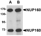 Description
Left: Western blot analysis of NUP160 in rat brain tissue lysate with NUP160 antibody at (A) 0.5 and (B) 1 µg/ml.
Below: Immunohistochemistry of NUP160 in human brain tissue with NUP160 antibody at 2.5 μg/ml.
Other Product Images
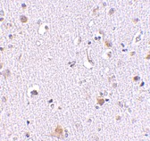 Source :NUP160 antibody was raised against a 14 amino acid peptide from near the carboxy terminus of human NUP160.
Purification : Affinity chromatography purified via peptide column
Clonality and Clone : This is a polyclonal antibody.
Host : NUP160 antibody was raised in rabbit. Please use anti-rabbit secondary antibodies.
Application : NUP160 antibody can be used for detection of NUP160 by Western blot at 0.5 – 1 µg/ml.
Tested Application(s) : E, WB
Buffer : Antibody is supplied in PBS containing 0.02% sodium azide.
Blocking Peptide :Cat.No. 4707P - NUP160 Peptide
Long-Term Storage : NUP160 antibody can be stored at 4ºC, stable for one year. As with all antibodies care should be taken to avoid repeated freeze thaw cycles. Antibodies should not be exposed to prolonged high temperatures.
Positive Control :
1. Cat. No. 1203 - A549 Cell Lysate
Species Reactivity : H, M, R
GI Number : 119588287
Accession Number : EAW67881
Short Description : (CT) Nucleoporin 160
References
1. Tran EJ and Wente SR. Dynamic nuclear pore complex: life on the edge. Cell 2006; 125:1041-53.
2. Boehmer T, Enninga J, Dales S, et al. Depletion of a single nucleoporin, Nup107, prevents the assembly of a subset of nucleoporins into the nuclear pore complex. Proc. Natl. Acad. Sci. USA 2003; 100:981-5.
3. Orjalo AV, Arnaoutov A, Shen Z, et al. The Nup107-160 nucleoporin complex is required for correct bipolar spindle assembly. Mol. Bio. Cell 2006; 17:3806-18.
4. Brass AL, Dykxhoorn DM, Benita Y, et al. Identification of host proteins required for HIV infection through a functional genomic screen. Science 2008; 319:921-6.
Catalog# :4707
The nuclear pore complex (NPC) is a protein assembly localized at the nuclear rim and mediates macromolecular transport between the nucleus and the cytoplasm. The mammalian nucleoporin (NUP)-160 is part of the hetero-oligomeric complex that also contains NUP107, NUP133, NUP96, and mammalian homolog of yeast sec13p. While the majority of the NUP107-160 nuclear pore sub-complex localizes to the nuclear pore, a small fraction is observed at kinetochores and pro-metaphase spindle poles in mitotic cells in association with proteins such as Mad1, Mad2, Bub3 and Cdc20. Immunodepletion of the NUP107-160 complex resulted in defective spindle assembly indicating that it has multiple functions. NUP160 has recently been identified as an HIV dependency factor (HDF), suggesting that NUP160 may be an important drug target in HIV treatment. Multiple isoforms of NUP160 are known to exist.
Additional Names : NUP160 (CT), Nucleoporin 160, nuclear pore complex 160
DescriptionLeft: Western blot analysis of NUP160 in rat brain tissue lysate with NUP160 antibody at (A) 0.5 and (B) 1 µg/ml.
Below: Immunohistochemistry of NUP160 in human brain tissue with NUP160 antibody at 2.5 μg/ml.
Other Product Images
Source :NUP160 antibody was raised against a 14 amino acid peptide from near the carboxy terminus of human NUP160.Purification : Affinity chromatography purified via peptide column
Clonality and Clone : This is a polyclonal antibody.
Host : NUP160 antibody was raised in rabbit. Please use anti-rabbit secondary antibodies.
Application : NUP160 antibody can be used for detection of NUP160 by Western blot at 0.5 – 1 µg/ml.
Tested Application(s) : E, WB
Buffer : Antibody is supplied in PBS containing 0.02% sodium azide.
Blocking Peptide :Cat.No. 4707P - NUP160 Peptide
Long-Term Storage : NUP160 antibody can be stored at 4ºC, stable for one year. As with all antibodies care should be taken to avoid repeated freeze thaw cycles. Antibodies should not be exposed to prolonged high temperatures.
Positive Control :
1. Cat. No. 1203 - A549 Cell Lysate
Species Reactivity : H, M, R
GI Number : 119588287
Accession Number : EAW67881
Short Description : (CT) Nucleoporin 160
References
1. Tran EJ and Wente SR. Dynamic nuclear pore complex: life on the edge. Cell 2006; 125:1041-53.
2. Boehmer T, Enninga J, Dales S, et al. Depletion of a single nucleoporin, Nup107, prevents the assembly of a subset of nucleoporins into the nuclear pore complex. Proc. Natl. Acad. Sci. USA 2003; 100:981-5.
3. Orjalo AV, Arnaoutov A, Shen Z, et al. The Nup107-160 nucleoporin complex is required for correct bipolar spindle assembly. Mol. Bio. Cell 2006; 17:3806-18.
4. Brass AL, Dykxhoorn DM, Benita Y, et al. Identification of host proteins required for HIV infection through a functional genomic screen. Science 2008; 319:921-6.
Subscribe to:
Posts (Atom)