Catalog# :4305
The six-transmembrane epithelial antigen of prostate 1 (STEAP1) was the first member of a family of metalloreductases identified as cell-surface antigens in prostate tissue. The normal function of STEAP is still uncertain; unlike other members of the STEAP family, STEAP1 does not promote iron or copper reduction or uptake and lacks the FNO-like reductase domain critical for activity. However, its expression is highly increased in multiple cancer cell lines, including prostate, bladder, colon, and ovarian cancers. Supporting this is evidence that STEAP1 peptides can be used to stimulate CD8+ T cells from healthy donors, enabling them to recognize STEAP1-positive human tumor cells, suggesting that STEAP1 may a potential target for cancer immunotherapy. At least three isoforms of STEAP1 are known to exist. This STEAP1 antibody does not cross-react with other STEAP proteins.
Additional Names : STEAP1 (IN), Six transmembrane epithelial antigen of prostate 1, STEAP
 Description
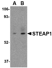
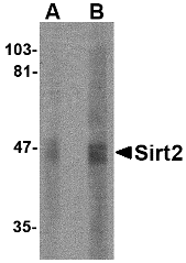
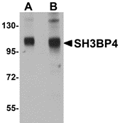
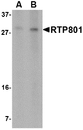
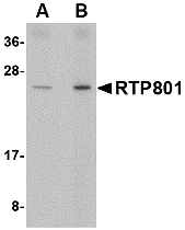
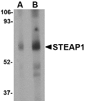
Left: Western blot analysis of STEAP1 in human spleen tissue lysate with STEAP1 antibody at (A) 1 and (B) 2 µg/ml.
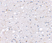
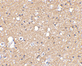
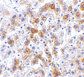
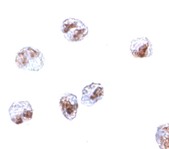
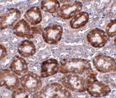
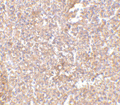
Below: Immunohistochemistry of STEAP1 in human spleen tissue with STEAP1 antibody at 2.5 µg/ml.
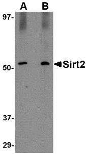
Other Product Images
 Source :STEAP1 antibody was raised against a 16 amino acid peptide from near the center of human STEAP1.
Purification : Affinity chromatography purified via peptide column
Clonality and Clone : This is a polyclonal antibody.
Host : STEAP1 antibody was raised in rabbit. Please use anti-rabbit secondary antibodies.
Application : STEAP1 antibody can be used for detection of STEAP1 by Western blot at 1 – 2 µg/ml.
Tested Application(s) : E, WB, IHC
Buffer : Antibody is supplied in PBS containing 0.02% sodium azide.
Blocking Peptide :Cat.No. 4305P - STEAP1 Peptide
Long-Term Storage : STEAP1 antibody can be stored at 4ºC, stable for one year. As with all antibodies care should be taken to avoid repeated freeze thaw cycles. Antibodies should not be exposed to prolonged high temperatures.
Positive Control :
1. Cat. No. 1306 - Human Spleen Tissue Lysate
Species Reactivity : H, M, R
GI Number : 51094921
Accession Number : EAL24166
Short Description : (IN) Six transmembrane epithelial antigen of prostate 1
References
1. Hubert RS, Vivanco I, Chen E, et al. STEAP: a prostate-specific cell-surface antigen highly expressed in human prostate tumors. Proc. Natl. Acad. Sci. USA 1999; 96:14523-8.
2. Ohgami RS, Campagna DR, McDonald A, et al. The Steap proteins are metalloreductases. Blood 2006; 108:1388-94.
3. Ohgami RS, Campagna DR, Greer EL, et al. Identification of a ferrireductase required for efficient transferrin-dependent iron uptake in erythroid cells. Nat. Genet. 2005; 37:1264-9.
4. Alves PM, Faure O, Graff-Dubois S, et al. STEAP, a prostate tumor antigen, is a target of human CD8+ T cells. Cancer Immunol. Immunother. 2006; 55:1515-23.