Thursday, March 31, 2011

Unc93b Antibody

Unc93b Antibody

Catalog# :4553

The endoplasmic reticulum (ER) protein Unc93b, a human homolog of the C. elegans Unc93 gene, was initially identified by a forward genetic screen using N-ethyl-N-nitrosourea where a histidine-to-arginine substitution in Unc93b caused defects in Toll-like receptor (TLR) 3, 7 and 9 signaling. Unlike Unc93a, another homolog of the C. elegans Unc93 gene whose function is unknown, Unc93b specifically interacts with TLR3, 7 and 9; the histidine-to-arginine point mutation used to identify Unc93b abolishes this interaction. Mice carrying this point mutation are highly susceptible to infection with a number of viruses, indicating that Unc93b plays an important role in innate immunity. Multiple isoforms of Unc93a are known to exist. This antibody will not cross-react with Unc93a.

Additional Names : Unc93b, Unc93b1, homolog of C. elegans Unc93
Description
Left:Western blot analysis of Unc93b in human heart tissue lysate with Unc93b antibody at (A) 0.5 and (B) 1 µg/ml.

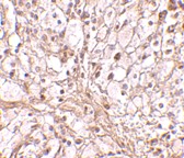
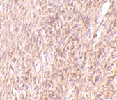
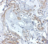
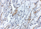
Below:Immunohistochemistry of Unc93b in human heart with Unc93b antibody at 2.5 µg/ml.

Other Product Images
Source :Unc93b antibody was raised against a 19 amino acid peptide from near the amino terminus of human Unc93b.
Purification : Affinity chromatography purified via peptide column
Clonality and Clone : This is a polyclonal antibody.
Host : Unc93b antibody was raised in rabbit.
Please use anti-rabbit secondary antibodies.
Application : Unc93b antibody can be used for the detection of Unc93b by Western blot at 0.5 – 1 µg/ml.
Tested Application(s) : E, WB, IHC
Buffer : Antibody is supplied in PBS containing 0.02% sodium azide.
Blocking Peptide :Cat.No. 4553P - Unc93b Peptide
Long-Term Storage : Unc93b antibody can be stored at 4ºC, stable for one year. As with all antibodies care should be taken to avoid repeated freeze thaw cycles. Antibodies should not be exposed to prolonged high temperatures.
Positive Control :
1. Cat. No. 1301 - Human Heart Tissue Lysate
Species Reactivity : H, M, R
GI Number : 116284350
Accession Number : AAH33623
Short Description : homolog of C. elegans UNC93
References
1. Tabeta K, Hoebe K, Janssen EM, et al. The Unc93b1 mutation 3d disrupts exogenous antigen presentation and signaling via Toll-like receptors 3, 7, and 9. Nat. Immunol. 2006; 7:156-64.
2. Liu Y, Dodds P, Emilion G, et al. The human homolog of unc-93 maps to chromosome 6q27 – characterisation and analysis in sporadic epithelial ovarian cancer. BioMed Central Genetics 2002; 3:20.
3. Brinkmann MM, Spooner E, Hoebe K, et al. The interaction between the ER membrane protein UNC93B and TLR3, 7, and 9 is crucial for TLR signaling. J. Cell Biol. 2007; 177:265-75.

TRIM30 Antibody

TRIM30 Antibody

Catalog# :4745

TRIM30 belongs to a family of the tripartite motif (TRIM) proteins involved in the regulation of cell proliferation, differentiation, development, oncogenesis, apoptosis and antiviral responses. The TRIM protein family is an expanding family of RING ('really interesting new gene') proteins, also known as RBCC proteins as they contain an RBCC motif, which comprises a RING domain, one or two B-boxes and a predicted coiled-coil region. Studies have shown that some TRIM family members are critical to innate immunity; TRIM5, TRIM19 and TRIM25, for example, have been shown to restrict viral infection. A recent study shows that TRIM30 functions as a negative modulator of the TLR signaling pathway, by targeting TAB2 and TAB3, and contributes to the inhibition of TLR-mediated NFkappa-B activation. The importance of TRIM30 in the attenuation or termination of NFkappa-B activation suggests that targeting of TAB2 and TAB3 by TRIM30 alpha may be a mechanism for modulating many types of immune responses.

Additional Names : TRIM30 (CT), Tripartite motif-containing 30, Rpt-1, Rpt1, down regulatory protein of interleukin 2 receptor
Description
Left:Western blot analysis of TRIM30 in mouse spleen tissue lysate with TRIM30 antibody at 1 µg/ml.

Other Product Images
Source :TRIM30 antibody was raised against a 16 amino acid peptide near the carboxy terminus of the mouse TRIM30.
Purification : Affinity chromatography purified via peptide column
Clonality and Clone : This is a polyclonal antibody.
Host : TRIM30 antibody was raised in rabbit.
Please use anti-rabbit secondary antibodies.
Application : TRIM30 antibody can be used for detection of TRIM30 by Western blot at 0.5 - 1 µg/ml.
Tested Application(s) : E, WB
Buffer : Antibody is supplied in PBS containing 0.02% sodium azide.
Blocking Peptide :Cat.No. 4745P - TRIM30 Peptide
Long-Term Storage : TRIM30 antibody can be stored at 4ºC, stable for one year. As with all antibodies care should be taken to avoid repeated freeze thaw cycles. Antibodies should not be exposed to prolonged high temperatures.
Positive Control :
1. Cat. No. 1406 - Mouse Spleen Tissue Lysate
Species Reactivity : M
GI Number : 125347379
Accession Number : NP_033125
Short Description : (CT) Tripartite motif-containing 30
References
1. Nisole S, Stoye JP, and Saib A. TRIM family proteins: retroviral restriction and antiviral defence. Nat. Rev. Microbiol. 2005; 3:799-808.
2. Sakuma R, Noser JA, Ohmine S, et al. Rhesus monkey TRIM5 alpha restricts HIV-1 production through rapid degradation of viral Gag polyproteins. Nat. Med. 2007; 13:631–635.
3. Gack MU, Shin YC, Joo CH, et al. TRIM25 RING-finger E3 ubiquitin ligase is essential for RIG-I-mediated antiviral activity. Nature 2007; 446:916–920.
4. Patarca R, Freeman GJ, Schwartz J, et al. rpt-1, an intracellular protein from helper/inducer T cells that regulates gene expression of interleukin 2 receptor and human immunodeficiency virus type 1. Proc. Natl. Acad. Sci. USA 1988; 85:2733-7.

TRIM30 Antibody

TRIM30 Antibody

Catalog# :4747

TRIM30 belongs to a family of the tripartite motif (TRIM) proteins involved in the regulation of cell proliferation, differentiation, development, oncogenesis, apoptosis and antiviral responses. The TRIM protein family is an expanding family of RING ('really interesting new gene') proteins, also known as RBCC proteins as they contain an RBCC motif, which comprises a RING domain, one or two B-boxes and a predicted coiled-coil region. Studies have shown that some TRIM family members are critical to innate immunity; TRIM5, TRIM19 and TRIM25, for example, have been shown to restrict viral infection. A recent study shows that TRIM30 functions as a negative modulator of the TLR signaling pathway, by targeting TAB2 and TAB3, and contributes to the inhibition of TLR-mediated NFkappa-B activation. The importance of TRIM30 in the attenuation or termination of NFkappa-B activation suggests that targeting of TAB2 and TAB3 by TRIM30 alpha may be a mechanism for modulating many types of immune responses.

Additional Names : TRIM30 (NT), Tripartite motif-containing 30, Rpt-1, Rpt1, down regulatory protein of interleukin 2 receptor
Description
Left: Western blot analysis of TRIM30 in mouse heart tissue lysate with TRIM30 antibody at (A) 1 and (B) 2 µg/ml.

Below:Immunohistochemistry of TRIM30 in mouse ovary tissue with TRIM30 antibody at 10 μg/ml.

Other Product Images

Source :TRIM30 antibody was raised against a 24 amino acid peptide near the amino terminus of the mouse TRIM30.
Purification : Affinity chromatography purified via peptide column
Clonality and Clone : This is a polyclonal antibody.
Host : TRIM30 antibody was raised in rabbit.
Please use anti-rabbit secondary antibodies.
Application : TRIM30 antibody can be used for detection of TRIM30 by Western blot at 1 – 2 µg/ml.
Tested Application(s) : E, WB, IHC
Buffer : Antibody is supplied in PBS containing 0.02% sodium azide.
Blocking Peptide :Cat.No. 4747P - TRIM30 Peptide
Long-Term Storage : TRIM30 antibody can be stored at 4ºC, stable for one year. As with all antibodies care should be taken to avoid repeated freeze thaw cycles. Antibodies should not be exposed to prolonged high temperatures.
Positive Control :
1. Cat. No. 1401 - Mouse Heart Tissue Lysate
Species Reactivity : M, R
GI Number : 125347379
Accession Number : NP_033125
Short Description : (NT) Tripartite motif-containing 30
References
1. Nisole S, Stoye JP, and Saib A. TRIM family proteins: retroviral restriction and antiviral defence. Nat. Rev. Microbiol. 2005; 3:799-808.
2. Sakuma R, Noser JA, Ohmine S, et al. Rhesus monkey TRIM5 alpha restricts HIV-1 production through rapid degradation of viral Gag polyproteins. Nat. Med. 2007; 13:631–635.
3. Gack MU, Shin YC, Joo CH, et al. TRIM25 RING-finger E3 ubiquitin ligase is essential for RIG-I-mediated antiviral activity. Nature 2007; 446:916–920.
4. Patarca R, Freeman GJ, Schwartz J, et al. rpt-1, an intracellular protein from helper/inducer T cells that regulates gene expression of interleukin 2 receptor and human immunodeficiency virus type 1. Proc. Natl. Acad. Sci. USA 1988; 85:2733-7.

Modified RNA


Biosyn
can provide RNA, RNA analogs and chimeras in large quantities for your diagnostic and therapeutic research. Biosyn continues the tradition of providing researchers with the highest quality RNA Oligonucleotide synthesis, in quantities ranging from multi-mg to several grams.

RNA

Ribonucleic acid or RNA is a nucleic acid made from a long chain of nucleotide units. Each nucleotide consists of a nitrogenous base, a ribose sugar, and a phosphate. RNA is very similar to DNA, but differs in a few important structural details: in the cell RNA is usually single stranded, while DNA is usually double stranded. RNA nucleotides contain ribose while DNA contains deoxyribose (a type of ribose that lacks one oxygen atom), and RNA has the nucleotide uracil rather than thymine which is present in DNA.

RNA Hybrid

RNA hybrid is a double-stranded polynucleic acids in which one strand is DNA and the other strand is the complementary RNA; formed during transcription and during multiplication of oncogenic RNA viruses.This form is also assumed by DNA-RNA hybrid helices and by regions of double-stranded RNA. It is a right-handed helix and is a more compact form than beta-DNA.

RNA Interference

RNA interference (RNAi) by double stranded RNA (dsRNAs) molecules of approximately 20-25 nucleotides termed short interfering (siRNAs) is a powerful method for preventing the expression of a particular gene. The dsRNA dominantly silences gene expression in a sequence-specific manner by causing the corresponding endogenous mRNA to be degraded.
Biosyn can provide RNA, RNA analogs and chimeras in large quantities for your diagnostic and therapeutic research. Biosyn continues the tradition of providing researchers with the highest quality RNA Oligonucleotide synthesis, in quantities ranging from multi-mg to several grams.

Tuesday, March 29, 2011

TNFAIP3 Antibody

TNFAIP3 Antibody

Catalog# :5131

TNFAIP3, also known as A20, is located in chromosome band 6q23, a region that is often deleted in B cell lymphomas. Recently, it was identified as a tumor suppressor gene in Hodgkin lymphoma and several subtypes of non-Hodgkin lymphomas. TNFAIP3 was initially identified as a zinc-finger protein that is rapidly and transiently induced by TNF-a, inhibiting NF-kB-dependent gene expression, and protecting cells from TNF-a-cytotoxicity. Overexpression of TNFAIP3 also inhibits the TLR2- and TLR4-mediated interleukin-8 synthesis in airway epithelial cells, suggesting that TNFAIP3 also acts as a negative regulator of TLR-mediated inflammatory responses, thereby protecting the host against harmful over-responses to pathogens. At least two isoforms of TNFAIP3 are known to exist.

Additional Names : TNFAIP3 (CT),Tumor necrosis factor alpha-induced protein 3, A20, OTUD7C, TNFA1P2
Description
Left:
Western blot analysis of TNFAIP3 in SK-N-SH cell lysate with TNFAIP3 antibody at 1 µg/ml.

Below:
Immunohistochemistry of TNFAIP3 in human lung tissue with TNFAIP3 antibody at 5 μg/ml.

Other Product Images
Source :TNFAIP3 antibody was raised against a 14 amino acid peptide near the carboxy terminus of human TNFAIP3.
Purification : Affinity chromatography purified via peptide column
Clonality and Clone : This is a polyclonal antibody.
Host : TNFAIP3 antibody was raised in rabbit.
Please use anti-rabbit secondary antibodies.
Application : TNFAIP3 antibody can be used for detection of TNFAIP3 by Western blot at 1 – 2 µg/ml.
Tested Application(s) : E, WB
Buffer : Antibody is supplied in PBS containing 0.02% sodium azide.
Blocking Peptide :Cat.No. 5131P - TNFAIP3 Peptide
Long-Term Storage : TNFAIP3 antibody can be stored at 4ºC, stable for one year. As with all antibodies care should be taken to avoid repeated freeze thaw cycles. Antibodies should not be exposed to prolonged high temperatures.
Positive Control :
1. Cat. No. 1210 - 293 Cell Lysate
Species Reactivity : H, M, R
GI Number : 2498097
Accession Number : Q60769
Short Description : (CT) Tumor necrosis factor alpha-induced protein 3
References
1. Zhang YP, Matthiesen S, Harder R, et al. A 3-cM commonly deleted region in 6q21 in leukemias and lymphomas delineated by fluorescence in situ hybridization. Genes Chromosomes Cancer 2000; 27:52-58.
2. Schmitz R, Hansmann M-L, Bohle V, et al. TNFAIP3 (A20) is a tumor suppressor gene in Hodgkin lymphoma and primary mediastinal B cell lymphoma. J. Exp. Med. 2009; 206:981-9.
3. Honma K, Tsuzuki S, Nakagawa M, et al. TNFAIP3/A20 functions as a novel tumor suppressor gene in several subtypes of non-Hodgkins lymphomas. Blood 2009; epub.
4. Opipari AW Jr, Hu MN, Yabkowitz R, et al. The A20 zinc finger protein protects cells from tumor necrosis factor cytotoxicity. J. Biol. Chem. 1992; 267:12424-7.

TNFAIP3 Antibody

TNFAIP3 Antibody

Catalog# :5145

TNFAIP3, also known as A20, is located in chromosome band 6q23, a region that is often deleted in B cell lymphomas. Recently, it was identified as a tumor suppressor gene in Hodgkin lymphoma and several subtypes of non-Hodgkin lymphomas. TNFAIP3 was initially identified as a zinc-finger protein that is rapidly and transiently induced by TNF-a, inhibiting NF-kB-dependent gene expression, and protecting cells from TNF-a-cytotoxicity. Overexpression of TNFAIP3 also inhibits the TLR2- and TLR4-mediated interleukin-8 synthesis in airway epithelial cells, suggesting that TNFAIP3 also acts as a negative regulator of TLR-mediated inflammatory responses, thereby protecting the host against harmful over-responses to pathogens. At least two isoforms of TNFAIP3 are known to exist.

Additional Names : TNFAIP3 (IN),Tumor necrosis factor alpha-induced protein 3, A20, OTUD7C, TNFA1P2

Description
Left:Western blot analysis of TNFAIP3 in rat lung tissue lysate with TNFAIP3 antibody at (A) 1 and (B) 2 µg/ml.

Other Product Images


Source :TNFAIP3 antibody was raised against a 17 amino acid peptide near the center of human TNFAIP3.
Purification : Affinity chromatography purified via peptide column
Clonality and Clone : This is a polyclonal antibody.
Host : TNFAIP3 antibody was raised in rabbit.
Please use anti-rabbit secondary antibodies.
Application : TNFAIP3 antibody can be used for detection of TNFAIP3 by Western blot at 1 – 2 µg/ml.
Tested Application(s) : E, WB, IHC
Buffer : Antibody is supplied in PBS containing 0.02% sodium azide.
Blocking Peptide :Cat.No. 5145P - TNFAIP3 Peptide
Long-Term Storage : TNFAIP3 antibody can be stored at 4ºC, stable for one year. As with all antibodies care should be taken to avoid repeated freeze thaw cycles. Antibodies should not be exposed to prolonged high temperatures.
Positive Control :
1. Cat. No. 1462 - Rat Lung Tissue Lysate
Species Reactivity : H, M, R
GI Number : 2498097
Accession Number : Q60769
Short Description : (IN) Tumor necrosis factor alpha-induced protein 3
References
1. Zhang YP, Matthiesen S, Harder R, et al. A 3-cM commonly deleted region in 6q21 in leukemias and lymphomas delineated by fluorescence in situ hybridization. Genes Chromosomes Cancer 2000; 27:52-58.
2. Schmitz R, Hansmann M-L, Bohle V, et al. TNFAIP3 (A20) is a tumor suppressor gene in Hodgkin lymphoma and primary mediastinal B cell lymphoma. J. Exp. Med. 2009; 206:981-9.
3. Honma K, Tsuzuki S, Nakagawa M, et al. TNFAIP3/A20 functions as a novel tumor suppressor gene in several subtypes of non-Hodgkins lymphomas. Blood 2009; epub.
4. Opipari AW Jr, Hu MN, Yabkowitz R, et al. The A20 zinc finger protein protects cells from tumor necrosis factor cytotoxicity. J. Biol. Chem. 1992; 267:12424-7.

SLPI Antibody

SLPI Antibody

Catalog# :4251

Secretory leukocyte protease inhibitor (SLPI) is produced at mucosal surfaces, primarily the upper respiratory tract and is thought to play an important role in the antiprotease defense mechanism of the lung. SLPI forms inhibitory complexes with numerous proteolytic enzymes such as neutrophil elastase, and has been shown to possess anti-inflammatory, anti-viral, and antibacterial activities. Its expression in oral epithelial cells is stimulated by HIV-1 gp120, suggesting that SLPI is a component of the oral mucosal response to HIV-1. In peripheral blood monocytes, SLPI can inhibit NF-kB activation by inhibiting IkB degradation in the cytoplasm and competing for NF-kB binding sites in the nucleus. This attenuation of the inflammatory response may also act to suppress liver metastases and other cancer cell invasions, but promote blood-borne metastasis via an invasion-independent pathway.

Additional Names : SLPI, mSLPI, Secretory leukocyte protease inhibitor, HUSI

Description
Left: Western blot analysis of SLPI in A-20 cell lysate with SLPI antibody at 2 µg/ml.


Source :SLPI antibody was raised against a 17 amino acid peptide from near the center of mouse SLPI.
Purification : Affinity chromatography purified via peptide column
Clonality and Clone : This is a polyclonal antibody.
Host : SLPI antibody was raised in rabbit.
Please use anti-rabbit secondary antibodies.
Application : SLPI antibody can be used for detection of SLPI by Western blot at 2 µg/ml.
Tested Application(s) : E, WB
Buffer : Antibody is supplied in PBS containing 0.02% sodium azide.
Blocking Peptide :Cat.No. 4251P - SLPI Peptide
Long-Term Storage : SLPI antibody can be stored at 4ºC, stable for one year. As with all antibodies care should be taken to avoid repeated freeze thaw cycles. Antibodies should not be exposed to prolonged high temperatures.
Positive Control :
1. Cat. No. 1288 - A20 Cell Lysate
Species Reactivity : M
GI Number : 6755574
Accession Number : NP_035544
Short Description : Mouse secretory leukocyte protease inhibitor
References
1. Abe T, Kobayashi N, Yoshimura K, et al. Expression of the secretory leukoprotease inhibitor gene in epithelial cells. J. Clin. Invest. 1991; 87:2207-15.
2. Hiemstra PS, Fernie-King BA, McMichael J, et al. Antimicrobial peptides: mediators of innate immunity as templates for the development of novel anti-infective and immune therapies. Curr. Pharm. Des. 2004; 10:2891-905.
3. Jana NK, Gray LR and Shugars DC. Human immunodeficiency virus type 1 stimulates the expression and production of secretory leukocyte protease inhibitor (SPLI) in oral epithelial cells: a role for SLPI in innate mucosal immunity. J. Virol. 2005; 79:6432-40.
4. Taggart CC, Cryan S-A, Weldon S, et al. Secretory leucoprotease inhibitor binds to NF-kB binding sites in monocytes and inhibits p65 binding. J. Exp. Med. 2005; 202:1659-68.

SLPI Antibody

SLPI Antibody

Catalog# :4249

Secretory leukocyte protease inhibitor (SLPI) is produced at mucosal surfaces, primarily the upper respiratory tract and is thought to play an important role in the antiprotease defense mechanism of the lung. SLPI forms inhibitory complexes with numerous proteolytic enzymes such as neutrophil elastase, and has been shown to possess anti-inflammatory, anti-viral, and antibacterial activities. Its expression in oral epithelial cells is stimulated by HIV-1 gp120, suggesting that SLPI is a component of the oral mucosal response to HIV-1. In peripheral blood monocytes, SLPI can inhibit NF-kB activation by inhibiting IkB degradation in the cytoplasm and competing for NF-kB binding sites in the nucleus. This attenuation of the inflammatory response may also act to suppress liver metastases and other cancer cell invasions, but promote blood-borne metastasis via an invasion-independent pathway.

Additional Names : SLPI, Secretory leukocyte protease inhibitor, HUSI
Description
Left: Western blot analysis of SLPI in Daudi cell lysate with SLPI antibody at (A) 1 and (B) 2 µg/ml.

Below:Immunohistochemistry of SLPI in mouse lung tissue with SLPI antibody at 10 µg/ml.

Other Product Images


Source :SLPI antibody was raised against a 17 amino acid peptide from near the center of human SLPI.
Purification : Affinity chromatography purified via peptide column
Clonality and Clone : This is a polyclonal antibody.
Host : SLPI antibody was raised in rabbit.
Please use anti-rabbit secondary antibodies.
Application : SLPI antibody can be used for detection of SLPI by Western blot at 0.5 – 1 µg/ml.
Tested Application(s) : E, WB, IHC
Buffer : Antibody is supplied in PBS containing 0.02% sodium azide.
Blocking Peptide :Cat.No. 4249P - SLPI Peptide
Long-Term Storage : SLPI antibody can be stored at 4ºC, stable for one year. As with all antibodies care should be taken to avoid repeated freeze thaw cycles. Antibodies should not be exposed to prolonged high temperatures.
Positive Control :
1. Cat. No. 1224 - Daudi Cell Lysate
2. Cat. No. 1402 - Mouse Lung Tissue Lysate
Species Reactivity : H, M
GI Number : 758101
Accession Number : CAA28187
Short Description : Secretory leukocyte protease inhibitor
References
1. Abe T, Kobayashi N, Yoshimura K, et al. Expression of the secretory leukoprotease inhibitor gene in epithelial cells. J. Clin. Invest. 1991; 87:2207-15.
2. Hiemstra PS, Fernie-King BA, McMichael J, et al. Antimicrobial peptides: mediators of innate immunity as templates for the development of novel anti-infective and immune therapies. Curr. Pharm. Des. 2004; 10:2891-905.
3. Jana NK, Gray LR and Shugars DC. Human immunodeficiency virus type 1 stimulates the expression and production of secretory leukocyte protease inhibitor (SPLI) in oral epithelial cells: a role for SLPI in innate mucosal immunity. J. Virol. 2005; 79:6432-40.
4. Taggart CC, Cryan S-A, Weldon S, et al. Secretory leucoprotease inhibitor binds to NF-kB binding sites in monocytes and inhibits p65 binding. J. Exp. Med. 2005; 202:1659-68.

Prosapip2 Antibody

Prosapip2 Antibody

Catalog# :5397

Prosapip2 (TBKBP1) is essential for signal transduction during viral infection thus plays a major role in the TNF/NF-kappaB pathway. It is an adaptor protein that constitutively binds TBK1 (TANK-binding kinase) and IKBKE and may play a role in antiviral innate immunity. Prosapip2 is a 615 amino acid adaptor protein belonging to the fibrillar collagen family, consisting of trimers of identical alpha 1 chains which are linked to each other by interchain disulfide bonds. It has a ubiquitous expression with highest levels in ovary, followed by the neuronal system. Prosapip2 binds to TBK1 and helps in the activation of IRF3 which controls the expression of antiviral genes during infection. Recent studies show that Prosapip2 is an interaction partner of ProSAP2/Shank3 and actin, suggesting a role as a linker molecule between postsynaptic density and the cytoskeleton.

Additional Names : Prosapip2 (CT), TANK-binding kinase 1-binding protein, TBK1-binding protein 1, TBKBP1, SINTBAD
Description
Left: Western blot analysis of Prosapip2 in rat liver tissue lysate with Prosapip2 antibody at 1 µg/ml.


Source :Prosapip2 antibody was raised against a 16 amino acid peptide from near the carboxy terminus of human Prosapip2.
Purification : Affinity chromatography purified via peptide column
Clonality and Clone : This is a polyclonal antibody.
Host : Prosapip2 antibody was raised in rabbit.
Please use anti-rabbit secondary antibodies
Application : Prosapip2 antibody can be used for detection of Prosapip2 by Western blot at 1 - 2 µg/ml.
Tested Application(s) : E, WB
Buffer : Antibody is supplied in PBS containing 0.02% sodium azide.
Blocking Peptide :Cat.No. 5397P - Prosapip2 Peptide
Long-Term Storage : Prosapip2 antibody can be stored at 4ºC, stable for one year. As with all antibodies care should be taken to avoid repeated freeze thaw cycles. Antibodies should not be exposed to prolonged high temperatures.
Positive Control :
1. Cat. No. 1464 - Rat Liver Tissue Lysate
Species Reactivity : H, M, R
GI Number : 7662302
Accession Number : NP_055541
Short Description : TANK-binding kinase 1-binding protein
References
1. Bouwmeester T, Bauch A, Ruffner H, et al. A physical and functional map of the human TNF-alpha/NF-kappa B signal transduction pathway. Nat. Cell Biol. 2004; 6:97–105
2. Ryzhakov G and Randow F. SINTBAD, a novel component of innate antiviral immunity, shares a TBK1-binding domain with NAP1 and TANK. EMBO J. 2007; 26:3180–90.
3. Meffert MK, Chang JM, Wiltgen BJ, et al. NF-kappa B functions in synaptic signaling and behavior. Nat. Neurosci. 2003; 6:1072–78.
4. Liebau S, Proepper C, Schmidt T, et al. ProSAPiP2, a novel postsynaptic density protein that interacts with ProSAP2/Shank3. Biochem. Biophys. Res Commun. 2009; 385:460-5.

Phospho Specific Antibodies


Bio-Synthesis offers the production of phosphorylation state-specific polyclonal antibodies. With over 20 years of experience in phosphorylated peptide synthesis and phosphor specific antibodies that recognize only the activated form of the protein target. We routinely assist clients with antigen peptide predicition using our sophisticated software to determine the best regions on the protein of interest based on antigenicity hydrophobicity and hydrophilicity, exibility, secondary structure and aggregation potential. The complexity of manufacturing the peptide is also considered. Once the sequence has been determined, peptide synthesis, conjugation, polyclonal or monclonal antibody production, affinity purifcation, fragmentation, and immunoassay development can all perform in our state-of-art facilities.

Phospho Specific

Protein phosphorylation-dephosphorylation is one of the major signaling mechanisms for modulating the functional properties of proteins involved in gene expression, cell adhesion, cell cycle, cell proliferation, and differentiation.

Peptide Modification

The most common peptide modification used for polyclonal antibody production is the phosphorylation of amino acids; which is accomplished immunizing host animals with a short synthetic peptide containing the phosphoryl (PO(3)) group on the residue of interest.

Monday, March 28, 2011

PKR Antibody

PKR Antibody

Catalog# :3949

The interferon-inducible, double-stranded RNA (dsRNA)-dependent protein kinase PKR is a member of the eukaryotic initiation factor-2 alpha (eIF2-alpha) kinase family, possessing serine-threonine kinase activity and two dsRNA-binding motifs that acts as part of the innate immune system. Upon binding dsRNA, PKR undergoes a conformational change leading to its activation and its phosphorylation of the translation factor eIF2, resulting in a general shutdown of protein synthesis and induction of apoptosis through upregulation of caspase-8 and capsase-9 activity in order to prevent the production of more viruses. To evade the antiviral effects of PKR, viruses have evolved multiple mechanisms, such as the inhibition of PKR by the non-structural protein (NS1) of the influenza virus. More recently, PKR has been implicated in several neurodegenerative diseases including Alzheimer, Huntington, and amyotrophic lateral sclerosis.

Additional Names : PKR (NT), Protein kinase RNA-activated, interferon-inducible RNA-dependent protein kinase
Description
Left: Western blot analysis of PKR in Caco-2 whole cell lysate with PKR antibody at (A) 1 and (B) 2 µg/ml.

Below:Immunohistochemistry of PKR in rat lung tissue with PKR antibody at 5 µg/ml.

Other Product Images
Source :PKR antibody was raised against a peptide corresponding to 14 amino acids near the amino terminus of human PKR.
Purification : Affinity chromatography purified via peptide column
Clonality and Clone : This is a polyclonal antibody.
Host : PKR antibody was raised in rabbit.
Please use anti-rabbit secondary antibodies.
Application : PKR antibody can be used for detection of PKR by Western blot at 1 µg/ml.
Tested Application(s) : E, WB, IHC
Buffer : Antibody is supplied in PBS containing 0.02% sodium azide.
Blocking Peptide :Cat.No. 3949P - PKR Peptide
Long-Term Storage : PKR antibody can be stored at 4ºC, stable for one year. As with all antibodies care should be taken to avoid repeated freeze thaw cycles. Antibodies should not be exposed to prolonged high temperatures.
Positive Control :
1. Cat. No. 1223 - Caco-2 Cell Lysate
2. Cat. No. 1462 - Rat Lung Tissue Lysate
Species Reactivity : H, M, R
GI Number : 31747519
Accession Number : AAP57628
Short Description : (NT) a dsRNA-activate protein kinase
References
1. Meurs E, Chong K, Galabru J, et al. Molecular cloning and characterization of the human double-stranded RNA-activated protein kinase induced by interferon. Cell 1990; 62 :379-90.
2. Saelens X, Kalai M, and Vandenabeele P. Translation inhibition in apoptosis: Caspase-dependent PKR activation and eIF2-alpha phosphorylation. J. Biol. Chem. 2001; 276:41620-8.
3. Gil J and Esteban M. Induction of apoptosis by the dsRNA-dependent protein kinase (PKR): Mechanism of action. Apoptosis 2000; 5:107-14.
4. Gil J, Garcia MA, and Esteban M. Caspase 9 activation by the dsRNA-dependent protein kinase, PKR: Molecular mechanism and relevance. FEBS Lett. 2002; 19:3665-74.

PKR Antibody

PKR Antibody

Catalog# :3947

The interferon-inducible, double-stranded RNA (dsRNA)-dependent protein kinase PKR is a member of the eukaryotic initiation factor-2 alpha (eIF2-alpha) kinase family, possessing serine-threonine kinase activity and two dsRNA-binding motifs that acts as part of the innate immune system. Upon binding dsRNA, PKR undergoes a conformational change leading to its activation and its phosphorylation of the translation factor eIF2, resulting in a general shutdown of protein synthesis and induction of apoptosis through upregulation of caspase-8 and capsase-9 activity in order to prevent the production of more viruses. To evade the antiviral effects of PKR, viruses have evolved multiple mechanisms, such as the inhibition of PKR by the non-structural protein (NS1) of the influenza virus. More recently, PKR has been implicated in several neurodegenerative diseases including Alzheimer, Huntington, and amyotrophic lateral sclerosis.

Additional Names : PKR (CT), Protein kinase RNA-activated, interferon-inducible RNA-dependent protein kinase
Description
Left:Western blot analysis of PKR in A431 whole cell lysate with PKR antibody at (A) 0.5, (B) 1 and (C) 2 µg/ml.

Below:Immunohistochemistry of PKR in rat lung tissue with PKR antibody at 2.5 µg/ml.

Other Product Images
Source :PKR antibody was raised against a peptide corresponding to 14 amino acids near the carboxy terminus of human PKR.
Purification : Affinity chromatography purified via peptide column
Clonality and Clone : This is a polyclonal antibody.
Host : PKR antibody was raised in rabbit.
Please use anti-rabbit secondary antibodies.
Application : PKR antibody can be used for detection of PKR by Western blot at 0.5 – 1 µg/ml.
Tested Application(s) : E, WB, IHC
Buffer : Antibody is supplied in PBS containing 0.02% sodium azide.
Blocking Peptide :Cat.No. 3947P - PKR Peptide
Long-Term Storage : PKR antibody can be stored at 4ºC, stable for one year. As with all antibodies care should be taken to avoid repeated freeze thaw cycles. Antibodies should not be exposed to prolonged high temperatures.
Positive Control :
1. Cat. No. 1202 - A431 Cell Lysate
2. Cat. No. 1462 - Rat Lung Tissue Lysate
Species Reactivity : H, M, R
GI Number : 31747519
Accession Number : AAP57628
Short Description : (CT) a dsRNA-activate protein kinase
References
1. Meurs E, Chong K, Galabru J, et al. Molecular cloning and characterization of the human double-stranded RNA-activated protein kinase induced by interferon. Cell 1990; 62 :379-90.
2. Saelens X, Kalai M, and Vandenabeele P. Translation inhibition in apoptosis: Caspase-dependent PKR activation and eIF2-alpha phosphorylation. J. Biol. Chem. 2001; 276:41620-8.
3. Gil J and Esteban M. Induction of apoptosis by the dsRNA-dependent protein kinase (PKR): Mechanism of action. Apoptosis 2000; 5:107-14.
4. Gil J, Garcia MA, and Esteban M. Caspase 9 activation by the dsRNA-dependent protein kinase, PKR: Molecular mechanism and relevance. FEBS Lett. 2002; 19:3665-74.

OTUD5 Antibody

OTUD5 Antibody

Catalog# :4811

OTUD5 is a member of the OTU (ovarian tumor) domain-containing cysteine protease superfamily. The OTU domain confers deubiquitinase activity and OTUD5 has been shown to suppress the type I interferon (IFN-I)-dependent innate immune response by cleaving the polyubiquitin chain from TRAF3, an essential type I interferon adaptor protein. Cleavage results in disassociation of TRAF3 from a downstream signaling complex containing TBK1 and the disruption of the IFN-I signaling cascade, indicating that OTUD5 acts as a negative regulator of innate immune responses. It has been suggested that by suppressing IFN-I production, OTUD5 may function to inhibit the emergence of certain autoimmune disorders such as systemic lupus erythematosus. Multiple isoforms of OTUD5 are known to exist.

Additional Names : OTUD5, OTU domain-containing protein 5, Deubiquitinating enzyme A, DUBA
Description
Left: Western blot analysis of OTUD5 in human kidney lysate with OTUD5 antibody at (A) 1 and (B) 2 µg/ml. Below: Immunohistochemistry of OTUD5 in mouse kidney tissue with OTUD5 antibody at 2.5 µg/ml.


Source :OTUD5 antibody was raised against a 13 amino acid peptide near the carboxy terminus of the human OTUD5.
Purification : Affinity chromatography purified via peptide column
Clonality and Clone : This is a polyclonal antibody.
Host : OTUD5 antibody was raised in rabbit.
Application : OTUD5 antibody can be used for detection of OTUD5 by Western blot at 1 – 2 µg/ml.
Tested Application(s) : E, WB, IHC
Buffer : Antibody is supplied in PBS containing 0.02% sodium azide.
Blocking Peptide :Cat.No. 4811P - OTUD5 Peptide
Long-Term Storage : OTUD5 antibody can be stored at 4ºC, stable for one year. As with all antibodies care should be taken to avoid repeated freeze thaw cycles. Antibodies should not be exposed to prolonged high temperatures.
Positive Control :
1. Cat. No. 1305 - Human Kidney Tissue Lysate
Species Reactivity : H, M, R
GI Number : 119571108
Accession Number : EAW50723
Short Description : OTU domain-containing protein 5
References

1. Kayagi N, Phung Q, Chan S, et al. DUBA: A deubiquitinase that regultes type I interferon production. Science 2007; 318:1628-32.
2. Borodovsky A, Ovaa H, Kolli N, et al. Chemistry-based functional genomics reveals novel members of the deubiquitinating enzyme family. Chem. Biol. 2002; 10:1149-59.

MyD88 Monoclonal Antibody

MyD88 Monoclonal Antibody

Catalog# :PM-4673

The pro-inflammatory cytokine IL-1 induced cellular response requires IL-1 receptor complex including IL-1RI and IL-1RAcP. MyD88 has been identified as an adapter molecule in the IL-1 signaling pathway. MyD88 associates with and recruits IRAK to the IL-1 receptor complex in response to IL-1 treatment and dominant negative form of MyD88 attenuates IL-1R-mediated NF-kB activation. MyD88 is also employed as a regulator molecule by IL-18 receptor and human Toll receptor, which are members in the Toll/IL-1R family of receptors. Targeted disruption of the MyD88 gene results in lose of cellular responses to IL-1 and IL-18, and MyD88-deficient mice lack responses to bacterial product LPS that employs Toll-like receptors 2 and 4 (TLR2 and TLR4) as the signaling receptors. MyD88 is a general adapter protein for the Toll/IL-1R family of receptors and plays an important role in the inflammatory response induced by cytokines IL-1 and IL-18 and endotoxin. MyD88 gene is expressed in many tissues.

Additional Names : MyD88 (2E9C2): Myeloid differentiation primary response gene 88
Description
Left: Western blot analysis of MyD88 in EL4 whole cell lysate with MyD88 antibody at 2 µg/ml. Below: Immunohistochemistry of MyD88 in human heart with MyD88 antibody at 2.5 µg/ml.


Source :Mouse monoclonal MyD88 antibody was raised against a recombinant protein corresponding to amino acids 176 to 280 of human MyD88.
Purification : Immunoaffinity chromotography purified IgG
Clonality and Clone : This is a monoclonal antibody. (Clone 2E9C2)
Host : MyD88 monoclonal antibody was raised in mouse.
Immunogen : Mouse monoclonal MyD88 antibody was raised against a recombinant protein corresponding to amino acids 176 to 280 of human MyD88.
Application : MyD88 antibody can be used for detection of MyD88 by Western blot at 2 – 4 µg/ml.
Tested Application(s) : E, WB, IHC
Buffer : Antibody is supplied in PBS containing 0.02% sodium azide.
Blocking Peptide :
Long-Term Storage : MyD88 monoclonal antibody can be stored at 4ºC, stable for one year.
Positive Control :
1. Cat. No. 1287 - EL4 Cell Lysate
2. Cat. No. 95-104 - MyD88 Recombinant Protein
Species Reactivity : H, M
Short Description : Myeloid differentiation primary response gene 88
References
1. Muzio M, Ni J, Feng P, et al. IRAK (Pelle) family member IRAK-2 and MyD88 as proximal mediators of IL-1 signaling. Science 1997; 278:1612-5.
2. Adachi O, Kawai T, Takeda K, et al. Targeted disruption of the MyD88 gene results in loss of IL-1- and IL-18-mediated function. Immunity 1998; 9:143-50.
3. Medzhitov R, Preston-Hurlburt P, Kopp E, et al. MyD88 is an adaptor protein in the hToll/IL-1 receptor family signaling pathways. Mol. Cell 1998; 2:253-8.
4. Kawai T, Adachi O, Ogawa T, et al. Unresponsiveness of MyD88-deficient mice to endotoxin. Immunity 1999; 11:115-22.

MyD88 Monoclonal Antibody

MyD88 Monoclonal Antibody

Catalog# :PM-4673

The pro-inflammatory cytokine IL-1 induced cellular response requires IL-1 receptor complex including IL-1RI and IL-1RAcP. MyD88 has been identified as an adapter molecule in the IL-1 signaling pathway. MyD88 associates with and recruits IRAK to the IL-1 receptor complex in response to IL-1 treatment and dominant negative form of MyD88 attenuates IL-1R-mediated NF-kB activation. MyD88 is also employed as a regulator molecule by IL-18 receptor and human Toll receptor, which are members in the Toll/IL-1R family of receptors. Targeted disruption of the MyD88 gene results in lose of cellular responses to IL-1 and IL-18, and MyD88-deficient mice lack responses to bacterial product LPS that employs Toll-like receptors 2 and 4 (TLR2 and TLR4) as the signaling receptors. MyD88 is a general adapter protein for the Toll/IL-1R family of receptors and plays an important role in the inflammatory response induced by cytokines IL-1 and IL-18 and endotoxin. MyD88 gene is expressed in many tissues.

Additional Names : MyD88 (2E9C2): Myeloid differentiation primary response gene 88
Description
Left: Western blot analysis of MyD88 in EL4 whole cell lysate with MyD88 antibody at 2 µg/ml. Below: Immunohistochemistry of MyD88 in human heart with MyD88 antibody at 2.5 µg/ml.


Source :Mouse monoclonal MyD88 antibody was raised against a recombinant protein corresponding to amino acids 176 to 280 of human MyD88.
Purification : Immunoaffinity chromotography purified IgG
Clonality and Clone : This is a monoclonal antibody. (Clone 2E9C2)
Host : MyD88 monoclonal antibody was raised in mouse.
Immunogen : Mouse monoclonal MyD88 antibody was raised against a recombinant protein corresponding to amino acids 176 to 280 of human MyD88.
Application : MyD88 antibody can be used for detection of MyD88 by Western blot at 2 – 4 µg/ml.
Tested Application(s) : E, WB, IHC
Buffer : Antibody is supplied in PBS containing 0.02% sodium azide.
Blocking Peptide :
Long-Term Storage : MyD88 monoclonal antibody can be stored at 4ºC, stable for one year.
Positive Control :
1. Cat. No. 1287 - EL4 Cell Lysate
2. Cat. No. 95-104 - MyD88 Recombinant Protein
Species Reactivity : H, M
Short Description : Myeloid differentiation primary response gene 88
References
1. Muzio M, Ni J, Feng P, et al. IRAK (Pelle) family member IRAK-2 and MyD88 as proximal mediators of IL-1 signaling. Science 1997; 278:1612-5.
2. Adachi O, Kawai T, Takeda K, et al. Targeted disruption of the MyD88 gene results in loss of IL-1- and IL-18-mediated function. Immunity 1998; 9:143-50.
3. Medzhitov R, Preston-Hurlburt P, Kopp E, et al. MyD88 is an adaptor protein in the hToll/IL-1 receptor family signaling pathways. Mol. Cell 1998; 2:253-8.
4. Kawai T, Adachi O, Ogawa T, et al. Unresponsiveness of MyD88-deficient mice to endotoxin. Immunity 1999; 11:115-22.

RNA Probe Labeling


Bio Synthesis has provided custom oligonucleotide production services worldwide, including researcher at university, biotechnology and pharmaceutical institutions. We offer wide variety of oligo modifications such as RNA probe labeling from small discovery research scale to multi-gram ASR/cGMP for clinical diagnostic production.

RNA Probe

RNA, usually prepared by transcription from cloned DNA, which complements a specific mRNA or DNA and is generally used for studies of virus genes, distribution of specific RNA in tissues and cells, integration of viral DNA into genomes, transcription, etc. Whereas DNA PROBES are preferred for use at a more macroscopic level for detection of the presence of DNA/RNA from specific species or subspecies, RNA probes are preferred for genetic studies.

RNA Molecules

RNA molecules may also function as enzymes. They do so either alone or in association with proteins. RNA molecules associate with proteins, for example, when they serve as components of machinery that helps make other, newly formed RNA molecules functional.

Phosphospecific Antibody Production

Phosphospecific Antibody is binds only the phosphorylated form of a protein or peptide. It is the assurance that it reacts with the proper phosphorylated protein in its proper form.

Sunday, March 27, 2011

MPYS Antibody

MPYS Antibody

Catalog# :5269

MPYS is a recently identified plasma membrane tetraspanner that is associated with major histocompatibility complex class II (MHC-II) and mediates its transduction of apoptotic signals. It has also been found to be associated with VISA, a mitochondrial protein that acts as an adaptor in virus-triggered signaling. MPYS also interacts with IRF3 and recruits the kinase TBK1 to the VISA-associated complex, acting as a critical mediator of virus-triggered IRF3 activation and interferon (IFN) expression. It is thought that the binding of nucleic acid to the innate immune protein RIG-I causes complex formation between RIG-I, VISA, and MPYS. This complex then recruits TBK1 to phosphorylate IRF3 which then directly activates IFN transcription. At least three isoforms of MPYS are known to exist.

Additional Names : MPYS, Transmembrane protein 173, TMEM173, ERIS, MITA, STING
Description
Left: Western blot analysis of MPYS in A-20 cell lysate with MPYS antibody at (A) 1 and (B) 2 µg/ml.


Source :MPYS antibody was raised against a 17 amino acid peptide near the carboxy terminus of human MPYS.
Purification : Affinity chromatography purified via peptide column
Clonality and Clone : Polyclonal
Host : Rabbit
Application : MPYS antibody can be used for detection of MPYS by Western blot at 1 - 2 µg/ml.
Tested Application(s) : E, WB
Buffer : Antibody is supplied in PBS containing 0.02% sodium azide.
Blocking Peptide :Cat.No. 5269P - MPYS Peptide
Long-Term Storage : MPYS antibody can be stored at 4ºC, stable for one year. As with all antibodies care should be taken to avoid repeated freeze thaw cycles. Antibodies should not be exposed to prolonged high temperatures.
Positive Control :
1. Cat. No. 1288 - A20 Cell Lysate
Species Reactivity : H, M
GI Number : 38093659
Accession Number : NP_938023
Short Description : Transmembrane protein 173
References
1. Jin L, Waterman PM, Jonscher KR, et al. MPYS, a novel membrane tetraspanner, is associated with major histocompatibility complex class II and mediates transduction of apoptotic signals. Mol. Cell. Biol. 2008; 28:5014-26.
2. Zhong B, Yang Y, Li S, et al. The adaptor protein MITA links virus-sensing receptors to IRF3 transcription factor activation. Immunity 2008; 29:538-50.

MDA5 Antibody

MDA5 Antibody

Catalog# :4039

The innate immune system detects viral infection by recognizing various viral components and triggers antiviral responses. Like the toll-like receptor 3 (TLR3), the melanoma differentiation-associated protein 5 (MDA5) recognizes double-stranded (ds) RNA, a molecular pattern associated with viral infection. MDA5, a member of the DEAD/DEAH-box RNA helicase family, consists of an amino-terminal caspase recruitment domain (CARD) and a carboxyl-terminal RNA helicase domain similar to that of the related protein RIG-1. When stimulated by dsRNA, MDA5 recruits the adaptor protein VISA and ultimately causes the activation of IRF-3 and NF-kB. MDA5 and RIG-1 recognize different types of dsRNA, with MDA5 recognizing poly (I:C). MDA5-null mice were highly susceptible to infection with picornaviruses, which possess such sequences, demonstrating the importance of MDA5 in innate immunity.

Additional Names : MDA5 (IN), Melanoma differentiation-associated protein 5, Interferon-induced with helicase C domain protein 1, IFIH1

Description
Left: Western blot analysis of MDA5 in Daudi cell lysate with MDA5 antibody at (A) 1, (B) 2 and (C) 4 µg/ml. Below: Immunohistochemistry of MDA5 in human lymph node tissue with MDA5 antibody at 5 µg/ml.

Source :MDA5 antibody was raised against a 16 amino acid peptide from near the center of human MDA5.
Purification : Affinity chromatography purified via peptide column
Clonality and Clone : This is a polyclonal antibody.
Host : MDA5 antibody was raised in rabbit.
Application : MDA5 antibody can be used for detection of MDA5 by Western blot at 1 – 2 µg/ml.
Tested Application(s) : E, WB, IHC
Buffer : Antibody is supplied in PBS containing 0.02% sodium azide.
Blocking Peptide :Cat.No. 4039P - MDA5 Peptide
Long-Term Storage : MDA5 antibody can be stored at 4ºC, stable for one year. As with all antibodies care should be taken to avoid repeated freeze thaw cycles. Antibodies should not be exposed to prolonged high temperatures.
Positive Control :
1. Cat. No. 1224 - Daudi Cell Lysate
2. Cat. No. 1369 - Human Lymph Node Tissue Lysate
Species Reactivity : H, M
GI Number : 27886568
Accession Number : NP_071451
Short Description : (IN) Melanoma differentiation-associated protein 5
References
1. Akira S, Uematsu S, and Takeuchi O. Pathogen recognition and innate immunity. Cell 2006; 124:783-801.
2. Hiscott J, Nguyen T-LA, Arguello M, et al. Manipulation of the nuclear factor-B pathway and the innate immune response by viruses. Oncogene 2006; 25:6844-67.
3. Kang D, Gopalrishnan RV, Lin L, et al. Expression analysis and genomic characterization of human melanoma differentiation associated gene-5, mda-5: a novel type I interferon-responsive apoptosis-inducing gene. Oncogene 2004; 23:1789-800.
4. Andrejeva J, Childs KS, Young DF, et al. The V proteins of the paramyxoviruses bind the IFN-inducible RNA helicase, mda-5, and inhibit its activation of the IFN-beta promoter. Proc. Natl. Acad. Sci. USA 2004; 101:17264-9.

MDA5 Antibody

MDA5 Antibody

Catalog# :4037

The innate immune system detects viral infection by recognizing various viral components and triggers antiviral responses. Like the toll-like receptor 3 (TLR3), the melanoma differentiation-associated protein 5 (MDA5) recognizes double-stranded (ds) RNA, a molecular pattern associated with viral infection. MDA5, a member of the DEAD/DEAH-box RNA helicase family, consists of an amino-terminal caspase recruitment domain (CARD) and a carboxyl-terminal RNA helicase domain similar to that of the related protein RIG-1. When stimulated by dsRNA, MDA5 recruits the adaptor protein VISA and ultimately causes the activation of IRF-3 and NF-kB. MDA5 and RIG-1 recognize different types of dsRNA, with MDA5 recognizing poly (I:C). MDA5-null mice were highly susceptible to infection with picornaviruses, which possess such sequences, demonstrating the importance of MDA5 in innate immunity.

Additional Names : MDA5 (CT), Melanoma differentiation-associated protein 5, Interferon-induced with helicase C domain protein 1, IFIH1

Description
Left: Western blot analysis of MDA5 in Daudi cell lysate with MDA5 antibody at (A) 1, (B) 2 and (C) 4 µg/ml.

Below:Immunohistochemistry of MDA5 in human lymph node tissue with MDA5 antibody at 5 µg/ml.

Other Product Images
Source :MDA5 antibody was raised against a 16 amino acid peptide from near the carboxy terminus of human MDA5.
Purification : Affinity chromatography purified via peptide column
Clonality and Clone : This is a polyclonal antibody.
Host : MDA5 antibody was raised in rabbit.
Please use anti-rabbit secondary antibodies.
Application : MDA5 antibody can be used for detection of MDA5 by Western blot at 1 – 2 µg/ml.
Tested Application(s) : E, WB, IHC
Buffer : Antibody is supplied in PBS containing 0.02% sodium azide.
Blocking Peptide :Cat.No. 4037P - MDA5 Peptide
Long-Term Storage : MDA5 antibody can be stored at 4ºC, stable for one year. As with all antibodies care should be taken to avoid repeated freeze thaw cycles. Antibodies should not be exposed to prolonged high temperatures.
Positive Control :
1. Cat. No. 1224 - Daudi Cell Lysate
2. Cat. No. 1369 - Human Lymph Node Tissue Lysate
Species Reactivity : H, M
GI Number : 27886568
Accession Number : NP_071451
Short Description : (CT) Melanoma differentiation-associated protein 5
References
1. Akira S, Uematsu S, and Takeuchi O. Pathogen recognition and innate immunity. Cell 2006; 124:783-801.
2. Hiscott J, Nguyen T-LA, Arguello M, et al. Manipulation of the nuclear factor-B pathway and the innate immune response by viruses. Oncogene 2006; 25:6844-67.
3. Kang D, Gopalrishnan RV, Lin L, et al. Expression analysis and genomic characterization of human melanoma differentiation associated gene-5, mda-5: a novel type I interferon-responsive apoptosis-inducing gene. Oncogene 2004; 23:1789-800.
4. Andrejeva J, Childs KS, Young DF, et al. The V proteins of the paramyxoviruses bind the IFN-inducible RNA helicase, mda-5, and inhibit its activation of the IFN-beta promoter. Proc. Natl. Acad. Sci. USA 2004; 101:17264-9.

MD-2 Monoclonal Antibody


Catalog# :PM-4847

MD-2 is a member of the Toll/interleukin-1 receptor (TIR) family, a group of proteins that include the Toll-like receptors (TLRs). TLRs are signaling molecules that recognize different pathogen-associated molecular patterns (PAMPs) and serve as an important link between the innate and adaptive immune responses. TLR4, the major signaling receptor for lipopolysaccharide (LPS), requires the binding of MD-2 to its extracellular region for maximal response to LPS. The specificity of this response is determined by the species of MD-2; e.g., human MD-2 transfected into mouse cells can cause mouse TLR4 to react to LPS analogs that are normally antagonistic to human but not mouse TLR4.

Additional Names : MD-2 (1A2E3): Lymphocyte antigen 96 precursor, ESOP-1

Description
Left: Western blot analysis of (A) 25 and (B) 125 ng of MD-2 recombinant protein with MD-2 antibody at 1 µg/ml.

Below:Immunohistochemistry of MD-2 in human spleen with MD-2 antibody at 2.5 µg/ml.

Other Product Images
Source :Mouse monoclonal MD-2 antibody was raised against a recombinant protein corresponding to amino acids 2 to 160 of human MD-2
Purification : Immunoaffinity chromotography purified IgG
Clonality and Clone : This is a monoclonal antibody. (Clone 1A2E3)
Host : MD-2 monoclonal antibody was raised in mouse.
Please use anti-rabbit secondary antibodies.
Immunogen : Mouse monoclonal MD-2 antibody was raised against a recombinant protein corresponding to amino acids 2 to 160 of human MD-2
Application : MD-2 antibody can be used for detection of MD-2 by Western blot at 0.5 – 1 µg/ml.
Tested Application(s) : E, WB, IHC
Buffer : Antibody is supplied in PBS containing 0.02% sodium azide.
Blocking Peptide :
Long-Term Storage : MD-2 monoclonal antibody can be stored at 4ºC, stable for one year.
Positive Control :
1. Cat. No. 95-108 - MD-2 Recombinant Protein
Species Reactivity : H, M, R
Short Description : Co-receptor with TLR4 for LPS
References
1. O’Neill LAJ, Fitzgerald FA, and Bowie AG. The Toll-IL-1 receptor adaptor family grows to five members. Trends in Imm. 2003; 24:286-9.
2. Vogel SN, Fitzgerald KA, and Fenton MJ. TLRs: differential adapter utilization by toll-like receptors mediates TLR-specific patterns of gene expression. Mol. Interv. 2003; 3:466-77.
3. Takeda K, Kaisho T, and Akira S. Toll-like receptors. Annu. Rev. Immunol. 2003; 21:335-76.
4. Janeway CA Jr and Medzhitov R. Innate immune recognition. Annu. Rev. Immunol. 2002; 20:197-216.

Friday, March 25, 2011

MD-2 Monoclonal Antibody

MD-2 Monoclonal Antibody

Catalog# :PM-4845

MD-2 is a member of the Toll/interleukin-1 receptor (TIR) family, a group of proteins that include the Toll-like receptors (TLRs). TLRs are signaling molecules that recognize different pathogen-associated molecular patterns (PAMPs) and serve as an important link between the innate and adaptive immune responses. TLR4, the major signaling receptor for lipopolysaccharide (LPS), requires the binding of MD-2 to its extracellular region for maximal response to LPS. The specificity of this response is determined by the species of MD-2; e.g., human MD-2 transfected into mouse cells can cause mouse TLR4 to react to LPS analogs that are normally antagonistic to human but not mouse TLR4.

Additional Names : MD-2 (9F1B1): Lymphocyte antigen 96 precursor, ESOP-1
Description
Left: Western blot analysis of (A) 25 and (B) 125 ng of MD-2 recombinant protein with MD-2 antibody at 1 µg/ml. Below: Immunohistochemistry of MD-2 in human spleen with MD-2 antibody at 2.5 µg/ml.


Source :Mouse monoclonal MD-2 antibody was raised against a recombinant protein corresponding to amino acids 2 to 160 of human MD-2
Purification : Immunoaffinity chromotography purified IgG
Clonality and Clone : This is a monoclonal antibody. (Clone 9F1B1)
Host : MD-2 monoclonal antibody was raised in mouse.
Immunogen : Mouse monoclonal MD-2 antibody was raised against a recombinant protein corresponding to amino acids 2 to 160 of human MD-2
Application : MD-2 antibody can be used for detection of MD-2 by Western blot at 0.5 – 1 µg/ml.
Tested Application(s) : E, WB, IHC
Buffer : Antibody is supplied in PBS containing 0.02% sodium azide.
Blocking Peptide :
Long-Term Storage : MD-2 monoclonal antibody can be stored at 4ºC, stable for one year.
Positive Control :
1. Cat. No. 95-108 - MD-2 Recombinant Protein
Species Reactivity : H, M
Short Description : Co-receptor with TLR4 for LPS
References
1. O’Neill LAJ, Fitzgerald FA, and Bowie AG. The Toll-IL-1 receptor adaptor family grows to five members. Trends in Imm. 2003; 24:286-9.
2. Vogel SN, Fitzgerald KA, and Fenton MJ. TLRs: differential adapter utilization by toll-like receptors mediates TLR-specific patterns of gene expression. Mol. Interv. 2003; 3:466-77.
3. Takeda K, Kaisho T, and Akira S. Toll-like receptors. Annu. Rev. Immunol. 2003; 21:335-76.
4. Janeway CA Jr and Medzhitov R. Innate immune recognition. Annu. Rev. Immunol. 2002; 20:197-216.