Friday, April 29, 2011

Hybrid Genetics

Hybrid Genetics

Bio-Synthesis continued to provide quality hybrid oligonucleotide products and services for the research community, biotech and pharmaceutical company. At Bio-Synthesis we offer disovery scale synthesis to mid-scale multi gram quantities of antisense oligos for clinical diagnostic applications, in addition to the synthesis of these modi­fied oligos, we routinely assist customers in the design of the oligos that are particularly suited to their applications.

Medical Genetics

Medical genetics is the application of genetics to medicine. Medical genetics is a broad and varied field. It encompasses many different individual fields, including clinical genetics, biochemical genetics, cytogenetics, molecular genetics, the genetics of common diseases (such as neural tube defects), and genetic counseling.

Nomenclature

Human genetics differs from medical genetics in that human genetics may or may not apply to medicine, but medical genetics always applies to medicine. The study of Huntington's disease (a progressive neurologic disease) is properly part of both human genetics and medical genetics, whereas the study of eye color (except in situations such as albinism) is part of human genetics but not medical genetics. Genetic medicine is a newer term for medical genetics.

Allelic architecture of disease

Sometimes the link between a disease and an unusual gene variant is more subtle. The genetic architecture of common diseases is an important factor in determining the extent to which patterns of genetic variation influence group differences in health outcomes. According to the common disease/common variant hypothesis, common variants present in the ancestral population before the dispersal of modern humans from Africa play an important role in human diseases.

Visit www.biosyn.com for more information

NPAS3 Antibody


Catalog# :4107

Neuronal PAS domain protein 3 (NPAS3) is a brain-enriched basic helix-loop-helix PAS domain transcription factor and is broadly expressed in the developing neuroepithelium and has recently found to be disrupted by genetic translocation in a family affected with schizophrenia. It was recently shown to be involved in the regulation of FGF signaling in the dentate gyrus by controlling the expression of the FGF receptor subtype 1 and in turn neurogenesis emanating from this region. NPAS3-null mice were growth-retarded and displayed brain defects that included reduced size of the anterior hippocampus, hypoplasia of the corpus callosum, and enlargement of the ventricles, as well as several behavioral abnormalities. Furthermore, these NPAS3-null mice also exhibited disruptions in several neurosignaling pathways involving glutamate, dopamine, and serotonin. These results demonstrate the essential role played by NPAS3 during structural and functional brain development. At least three isoforms of NPAS3 are known to exist.

Additional Names : NPAS3 (NT), Neuronal PAS domain-containing protein 3, member of Pas protein 6, MOP6
Description
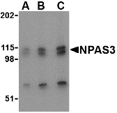
Left: Western blot analysis of NPAS3 in SK-N-SH cell lysate with NPAS3 antibody at (A) 0.5, (B) 1 and (C) 2 µg/ml.

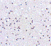
Below:Immunohistochemistry of NPAS3 in human brain tissue with NPAS3 antibody at 5 µg/ml.

Other Product Images

Source :NPAS3 antibody was raised against a 28 amino acid peptide from near the amino terminus of human NPAS3.
Purification : Affinity chromatography purified via peptide column
Clonality and Clone : This is a polyclonal antibody.
Host : NPAS3 antibody was raised in rabbit.
Application : NPAS3 antibody can be used for detection of NPAS3 by Western blot at 0.5 – 1 µg/ml.
Tested Application(s) : E, WB, IHC
Buffer : Antibody is supplied in PBS containing 0.02% sodium azide.
Blocking Peptide :Cat.No. 4107P - NPAS3 Peptide
Long-Term Storage : NPAS3 antibody can be stored at 4ºC, stable for one year. As with all antibodies care should be taken to avoid repeated freeze thaw cycles. Antibodies should not be exposed to prolonged high temperatures.
Positive Control :
1. Cat. No. 1220 - SK-N-SH Cell Lysate
2. Cat. No. 1303 - Human Brain Tissue Lysate
Species Reactivity : H, M, R
GI Number : 38604888
Accession Number : Q8IXF0
Short Description : (NT) Neuronal PAS domain-containing protein 3
References
1. Brunskill EW, Witte DP, Shreiner AB et al. Characterization of npas3, a novel basic helix-loop-helix PAS gene expressed in the developing mouse nervous system. Mech. Dev. 1999; 88:237-41.
2. Kamnasaran D, Muir WJ, Ferguson-Smith MA, et al. Disruption of the neuronal PAS3 gene in a family affected with schizophrenia. J. Med. Genet. 40:325-32.
3. Brunskill EW, Ehrman LA, Williams MT, et al. Abnormal neurodevelopment, neurosignaling and behaviour in Npas3-deficient mice. Euro. J. Neurosci. 2005; 22:1265-76.

NogoA Antibody


Catalog# :4089

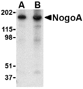
NogoA is a member of a family of integral membrane proteins termed reticulons that are thought to be involved in numerous disorders including neurodegenerative diseases. Reticulon proteins are known to regulate many cellular processes and interact with multiple proteins and receptors such as BACE. NogoA was initially identified as a myelin-associated neurite outgrowth inhibitor. It is highly expressed in oligodendrocytes in the white matter of the CNS; blocking its activity with antibodies or other factors results in improved axon regrowth and functional recovery in experimental CNS lesion models. NogoA has also been suggested to play a role in neurodegenerative diseases such as Amyotrophic lateral sclerosis, in which case NogoA is found at elevated levels in postmortem muscular samples, and multiple sclerosis (MS), in which case autoantibodies to NogoA have been found in serum and cerebrospinal fluid in MS patients. Despite its predicted molecular weight, NogoA typically migrates at 180kDa in an SDS-PAGE. At least five isoforms of Nogo are known to exist; this antibody is specific for NogoA and NogoE.

Additional Names : NogoA (IN), Neurite outgrowth inhibitor A, reticulon 4, RTN4

Description

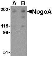
Left:Western blot analysis of NogoA in human brain tissue lysate with NogoA antibody at (A) 0.5 and (B) 1 µg/ml.

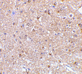
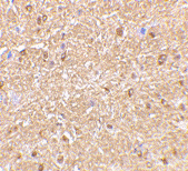
Below:Immunohistochemistry of NogoA in mouse brain tissue with NogoA antibody at 2.5 µg/ml.

Other Product Images
Source :NogoA antibody was raised against a 19 amino acid peptide from near the center of human NogoA.
Purification : Affinity chromatography purified via peptide column
Clonality and Clone : This is a polyclonal antibody.
Host : NogoA antibody was raised in rabbit.
Application : NogoA antibody can be used for detection of NogoA by Western blot at 0.5 – 1 µg/ml.
Tested Application(s) : E, WB, IHC
Buffer : Antibody is supplied in PBS containing 0.02% sodium azide.
Blocking Peptide :Cat.No. 4089P - NogoA Peptide
Long-Term Storage : NogoA antibody can be stored at 4ºC, stable for one year. As with all antibodies care should be taken to avoid repeated freeze thaw cycles. Antibodies should not be exposed to prolonged high temperatures.
Positive Control :
1. Cat. No. 1303 - Human Brain Tissue Lysate
2. Cat. No. 1403 - Mouse Brain Tissue Lysate
Species Reactivity : H, M, R
GI Number : 24431935
Accession Number : NP_065393
Short Description : (IN) a myelin-associated neurite outgrowth inhibitor
References
1. Yan R, ShiQ, Hu X, et al. Reticulon proteins: emerging players in neurodegenerative diseases. Cell. Mol. Life Sci. 2006; 63:877-889.
2. Chen MS, Huber AB, van der Haar ME, et al. Nogo-A is a myelin-associated neurite outgrowth inhibitor and an antigen for monoclonal antibody IN-1. Nature 2000; 403:434-9.
3. Schweigreiter R and Bandtlow CE. Nogo in the injured spinal cord. J. Neurotrauma 2006; 3-4:384-96.
4. Dupuis L, Gonzalez de Aguilar JL, di Scala F, et al. Nogo provides a molecular marker for diagnosis of amyloid lateral sclerosis. Neurobiol. Dis. 2002; 10:358-65.

NogoA Antibody


Catalog# :4087

NogoA is a member of a family of integral membrane proteins termed reticulons that are thought to be involved in numerous disorders including neurodegenerative diseases. Reticulon proteins are known to regulate many cellular processes and interact with multiple proteins and receptors such as BACE. NogoA was initially identified as a myelin-associated neurite outgrowth inhibitor. It is highly expressed in oligodendrocytes in the white matter of the CNS; blocking its activity with antibodies or other factors results in improved axon regrowth and functional recovery in experimental CNS lesion models. NogoA has also been suggested to play a role in neurodegenerative diseases such as Amyotrophic lateral sclerosis, in which case NogoA is found at elevated levels in postmortem muscular samples, and multiple sclerosis (MS), in which case autoantibodies to NogoA have been found in serum and cerebrospinal fluid in MS patients. Despite its predicted molecular weight, NogoA typically migrates at 180kDa in an SDS-PAGE. At least five isoforms of Nogo are known to exist; this antibody is specific for NogoA.

Additional Names : NogoA (NT), Neurite outgrowth inhibitor A, reticulon 4, RTN4

Description

Left: Western blot analysis of NogoA in mouse brain tissue lysate with NogoA antibody at (A) 0.5 and (B) 1 µg/ml.

Below:Immunohistochemistry of NogoA in mouse brain tissue with NogoA antibody at 2.5 µg/ml.

Other Product Images

Source :NogoA antibody was raised against a 23 amino acid peptide from near the amino terminus of human NogoA.
Purification : Affinity chromatography purified via peptide column
Clonality and Clone : This is a polyclonal antibody.
Host : NogoA antibody was raised in rabbit.
Please use anti-rabbit secondary antibodies.
Application : NogoA antibody can be used for detection of NogoA by Western blot at 0.5 – 1 µg/ml.
Tested Application(s) : E, WB, IHC
Buffer : Antibody is supplied in PBS containing 0.02% sodium azide.
Blocking Peptide :Cat.No. 4087P - NogoA Peptide
Long-Term Storage : NogoA antibody can be stored at 4ºC, stable for one year. As with all antibodies care should be taken to avoid repeated freeze thaw cycles. Antibodies should not be exposed to prolonged high temperatures.
Positive Control :
1. Cat. No. 1403 - Mouse Brain Tissue Lysate
Species Reactivity : H, M, R
GI Number : 24431935
Accession Number : NP_065393
Short Description : (NT) a myelin-associated neurite outgrowth inhibitor
References
1. Yan R, ShiQ, Hu X, et al. Reticulon proteins: emerging players in neurodegenerative diseases. Cell. Mol. Life Sci. 2006; 63:877-889.
2. Chen MS, Huber AB, van der Haar ME, et al. Nogo-A is a myelin-associated neurite outgrowth inhibitor and an antigen for monoclonal antibody IN-1. Nature 2000; 403:434-9.
3. Schweigreiter R and Bandtlow CE. Nogo in the injured spinal cord. J. Neurotrauma 2006; 3-4:384-96.
4. Dupuis L, Gonzalez de Aguilar JL, di Scala F, et al. Nogo provides a molecular marker for diagnosis of amyloid lateral sclerosis. Neurobiol. Dis. 2002; 10:358-65.

NK3R Antibody


Catalog# :4255

The tachykinins are a family of small peptides that include the neurotransmitters substance P, neurokinin A, and neurokinin B, which can act on three related but distinct seven transmembrane G-proteins coupled receptors, albeit at different concentrations. The NK-3 receptor (NK3R) has greatest affinity for neurokinin B and is highly expressed in the supraoptic and paraventricular nuclei. Following binding of its ligand, NK3R activates a phosphatidylinositol-calcium second messenger system. It is likely these signals lead to the release of vasopressin and oxytocin into the circulation. NK3R may be involved in learning and memory as mice lacking this gene expressed cognitive deficits compared to normal mice. Although it has been suggested that NK3R plays a role in the regulation of vagal afferent relay neurons, it is likely that these receptors are activated by substance P or neurokinin A, as the airway nerves do not express neurokinin B. NK3R antibody does not recognize NK1R or NK2R.

Additional Names : NK3R (IN), Neurokinin B receptor, NK-3 receptor, NK-3R, Tachykinin receptor 3, TACR3


Description

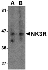
Left:Western blot analysis of NK3R in RAW264.7 cell lysate with NK3R antibody at (A) 0.5 and (B) 2 µg/ml.

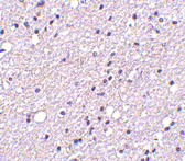
Below:Immunohistochemistry of NK3R in human brain tissue with NK3R antibody at 5 µg/ml.


Other Product Images

Source :NK3R antibody was raised against a 18 amino acid peptide from near the center of human NK3R.
Purification : Affinity chromatography purified via peptide column
Clonality and Clone : This is a polyclonal antibody.
Host : NK3R antibody was raised in rabbit.
Please use anti-rabbit secondary antibodies.
Application : NK3R antibody can be used for detection of NK3R by Western blot at 0.5 – 1 µg/ml.
Tested Application(s) : E, WB, IHC
Buffer : Antibody is supplied in PBS containing 0.02% sodium azide.
Blocking Peptide :Cat.No. 4255P - NK3R Peptide
Long-Term Storage : NK3R antibody can be stored at 4ºC, stable for one year. As with all antibodies care should be taken to avoid repeated freeze thaw cycles. Antibodies should not be exposed to prolonged high temperatures.
Positive Control :
1. Cat. No. 1283 - RAW264.7 Cell Lysate
2. Cat. No. 1303 - Human Brain Tissue Lysate
Species Reactivity : H, M
GI Number : 128364
Accession Number : P29371
Short Description : (IN) Neurokinin B receptor
References
1. Maggi CA. The mammalian tachykinin receptors. Gen. Pharmacol. 1995; 26:911-44.
2. Ding Y-Q, Shi J, Su L-Y, et al. Receptor (NK3)-containing neurons in the paraventricular and supraoptic nuclei of the rat hypothalamus synthesize vasopressin and express fos following intravenous injection of hypotonic saline. Neurosci. 1999; 91:1077-85.
3. Nakajima Y, Tsuchida K, Negishi M, et al. Direct linkage of three tachykinin receptors to stimulation of both phosphatidylinositol hydrolysis and cyclic AMP cascades in transfected Chinese hamster ovary cells. J. Biol. Chem. 1992; 267:2437-42.
4. Haley GE and Flynn FW. Tachykinin NK3 receptor contribution to systemic release of vasopressin and oxytocin in response to osmotic and hypotensive challenge. Am. J. Regul. Integr. Comp. Physiol. 2007; 293:R931-7.

Thursday, April 28, 2011

Ribsomal RNA Services


Biosyn Serving the DNA scientific community since 1984. It provides great resource of Ribosomal RNA services, RNA Service and RNA resources. Our every RNA product is designed and synthesized by professional scientists. Ribosomal RNA (rRNA) is the central component of the ribosome, the protein manufacturing machinery of all living cells.

Microarray Analysis


The quality of the RNA is essential to the overall success of the gene expression analysis using microarray technology. Once you submit your samples, the Project Scientist will take an aliquot of your RNA sample and assess its quality. We will proceed with labeling process only when RNA is confirmed to be suitable.

Ribosomal RNA


Ribosomal RNA (rRNA) is the central component of the ribosome, the protein manufacturing machinery of all living cells. The function of the rRNA is to provide a mechanism for decoding mRNA into amino acids and to interact with the tRNAs during translation by providing peptidyl transferase activity. The ribosome is composed of two subunits, named for how rapidly they sediment when subject to centrifugation.

Eukaryotes

In contrast, eukaryotes generally have many copies of the rRNA genes organized in tandem repeats; in humans approximately 300–400
rDNA repeats are present in five clusters (on chromosomes 13, 14, 15, 21 and 22).The 18S rRNA in most eukaryotes is in the small ribosomal subunit, and the large subunit contains three rRNA species (the 5S, 5.8S and 28S rRNAs).

For More information Visit: www.biosyn.com
NIPSNAP3B Antibody


Catalog# :4957

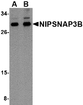
The NIPSNAP proteins comprise a family of evolutionarily well-conserved proteins with strong sequence similarity to the central portion of a protein encoded by C. elegans chromosome III between a 4-nitrophenylphosphatase (NIP) domain and non-neuronal SNAP25-like protein. NIPSNAP2, a novel gene encoding a protein with tyrosine phosphorylation sites and a transmembrane domain, is co-amplified with EGFR in approximately 40% of glioblastomas, the most common and malignant form of central nervous system tumors. While NIPSNAP3B is highly expressed skeletal muscle, NIPSNAP3A mRNA levels are low. NIPSNAP proteins have been suggested to be important in vesicular transport. NIPSNAP3B antibody is predicted to not cross-react with any other members of the NIPSNAP protein family.

Additional Names : NIPSNAP3B, Non-neuronal SNAP25-like protein 3B, NIPSNAP3

Description

Left: Western blot analysis of NIPSNAP3B in mouse brain tissue lysate with NIPSNAP3B antibody at (A) 1 and (B) 2 μg/ml.

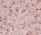
Below:Immunohistochemistry of NIPSNAP3B in mouse brain tissue with NIPSNAP3B antibody at 2.5 μg/ml.

Other Product Images


Source :NIPSNAP3B antibody was raised against a 16 amino acid peptide near the center of human NIPSNAP3B.
Purification : Affinity chromatography purified via peptide column
Clonality and Clone : This is a polyclonal antibody.
Host : NIPSNAP3B antibody was raised in rabbit.
Please use anti-rabbit secondary antibodies.
Application : NIPSNAP3B antibody can be used for detection of NIPSNAP3B by Western blot at 1 – 2 µg/ml.
Tested Application(s) : E, WB, IHC
Buffer : Antibody is supplied in PBS containing 0.02% sodium azide.
Blocking Peptide :Cat.No. 4957P - NIPSNAP3B Peptide
Long-Term Storage : NIPSNAP3B antibody can be stored at 4ºC, stable for one year. As with all antibodies care should be taken to avoid repeated freeze thaw cycles. Antibodies should not be exposed to prolonged high temperatures.
Positive Control :
1. Cat. No. 1403 - Mouse Brain Tissue Lysate
Species Reactivity : H, M
GI Number : 17380145
Accession Number : Q9BS92
Short Description : Non-neuronal SNAP25-like protein 3B
References
1. Seroussi E, Pan HQ, Kedra D, et al. Characterization of the human NIPSNAP1 gene from 22q12: a member of a novel gene family. Gene 1998; 212:13-20.
2. Wang X-Y, Smith DI, Liu W, et al. GBAS, a novel gene encoding a protein with tyrosine phosphorylation sites and a transmembrane domain, is co-amplified with EGFR. Genomics 1998; 49:448-51.
3. Buechler C, Bodzioch M, Bared SM, et al. Expression pattern and raft association of NIPSNAP3 and NIPSNAP4, highly homologous proteins encoded by genes in close proximity to the ATP-binding cassette transporter A1. Genomics 2004; 83:1116-24.

NIPSNAP3A Antibody


Catalog# :4947

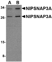
The NIPSNAP proteins comprise a family of evolutionarily well-conserved proteins with strong sequence similarity to the central portion of a protein encoded by C. elegans chromosome III between a 4-nitrophenylphosphatase (NIP) domain and non-neuronal SNAP25-like protein. NIPSNAP2, a novel gene encoding a protein with tyrosine phosphorylation sites and a transmembrane domain, is co-amplified with EGFR in approximately 40% of glioblastomas, the most common and malignant form of central nervous system tumors. While NIPSNAP3B is highly expressed skeletal muscle, NIPSNAP3A mRNA levels are low. NIPSNAP3A protein is associated with plasma membrane and partially localized in rafts. NIPSNAP proteins have been suggested to be important in vesicular transport. At least two isoforms of NIPSNAP3A are known to exist. NIPSNAP3A antibody is predicted to not cross-react with any other members of the NIPSNAP protein family.

Additional Names : NIPSNAP3A, Non-neuronal SNAP25-like protein 3A, NIPSNAP4, TASSC

Description

Left: Western blot analysis of NIPSNAP3A in mouse brain tissue lysate with NIPSNAP3A antibody at (A) 0.5 and (B) 1 µg/ml.

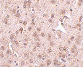
Below:Immunohistochemistry of NIPSNAP3A in mouse brain tissue with NIPSNAP3A antibody at 2.5 µg/ml.


Other Product Images


Source :NIPSNAP3A antibody was raised against a 16 amino acid peptide near the center of human NIPSNAP3A.
Purification : Affinity chromatography purified via peptide column
Clonality and Clone : This is a polyclonal antibody.
Host : NIPSNAP3A antibody was raised in rabbit.
Please use anti-rabbit secondary antibodies.
Application : NIPSNAP3A antibody can be used for detection of NIPSNAP3A by Western blot at 0.5 – 1 µg/ml.
Tested Application(s) : E, WB, IHC
Buffer : Antibody is supplied in PBS containing 0.02% sodium azide.
Blocking Peptide :Cat.No. 4947P - NIPSNAP3A Peptide
Long-Term Storage : NIPSNAP3A antibody can be stored at 4ºC, stable for one year. As with all antibodies care should be taken to avoid repeated freeze thaw cycles. Antibodies should not be exposed to prolonged high temperatures.
Positive Control :
1. Cat. No. 1403 - Mouse Brain Tissue Lysate
Species Reactivity : H, M
GI Number : 22267436
Accession Number : NP_056284
Short Description : Non-neuronal SNAP25-like protein 3A
References
1. Seroussi E, Pan HQ, Kedra D, et al. Characterization of the human NIPSNAP1 gene from 22q12: a member of a novel gene family. Gene 1998; 212:13-20.
2. Wang X-Y, Smith DI, Liu W, et al. GBAS, a novel gene encoding a protein with tyrosine phosphorylation sites and a transmembrane domain, is co-amplified with EGFR. Genomics 1998; 49:448-51.
3. Buechler C, Bodzioch M, Bared SM, et al. Expression pattern and raft association of NIPSNAP3 and NIPSNAP4, highly homologous proteins encoded by genes in close proximity to the ATP-binding cassette transporter A1. Genomics 2004; 83:1116-24.

NIPSNAP2 Antibody


Catalog# :4953

The NIPSNAP proteins comprise a family of evolutionarily well-conserved proteins with strong sequence similarity to the central portion of a protein encoded by C. elegans chromosome III between a 4-nitrophenylphosphatase (NIP) domain and non-neuronal SNAP25-like protein. NIPSNAP2, a novel gene encoding a protein with tyrosine phosphorylation sites and a transmembrane domain, is co-amplified with EGFR in approximately 40% of glioblastomas, the most common and malignant form of central nervous system tumors. It is widely expressed and most abundant in heart and skeletal muscle. NIPSNAP proteins have been suggested to be important in vesicular transport. NIPSNAP2 antibody is predicted to not cross-react with any other members of the NIPSNAP protein family.

Additional Names : NIPSNAP2 (IN), Non-neuronal SNAP25-like protein 2, Glioblastoma-amplified sequence, GBAS

Description
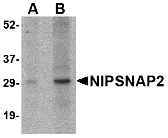
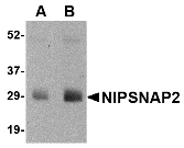
Left: Western blot analysis of NIPSNAP2 in human skeletal muscle tissue lysate with NIPSNAP2 antibody at (A) 1 and (B) 2 µg/ml.

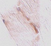
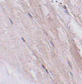
Below:Immunohistochemistry of NIPSNAP2 in mouse skeletal muscle tissue with NIPSNAP2 antibody at 2.5 µg/ml.

Other Product Images

Source :NIPSNAP2 antibody was raised against a 14 amino acid peptide near the center of human NIPSNAP2.
Purification : Affinity chromatography purified via peptide column
Clonality and Clone : This is a polyclonal antibody.
Host : NIPSNAP2 antibody was raised in rabbit.
Please use anti-rabbit secondary antibodies.
Application : NIPSNAP2 antibody can be used for detection of NIPSNAP2 by Western blot at 1 – 2 µg/ml.
Tested Application(s) : E, WB, IHC
Buffer : Antibody is supplied in PBS containing 0.02% sodium azide.
Blocking Peptide :Cat.No. 4953P - NIPSNAP2 Peptide
Long-Term Storage : NIPSNAP2 antibody can be stored at 4ºC, stable for one year. As with all antibodies care should be taken to avoid repeated freeze thaw cycles. Antibodies should not be exposed to prolonged high temperatures.
Positive Control :
1. Cat. No. 1375 - Human Skeletal Muscle Tissue Lysate
Species Reactivity : H, M, R
GI Number : 17380133
Accession Number : O75323
Short Description : (IN) Non-neuronal SNAP25-like protein 2
References
1. Seroussi E, Pan HQ, Kedra D, et al. Characterization of the human NIPSNAP1 gene from 22q12: a member of a novel gene family. Gene 1998; 212:13-20.
2. Wang X-Y, Smith DI, Liu W, et al. GBAS, a novel gene encoding a protein with tyrosine phosphorylation sites and a transmembrane domain, is co-amplified with EGFR. Genomics 1998; 49:448-51.

NIPSNAP2 Antibody


Catalog# :4943

The NIPSNAP proteins comprise a family of evolutionarily well-conserved proteins with strong sequence similarity to the central portion of a protein encoded by C. elegans chromosome III between a 4-nitrophenylphosphatase (NIP) domain and non-neuronal SNAP25-like protein. NIPSNAP2, a novel gene encoding a protein with tyrosine phosphorylation sites and a transmembrane domain, is co-amplified with EGFR in approximately 40% of glioblastomas, the most common and malignant form of central nervous system tumors. It is widely expressed and most abundant in heart and skeletal muscle. NIPSNAP proteins have been suggested to be important in vesicular transport. NIPSNAP2 antibody is predicted to not cross-react with any other members of the NIPSNAP protein family.

Additional Names : NIPSNAP2 (NT), Non-neuronal SNAP25-like protein 2, Glioblastoma-amplified sequence, GBAS

Description

Left: Western blot analysis of NIPSNAP2 in human skeletal muscle tissue lysate with NIPSNAP2 antibody at (A) 0.5 and (B) 1 µg/ml.

Below:Immunohistochemistry of NIPSNAP2 in mouse skeletal muscle tissue with NIPSNAP2 antibody at 2.5µg/ml.

Other Product Images

Source :NIPSNAP2 antibody was raised against a 14 amino acid peptide near the amino terminus of human NIPSNAP2.
Purification : Affinity chromatography purified via peptide column
Clonality and Clone : This is a polyclonal antibody.
Host : NIPSNAP2 antibody was raised in rabbit.
Please use anti-rabbit secondary antibodies.
Application : NIPSNAP2 antibody can be used for detection of NIPSNAP2 by Western blot at 1 – 2 µg/ml.
Tested Application(s) : E, WB, IHC
Buffer : Antibody is supplied in PBS containing 0.02% sodium azide.
Blocking Peptide :Cat.No. 4943P - NIPSNAP2 Peptide
Long-Term Storage : NIPSNAP2 antibody can be stored at 4ºC, stable for one year. As with all antibodies care should be taken to avoid repeated freeze thaw cycles. Antibodies should not be exposed to prolonged high temperatures.
Positive Control :
1. Cat. No. 1375 - Human Skeletal Muscle Tissue Lysate
Species Reactivity : H, M, R
GI Number : 17380133
Accession Number : O75323
Short Description : (NT) Non-neuronal SNAP25-like protein 2
References
1. Seroussi E, Pan HQ, Kedra D, et al. Characterization of the human NIPSNAP1 gene from 22q12: a member of a novel gene family. Gene 1998; 212:13-20.
2. Wang X-Y, Smith DI, Liu W, et al. GBAS, a novel gene encoding a protein with tyrosine phosphorylation sites and a transmembrane domain, is co-amplified with EGFR. Genomics 1998; 49:448-51.

Wednesday, April 27, 2011

NIPSNAP Antibody


Catalog# :4537

NIPSNAP is a member of an evolutionarily well conserved gene family and has a strong sequence similarity to the central portion of a protein encoded by C. elegans chromosome III between a 4-nitrophenylphosphatase (NIP) domain and non-neuronal SNAP25-like protein. Recent studies have indicated that NIPSNAP is involved in the regulation of the Ca2+-selective transient receptor potential vanilloid channel 6 (TRPV6). NIPSNAP1 associates with TRPV6 at the plasma membrane and inhibits TRPV6 currents. Other studies show that NIPSNAP’s expression is reduced in the phenylketonuria (PKU) mouse brain, suggesting that NIPSNAP may play a role in memory.

Additional Names :
NIPSNAP (IN), Non-neuronal SNAP25-like protein, NIPSNAP1

Description

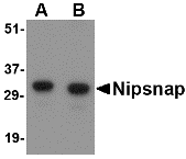
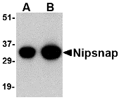
Left: Western blot analysis of NIPSNAP in human brain tissue lysate with NIPSNAP antibody at (A) 0.5 and (B) 1 µg/ml.

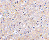
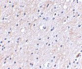
Below:Immunohistochemical staining of human brain tissue using Nipsnap antibody at 2.5 µg/ml.

Other Product Images
Source :NIPSNAP antibody was raised against a 19 amino acid peptide from near the center of human NIPSNAP.
Purification : Affinity chromatography purified via peptide column
Clonality and Clone : This is a polyclonal antibody.
Host : NIPSNAP antibody was raised in rabbit.
Please use anti-rabbit secondary antibodies
Application : NIPSNAP antibody can be used for the detection of NIPSNAP by Western blot at 0.5 – 1 µg/ml.
Tested Application(s) : E, WB, IHC
Buffer : Antibody is supplied in PBS containing 0.02% sodium azide.
Blocking Peptide :Cat.No. 4537P - NIPSNAP Peptide
Long-Term Storage : NIPSNAP antibody can be stored at 4ºC, stable for one year. As with all antibodies care should be taken to avoid repeated freeze thaw cycles. Antibodies should not be exposed to prolonged high temperatures.
Positive Control :
1.Cat. No. 1303 - Human Brain Tissue Lysate
Species Reactivity : H, M, R
GI Number : 193211616
Accession Number : NP_003625
Short Description : (IN) Non-neuronal SNAP25-like protein
References
1. Seroussi E, Pan H-Q, Kedra D, et al. Characterization of the human NIPSNAP1 gene from 22q12: a member of a novel gene family. Gene 1998; 212:13-20.
2. Schoeber JP, Topala CN, Lee KP, et al. Identification of Nipsnap1 as a novel auxiliary protein inhibiting TRPV6 activity. Pflugers Arch. 2008; epub.
3. Surendran S, Tyring SK and Matalon R. Expression of calpastatin, minopontin, NIPSNAP1, rabaptin-5 and neuronatin in the phenylketonuria (PKU) mouse brain: possible role on cognitive defect seen in PKU. Neurochem. Int. 2005; 46:595-9.

NIPSNAP Antibody


Catalog# :4535

NIPSNAP is a member of an evolutionarily well conserved gene family and has a strong sequence similarity to the central portion of a protein encoded by C. elegans chromosome III between a 4-nitrophenylphosphatase (NIP) domain and non-neuronal SNAP25-like protein. Recent studies have indicated that NIPSNAP is involved in the regulation of the Ca2+-selective transient receptor potential vanilloid channel 6 (TRPV6). NIPSNAP1 associates with TRPV6 at the plasma membrane and inhibits TRPV6 currents. Other studies show that NIPSNAP’s expression is reduced in the phenylketonuria (PKU) mouse brain, suggesting that NIPSNAP may play a role in memory.

Additional Names : NIPSNAP (CT), Non-neuronal SNAP25-like protein, NIPSNAP1

Description
Left: Western blot analysis of NIPSNAP in human brain tissue lysate with NIPSNAP antibody at (A) 0.5 and (B) 1 µg/ml.

Right: Immunohistochemical staining of human brain tissue using Nipsnap antibody at 2.5 µg/ml.

Other Product Images

Source :NIPSNAP antibody was raised against a 18 amino acid peptide from near the carboxy terminus of human NIPSNAP.
Purification : Affinity chromatography purified via peptide column
Clonality and Clone : This is a polyclonal antibody.
Host : NIPSNAP antibody was raised in rabbit.
Please use anti-rabbit secondary antibodies.
Application : NIPSNAP antibody can be used for the detection of NIPSNAP by Western blot at 0.5 – 1 µg/ml.
Tested Application(s) : E, WB, IHC
Buffer : Antibody is supplied in PBS containing 0.02% sodium azide.
Blocking Peptide :Cat.No. 4535P - NIPSNAP Peptide
Long-Term Storage : NIPSNAP antibody can be stored at 4ºC, stable for one year. As with all antibodies care should be taken to avoid repeated freeze thaw cycles. Antibodies should not be exposed to prolonged high temperatures.
Positive Control :
1. Cat. No. 1303 - Human Brain Tissue Lysate
Species Reactivity : H, M, R
GI Number : 193211616
Accession Number : NP_003625
Short Description : (CT) Non-neuronal SNAP25-like protein
References
1. Seroussi E, Pan H-Q, Kedra D, et al. Characterization of the human NIPSNAP1 gene from 22q12: a member of a novel gene family. Gene 1998; 212:13-20.
2. Schoeber JP, Topala CN, Lee KP, et al. Identification of Nipsnap1 as a novel auxiliary protein inhibiting TRPV6 activity. Pflugers Arch. 2008; epub.
3. Surendran S, Tyring SK and Matalon R. Expression of calpastatin, minopontin, NIPSNAP1, rabaptin-5 and neuronatin in the phenylketonuria (PKU) mouse brain: possible role on cognitive defect seen in PKU. Neurochem. Int. 2005; 46:595-9.

Nicastrin Antibody


Catalog# :3985

Nicastrin, in addition to presenilin, PEN2, and APH-1 forms the gamma-secretase protein complex, a membrane-bound aspartyl protease that can cleave certain proteins at peptide bonds buried within the hydrophobic environment of the lipid bilayer. This cleavage is responsible for a key step in signaling from several cell-surface receptors and is thought to be required for the generation of the neurotoxic amyloid peptides that are central to the pathogenesis of Alzheimer’s disease. Like the tumor necrosis factor-a-converting enzyme (TACE) and the b-site cleavage enzyme (BACE) protease families, gamma-secretase will cleave the amyloid precursor protein (APP), but within the intramembrane region of APP, resulting in either the non-toxic p3 (from the alpha and gamma cleavage site) or the toxic Abeta amyloid peptide (from the beta and gamma cleavage site). It is thought that accumulation of the Abeta peptide is the precursor to Alzheimer’s disease. Nicastrin is also thought to be involved in cell proliferation and signaling, especially in regards to activation of Notch receptors as loss of Nicastrin expression results in mouse embryonic lethality.

Additional Names : Nicastrin (IN), Anterior pharynx defective 2, APH-2


Description

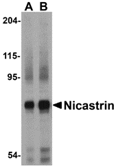
Left: Western blot analysis of Nicastrin in human brain tissue lysate with Nicastrin antibody at (A) 0.5 and (B) 1µg/ml.

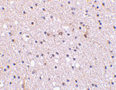
Right:Immunohistochemistry of Nicastrin in human brain tissue with Nicastrin antibody at 2.5 µg/ml.

Other Product Images

Source :Nicastrin antibody was raised against a 18 amino acid peptide from near the center of human Nicastrin.
Purification : Affinity chromatography purified via peptide column
Clonality and Clone : This is a polyclonal antibody.
Host : Nicastrin antibody was raised in rabbit.
Please use anti-rabbit secondary antibodies
Application : Nicastrin antibody can be used for detection of Nicastrin by Western blot at 0.5 – 1 µg/ml.
Tested Application(s) : E, WB, IHC
Buffer : Antibody is supplied in PBS containing 0.02% sodium azide.
Blocking Peptide :Cat.No. 3985P - Nicastrin Peptide
Long-Term Storage : Nicastrin antibody can be stored at 4ºC, stable for one year. As with all antibodies care should be taken to avoid repeated freeze thaw cycles. Antibodies should not be exposed to prolonged high temperatures.
Positive Control :
1.Cat. No. 1303 - Human Brain Tissue Lysate
2. Cat. No. 1303 - Human Brain Tissue Lysate
Species Reactivity : H, M, R
GI Number : 24638433
Accession Number : NP_056146
Short Description : (IN) a member of the gamma-secretase complex
References
1. Weihofen A and Martoglio B. Intramembrane-cleaving proteases: controlled liberation of proteins and bioactive peptides. Trends Cell Biol. 2003; 13:71-8.
2. Periz G and Fortini ME. Functional reconstitution of g-secretase through coordinated expression of presenilin, Nicastrin, aph-1, and pen-2. J. Neurosci. Res. 2004; 77:309-22.
3. Selkoe DJ. The cell biology of b-amyloid precursor protein and presenilin in Alzheimer’s disease. Trends Cell Biol. 1998; 8:447-53.
4. Nguyen V, Hawkins C, Bergeron C, et al. Loss of nicastrin elicits an apoptotic phenotype in mouse embryos. Brain Res. 2006; 1086:76-84.

Nicastrin Antibody


Catalog# :3983

Nicastrin, in addition to presenilin, PEN2, and APH-1 forms the gamma-secretase protein complex, a membrane-bound aspartyl protease that can cleave certain proteins at peptide bonds buried within the hydrophobic environment of the lipid bilayer. This cleavage is responsible for a key step in signaling from several cell-surface receptors and is thought to be required for the generation of the neurotoxic amyloid peptides that are central to the pathogenesis of Alzheimer’s disease. Like the tumor necrosis factor-a-converting enzyme (TACE) and the b-site cleavage enzyme (BACE) protease families, gamma-secretase will cleave the amyloid precursor protein (APP), but within the intramembrane region of APP, resulting in either the non-toxic p3 (from the alpha and gamma cleavage site) or the toxic Abeta amyloid peptide (from the beta and gamma cleavage site). It is thought that accumulation of the Abeta peptide is the precursor to Alzheimer’s disease. Nicastrin is also thought to be involved in cell proliferation and signaling, especially in regards to activation of Notch receptors as loss of Nicastrin expression results in mouse embryonic lethality.

Additional Names : Nicastrin (CT), Anterior pharynx defective 2, APH-2


Description

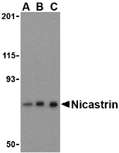
Left: Western blot analysis of Nicastrin in mouse brain tissue lysate with Nicastrin antibody at (A) 0.5, (B) 1, and (C) 2 µg/ml.

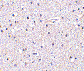
b>Below: Immunohistochemistry of Nicastrin in human brain tissue with Nicastrin antibody at 5 µg/ml.


Other Product Images

Source :Nicastrin antibody was raised against a 17 amino acid peptide from near the carboxy terminus of human Nicastrin.
Purification : Affinity chromatography purified via peptide column
Clonality and Clone : This is a polyclonal antibody.
Host : Nicastrin antibody was raised in rabbit.
Application : Nicastrin antibody can be used for detection of Nicastrin by Western blot at 0.5 – 1 µg/ml.
Tested Application(s) : E, WB, IHC
Buffer : Antibody is supplied in PBS containing 0.02% sodium azide.
Blocking Peptide :Cat.No. 3983P - Nicastrin Peptide
Long-Term Storage : Nicastrin antibody can be stored at 4ºC, stable for one year. As with all antibodies care should be taken to avoid repeated freeze thaw cycles. Antibodies should not be exposed to prolonged high temperatures.
Positive Control :
1. Cat. No. 1403 - Mouse Brain Tissue Lysate
2. Cat. No. 1303 - Human Brain Tissue Lysate
Species Reactivity : H, M, R
GI Number : 24638433
Accession Number : NP_056146
Short Description : (CT) a member of the gamma-secretase complex
References
1. Weihofen A and Martoglio B. Intramembrane-cleaving proteases: controlled liberation of proteins and bioactive peptides. Trends Cell Biol. 2003; 13:71-8.
2. Periz G and Fortini ME. Functional reconstitution of g-secretase through coordinated expression of presenilin, Nicastrin, aph-1, and pen-2. J. Neurosci. Res. 2004; 77:309-22.
3. Selkoe DJ. The cell biology of b-amyloid precursor protein and presenilin in Alzheimer’s disease. Trends Cell Biol. 1998; 8:447-53.
4. Nguyen V, Hawkins C, Bergeron C, et al. Loss of nicastrin elicits an apoptotic phenotype in mouse embryos. Brain Res. 2006; 1086:76-84.

Tuesday, April 26, 2011

RNA DNA Duplex


Biosyn can provide RNA, RNA analogs and chimeras in large quantities for your diagnostic and therapeutic research. Biosyn continues the tradition of providing researchers with the highest quality RNA Oligonucleotide synthesis, in quantities ranging from multi-mg to several grams.

RNA Oligonucleotide Synthesis

Oligonucleotide synthesis is the non-biological, chemical synthesis of defined short sequences of nucleic acids. It is extremely useful in laboratory procedures covering a wide range of molecular biology applications. Automated synthesizers allow the synthesis of oligonucleotides up to 200 bases even longer.

RNA Replication

It is therefore likely that the RNA replication reactions of these two picornaviruses involve an equivalent set of protein-protein and protein-RNA interactions and perhaps utilize the same host cell proteins. By targeting small, synthetic oligos, typically 19-23 bases in length against a specific gene of interest researchers are identifying gene function, elucidating pathways, and screening for potential new drug targets.

RNA Hybrid

RNA hybrid is a double-stranded polynucleic acids in which one strand is DNA and the other strand is the complementary RNA; formed during transcription and during multiplication of oncogenic RNA viruses.

Nhe-1 Antibody


Catalog# :4379

The Na+/H+ antiporter (Nhe-1) is a ubiquitous membrane-bound enzyme involved in pH regulation of vertebrate cells and is specifically inhibited by the diuretic drug amiloride and activated by a variety of signals including growth factors, mitogens, neurotransmitters, and tumor promoters. Nhe-1 acts as an anchor for actin filaments to control the integrity of the cortical cytoskeleton. This occurs through a previously unrecognized structural link between Nhe-1 and the actin-binding proteins ezrin, radixin, and moesin, collectively referred to as ERM proteins. A structural role for Nhe-1 has been proposed in regulating the cortical cytoskeleton that is independent of its function as an ion exchanger. It is also thought that Nhe-1 play a role in hypertension. At least two isoforms of Nhe-1 are known to exist.

Additional Names : Nhe-1 (IN), Sodium-hydrogen exchanger 1, solute carrier family 9 member 1, SLC9A1, Na+/H+ antiporter


Description

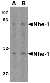
Left: Western blot analysis of Nhe-1 in MOLT4 cell lysate with in with Nhe-1 antibody at (A) 1 and (B) 2 µg/ml.

Below: Immunohistochemical staining of human brain tissue using Nhe-1 antibody at 2.5 µg/ml.


Other Product Images

Source :Nhe-1 antibody was raised against a 17 amino acid peptide near the center of the human Nhe-1.
Purification : Affinity chromatography purified via peptide column
Clonality and Clone : This is a polyclonal antibody.
Host : Nhe-1 antibody was raised in rabbit.
Please use anti-rabbit secondary antibodies.
Application : Nhe-1 antibody can be used for detection of Nhe-1 by Western blot at 1 – 2 µg/ml.
Tested Application(s) : E, WB, IHC
Buffer : Antibody is supplied in PBS containing 0.02% sodium azide.
Blocking Peptide :Cat.No. 4379P - Nhe-1 Peptide
Long-Term Storage : Nhe-1 antibody can be stored at 4ºC, stable for one year. As with all antibodies care should be taken to avoid repeated freeze thaw cycles. Antibodies should not be exposed to prolonged high temperatures.
Positive Control :
1. Cat. No. 1206 - MOLT4 Cell Lysate
Species Reactivity : H, M, R
GI Number : 127809
Accession Number : P19634
Short Description : (IN) Sodium-hydrogen exchanger 1
References
1. Mendoza SA. The Na+-H+ antiport is a mediator of cell proliferation. Acta Paediatr. Scand. 1987; 76:545-7.
2. Denker SP, Huang DC, Orlowski J, et al. Direct binding of the NA—H exchanger NHE1 to ERM proteins regulates the cortical cytoskeleton and cell shape independently of H(+) translocation. Mol. Cell. 2000; 6:1425-36.
3. Cingolani HE, Rebolledo OR, Portiansky EL, et al. Regression of hypertensive myocardial fibrosis by NA (+)/H(+) exchange inhibition. Hypertension 2003; 41:373-7.

Nhe-1 Antibody


Catalog# :4377

The Na+/H+ antiporter (Nhe-1) is a ubiquitous membrane-bound enzyme involved in pH regulation of vertebrate cells and is specifically inhibited by the diuretic drug amiloride and activated by a variety of signals including growth factors, mitogens, neurotransmitters, and tumor promoters. Nhe-1 acts as an anchor for actin filaments to control the integrity of the cortical cytoskeleton. This occurs through a previously unrecognized structural link between Nhe-1 and the actin-binding proteins ezrin, radixin, and moesin, collectively referred to as ERM proteins. A structural role for Nhe-1 has been proposed in regulating the cortical cytoskeleton that is independent of its function as an ion exchanger. It is also thought that Nhe-1 play a role in hypertension. At least two isoforms of Nhe-1 are known to exist.

Additional Names :
Nhe-1 (CT), Sodium-hydrogen exchanger 1, solute carrier family 9 member 1, SLC9A1, Na+/H+ antiporter


Description

Left: Western blot analysis of Nhe-1 in rat kidney tissue lysate with in with Nhe-1 antibody at (A) 1 and (B) 2 µg/ml.

Below: Immunohistochemical staining of human brain tissue using Nhe-1 antibody at 2.5 µg/ml.


Other Product Images

Source :Nhe-1 antibody was raised against a 20 amino acid peptide near the carboxy terminus of the human Nhe-1.
Purification : Affinity chromatography purified via peptide column
Clonality and Clone : This is a polyclonal antibody.
Host : Nhe-1 antibody was raised in rabbit.
Please use anti-rabbit secondary antibodies
Application : Nhe-1 antibody can be used for detection of Nhe-1 by Western blot at 1 – 2 µg/ml.
Tested Application(s) : E, WB, IHC
Buffer : Antibody is supplied in PBS containing 0.02% sodium azide.
Blocking Peptide :Cat.No. 4377P - Nhe-1 Peptide
Long-Term Storage : Nhe-1 antibody can be stored at 4ºC, stable for one year. As with all antibodies care should be taken to avoid repeated freeze thaw cycles. Antibodies should not be exposed to prolonged high temperatures.
Positive Control :
1. Cat. No. 1465 - Rat Kidney Tissue Lysate
Species Reactivity : H, M, R
GI Number : 127809
Accession Number : P19634
Short Description : (NT) Sodium-hydrogen exchanger 1
References
1. Mendoza SA. The Na+-H+ antiport is a mediator of cell proliferation. Acta Paediatr. Scand. 1987; 76:545-7.
2. Denker SP, Huang DC, Orlowski J, et al. Direct binding of the NA—H exchanger NHE1 to ERM proteins regulates the cortical cytoskeleton and cell shape independently of H(+) translocation. Mol. Cell. 2000; 6:1425-36.
3. Cingolani HE, Rebolledo OR, Portiansky EL, et al. Regression of hypertensive myocardial fibrosis by NA (+)/H(+) exchange inhibition. Hypertension 2003; 41:373-7.

Neurotrypsin Antibody


Catalog# :4105

Neurotrypsin is a central nervous system-expressed serine protease whose truncation or absence causes nonsyndromic mental retardation. It is most prominently expressed in structures that are involved in the processing and storage of learned behavior and memory, such as the cerebral cortex, the hippocampus, and amygdala. Evidence suggests that neurotrypsin has multiple functions, including axonal outgrowth, maintaining neuronal plasticity, and arranging the perineuronal environment, partly in coordination with other proteases including tissue plasminogen activator. At least two isoforms of neurotrypsin are known to exist.

Additional Names : Neurotrypsin (NT), Motopsin, Protease serine 12, PRSS12, MRT1, BSSP3

Description
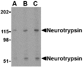
Left: Western blot analysis of neurotrypsin in SK-N-SH cell lysate with neurotrypsin antibody at (A) 0.5, (B) 1 and (C) 2 µg/ml.

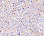
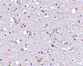
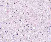
Below:Immunohistochemistry of Neurotrypsin in human brain tissue with Neurotrypsin antibody at 2.5 µg/ml.

Other Product Images

Source :Neurotrypsin antibody was raised against a 23 amino acid peptide from near the amino terminus of human neurotrypsin.
Purification : Affinity chromatography purified via peptide column
Clonality and Clone : This is a polyclonal antibody.
Host : Neurotrypsin antibody was raised in rabbit.
Please use anti-rabbit secondary antibodies.
Application : Neurotrypsin antibody can be used for detection of neurotrypsin by Western blot at 0.5 – 1 µg/ml.
Tested Application(s) : E, WB, IHC
Buffer : Antibody is supplied in PBS containing 0.02% sodium azide.
Blocking Peptide :Cat.No. 4105P - Neurotrypsin Peptide
Long-Term Storage : Neurotrypsin antibody can be stored at 4ºC, stable for one year. As with all antibodies care should be taken to avoid repeated freeze thaw cycles. Antibodies should not be exposed to prolonged high temperatures.
Positive Control :
1. Cat. No. 1220 - SK-N-SH Cell Lysate
Species Reactivity : H, M, R
GI Number : 2661424
Accession Number : CAA04816
Short Description : (NT) a serine protease of the nervous system
References
1. Gschwend TP, Krueger SR, Kozlov SV, et al. Neurotrypsin, a novel multidomain serine protease expressed in the nervous system. Mol. Cell. Neurosci. 1997; 9:207-19.
2. Molinari F, Rio M, Meskanaite V, et al. Truncating neurotrypsin mutation in autosomal recessive nonsyndromic mental retardation. Science 2002; 1779-81.
3. Mitsui S, Yamaguchi N, Osako Y, et al. Enzymatic properties and localization of motopsin (PRSS12), a protease who absence causes mental retardation. Brain Res. 2007; 1136:1-12.