Thursday, June 30, 2011
DNA Sequence Analysis
For over 20 years, BioSynthesis has provided custom oligonucleotide production services worldwide, including researcher at university, biotechnology and pharmaceutical institutions. We offer wide variety of oligo modifications and labelings from small discovery research scale to multi-gram ASR/cGMP for clinical diagnostic production. We offer design and synthesis of oligonucleotide primers and probes, also provides full range of DNA microarray printing, processing and analysis.
DNA Sequencing
The term DNA sequencing encompasses biochemical methods for determining the order of the nucleotide bases, adenine, guanine, cytosine, and thymine, in a DNA oligonucleotide. The sequence of DNA constitutes the heritable genetic information in nuclei, plasmids, mitochondria, and chloroplasts that forms the basis for the developmental programs of all living organisms.
Chain-termination Methods
While the chemical sequencing method of Maxam and Gilbert, and the plus-minus method of Sanger and Coulson were orders of magnitude faster than previous methods, the chain-terminator method developed by Sanger was even more efficient, and rapidly became the method of choice. The Maxam-Gilbert technique requires the use of highly toxic chemicals, and large amounts of radiolabeled DNA, whereas the chain-terminator method uses fewer toxic chemicals and lower amounts of radioactivity.
Recombinant DNA
Recombinant DNA is a man-made DNA sequence that has been assembled from other DNA sequences. They can be transformed into organisms in the form of plasmids or in the appropriate format, by using a viral vector.
IL-32 Antibody
Catalog#:3749
Interleukin-32 (IL-32) was initially identified as a transcript (NK4) that is selectively expressed in lymphocytes and NK cells and whose expression is increased following activation by IL-2 . It was later re-isolated from an IL-18-treated lung carcinoma cell line and re-named IL-32 (2). IL-32 is unusual in that it does not share sequence homology with known cytokine families and is highly expressed in immune tissues, existing in at least four differentially spliced isoforms. Because treatment of human monocytic and mouse macrophage cells with IL-32 induces several proinflammatory cytokines such as TNF-alpha, IL-8 and MIP-2, and because it is also induced in human peripheral lymphocyte cells after mitogen stimulation and in epithelial cells by IFNgamma, it has been suggested that IL-32 may play a role in autoimmune and inflammatory diseases such as rheumatoid arthritis (3).
Additional Names: IL-32 (CT), Interelukin 32
Description
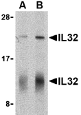
Left: Western blot analysis of IL-32 in human spleen cell lysate with IL-32 antibody at (A) 5 and (B) 10 µg/ml shows two isoforms of IL-32. Below: Immunohistochemistry of IL-32 in human spleen tissue with IL-32 antibody at 10 µg/ml.
Source:IL-32 antibody was raised against a 15 amino acid peptide from near the carboxy terminus of human IL-32.
Purification: Affinity chromatography purified via peptide column
Clonality and Clone: This is a polyclonal antibody.
Host: IL-32 antibody was raised in rabbit.
Immunogen: Human IL-32 (C-Terminus) Peptide (Cat. No. 3749P)
Application: IL-32 antibody can be used for the detection of IL-32 by Western blot at 5 – 10 µg/ml.This antibody detects all isoforms of IL-32.
Tested Application(s): E, WB, IHC
Buffer: Antibody is supplied in PBS containing 0.02% sodium azide.
Blocking Peptide:Cat. No. 3749P - IL-32 Peptide
Long-Term Storage: IL-32 antibody can be stored at 4ºC, stable for one year. As with all antibodies care should be taken to avoid repeated freeze thaw cycles. Antibodies should not be exposed to prolonged high temperatures.
Positive Control:
1. Cat. No. 1306 - Human Spleen Tissue Lysate
Species Reactivity: H, M
GI Number: 14424787
Accession Number: AAH09401
Short Description: (CT) Interleukin 32
References
1. Dahl CA, Schall RP, He HL, et al. Identification of a novel gene expressed in activated natural killer cells and T cells. J. Immunol. 1992; 148:597-603.
2. Kim S-H, Han S-Y, Azam T, et al. Interleukin-32: a cytokine and inducer of TNF-a. Immunity 2005; 22:131-42.
3. Cagnard N, Letourneur F, Essabbani A, et al. Interleukin-32, CCL2, PF4F1 and GFD10 are the only cytokine/chemokine genes differentially expressed by in vitro cultured rheumatoid and osteoarthritis fibroblast-like synoviocytes. Eur. Cyto. Network 2005; 16:289-92.
IL-31 Antibody
IL-31 Antibody
Catalog#:3747
Interleukin-31 (IL-31) is a recently discovered T-cell cytokine closely related to IL-6 type cytokines and is preferentially produced by T helper type 2 cells (1,2). IL-31 activity is mediated through the ligand-induced oligomerization of a dimeric receptor complex containing IL-31 receptor A and oncostatin M receptor (2). In response to IL-31 binding, these proteins activate the JAK/STAT and the AKT signaling pathways (3). RNA levels of IL-31 receptor A and oncostatin M receptor are induced in activated monocytes but are expressed constitutively in epithelial cells . IL-31, when overexpressed in transgenic mice, results in the development of pruritis, alopecia, and skin lesions and in humans may result in atopic dermatitis, suggesting that IL-31 may represent a novel target for antipruritic drug development (1,4).
Additional Names: IL-31 (IN), Interleukin 31, IL31
Description
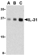
Left: Western blot analysis of IL-31 in human skeletal muscle tissue lysate with IL-31 antibody at (A) 2.5, (B) 5 and (C) 10 µg/ml.
Source:IL-31 antibody was raised against a 18 amino acid peptide from near the center of human IL-31.
Purification: Affinity chromatography purified via peptide column
Clonality and Clone: This is a polyclonal antibody.
Host: IL-31 antibody was raised in rabbit.
Please use anti-rabbit secondary antibodies.
Immunogen: Human IL-31 (Intermediate Domain) Peptide (Cat. No. 3747P)
Application: IL-31 antibody can be used for the detection of IL-31 by Western blot at 2.5 – 5 µg/ml.Despite its predicted size, IL-31 runs at approximately 27 - 30 kDa in SDS-PAGE.
Tested Application(s): E, WB
Buffer: Antibody is supplied in PBS containing 0.02% sodium azide.
Blocking Peptide:Cat. No. 3747P - IL-31 Peptide
Long-Term Storage: IL-31 antibody can be stored at 4ºC, stable for one year. As with all antibodies care should be taken to avoid repeated freeze thaw cycles. Antibodies should not be exposed to prolonged high temperatures.
Positive Control:
1. Cat. No. 1375 - Human Skeletal Muscle Tissue Lysate
Species Reactivity: H, M
GI Number: 46241139
Accession Number: AAS82846
Short Description: (IN) Interleukin 31
References
1. Dillon SR, Sprecher C, Hammond A, et al. Interleukin 31, a cytokine produced by activated T cells, induces dermatitis in mice. Nat. Immunol. 2004; 5:752-60.
2. Dreuw A, Radtke S, Pflanz S, et al. Characterization of the signaling capacities of the novel gp130-like cytokine receptor. J. Biol. Chem. 2004; 279:36112-20.
3. Diveu C, Lak-Hal AH, Froger J, et al. Predominant expression of the long isoform of GP130-like (GPL) receptor is required for interleukin-31 signaling. Eur. Cytokine Netw. 2004; 15:291-302.
4. Bilsborough J, Leung DY, Maurer M, et al. IL-31 is associated with cutaneous lymphocyte antigen-positive skin homing T cells in patients with atopic dermatitis. J. Allergy Clin. Immunol. 2006; 117:418-25.
IL-31 Antibody
IL-31 Antibody
Catalog#:3745
Interleukin-31 (IL-31) is a recently discovered T-cell cytokine closely related to IL-6 type cytokines and is preferentially produced by T helper type 2 cells (1,2). IL-31 activity is mediated through the ligand-induced oligomerization of a dimeric receptor complex containing IL-31 receptor A and oncostatin M receptor (2). In response to IL-31 binding, these proteins activate the JAK/STAT and the AKT signaling pathways (3). RNA levels of IL-31 receptor A and oncostatin M receptor are induced in activated monocytes but are expressed constitutively in epithelial cells . IL-31, when overexpressed in transgenic mice, results in the development of pruritis, alopecia, and skin lesions and in humans may result in atopic dermatitis, suggesting that IL-31 may represent a novel target for antipruritic drug development (1,4).
Additional Names: IL-31 (NT), Interleukin 31, IL31
Description
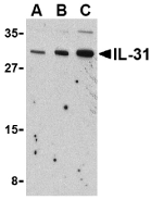
Left: Western blot analysis of IL-31 in RAW264.7 cell lysate with IL-31 antibody at (A) 2.5, (B) 5 and (C) 10 µg/ml.
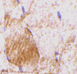
Below: Immunohistochemistry of IL-31 in rat skeletal muscle tissue with IL-31 antibody at 10 µg/ml.
Other Product Images
 Source:IL-31 antibody was raised against a 16 amino acid peptide from near the amino terminus of mouse IL-31.
Purification: Affinity chromatography purified via peptide column
Clonality and Clone: This is a polyclonal antibody.
Host: IL-31 antibody was raised in rabbit.
Please use anti-rabbit secondary antibodies.
Immunogen: Murine IL-31 (N-Terminus) Peptide (Cat. No. 3745P)
Application: IL-31 antibody can be used for the detection of IL-31 by Western blot at 2.5 – 5 µg/ml.
Tested Application(s): E, WB, IHC
Buffer: Antibody is supplied in PBS containing 0.02% sodium azide.
Blocking Peptide:Cat. No. 3745P - IL-31 Peptide
Long-Term Storage: IL-31 antibody can be stored at 4ºC, stable for one year. As with all antibodies care should be taken to avoid repeated freeze thaw cycles. Antibodies should not be exposed to prolonged high temperatures.
Positive Control:
1. Cat. No. 1283 - RAW264.7 Cell Lysate
2. Cat. No. 1467 - Rat Skeletal Muscle Tissue Lysate
Species Reactivity: M
GI Number: 46241139
Accession Number: AAS82846
Short Description: (NT) Interleukin 31
References
1. Dillon SR, Sprecher C, Hammond A, et al. Interleukin 31, a cytokine produced by activated T cells, induces dermatitis in mice. Nat. Immunol. 2004; 5:752-60.
2. Dreuw A, Radtke S, Pflanz S, et al. Characterization of the signaling capacities of the novel gp130-like cytokine receptor. J. Biol. Chem. 2004; 279:36112-20.
3. Diveu C, Lak-Hal AH, Froger J, et al. Predominant expression of the long isoform of GP130-like (GPL) receptor is required for interleukin-31 signaling. Eur. Cytokine Netw. 2004; 15:291-302.
4. Bilsborough J, Leung DY, Maurer M, et al. IL-31 is associated with cutaneous lymphocyte antigen-positive skin homing T cells in patients with atopic dermatitis. J. Allergy Clin. Immunol. 2006; 117:418-25.

Description
Left: Western blot analysis of IL-31 in RAW264.7 cell lysate with IL-31 antibody at (A) 2.5, (B) 5 and (C) 10 µg/ml.
Below: Immunohistochemistry of IL-31 in rat skeletal muscle tissue with IL-31 antibody at 10 µg/ml.
Other Product Images
 Source:IL-31 antibody was raised against a 16 amino acid peptide from near the amino terminus of mouse IL-31.
Purification: Affinity chromatography purified via peptide column
Clonality and Clone: This is a polyclonal antibody.
Host: IL-31 antibody was raised in rabbit.
Please use anti-rabbit secondary antibodies.
Immunogen: Murine IL-31 (N-Terminus) Peptide (Cat. No. 3745P)
Application: IL-31 antibody can be used for the detection of IL-31 by Western blot at 2.5 – 5 µg/ml.
Tested Application(s): E, WB, IHC
Buffer: Antibody is supplied in PBS containing 0.02% sodium azide.
Blocking Peptide:Cat. No. 3745P - IL-31 Peptide
Long-Term Storage: IL-31 antibody can be stored at 4ºC, stable for one year. As with all antibodies care should be taken to avoid repeated freeze thaw cycles. Antibodies should not be exposed to prolonged high temperatures.
Positive Control:
1. Cat. No. 1283 - RAW264.7 Cell Lysate
2. Cat. No. 1467 - Rat Skeletal Muscle Tissue Lysate
Species Reactivity: M
GI Number: 46241139
Accession Number: AAS82846
Short Description: (NT) Interleukin 31
References
1. Dillon SR, Sprecher C, Hammond A, et al. Interleukin 31, a cytokine produced by activated T cells, induces dermatitis in mice. Nat. Immunol. 2004; 5:752-60.
2. Dreuw A, Radtke S, Pflanz S, et al. Characterization of the signaling capacities of the novel gp130-like cytokine receptor. J. Biol. Chem. 2004; 279:36112-20.
3. Diveu C, Lak-Hal AH, Froger J, et al. Predominant expression of the long isoform of GP130-like (GPL) receptor is required for interleukin-31 signaling. Eur. Cytokine Netw. 2004; 15:291-302.
4. Bilsborough J, Leung DY, Maurer M, et al. IL-31 is associated with cutaneous lymphocyte antigen-positive skin homing T cells in patients with atopic dermatitis. J. Allergy Clin. Immunol. 2006; 117:418-25.
Thursday, June 23, 2011
DNA Strand
Bio-Synthesis continued to provide quality DNA products and services for the research community, but it has also become a world leader in providing custom peptide products and services. We offer comprehensive services for polyclonal antibody production with large selection for host animals.
DNA Strand Hypothesis
The immortal DNA strand hypothesis was proposed in 1975 by John Cairns as a mechanism for adult stem cells to minimize mutations in their genomes. This hypothesis proposes that instead of segregating their DNA during mitosis in a random manner, adult stem cells divide their DNA asymmetrically, and retain a distinct template set of DNA strands (parental strands) in each division. By retaining the same set of template DNA strands, adult stem cells would pass mutations arising from errors in DNA replication on to non-stem cell daughters that soon terminally differentiate (end mitotic divisions and become a functional cell).
DNA Strands
In the label-retention assay, the goal is to mark 'immortal' or parental DNA strands with a DNA label such as tritiated thymidine or bromodeoxyuridine (BrdU). These types of DNA labels will incorporate into the newly-synthesized DNA of dividing cells during S phase. A pulse of DNA label is given to adult stem cells under conditions where they have not yet delineated an immortal DNA strand.
For more information visit: www.biosyn.com
IL-17 Antibody
Catalog#:4877
Interleukin 17 (IL-17) is a family of pro-inflammatory cytokines produced by activated T cells and is thought to have a major role in the initiation and perpetuation of rheumatoid arthritis. IL-17 regulates the activities of NF-kB and mitogen-activated protein kinases such as ERK and JNK. In addition, IL-17 stimulates the expression of IL-6 and cyclooxygenase-2 and enhances the production of nitric oxide. IL-17-producing T helper cells (TH-17 cells) have been the subject of much attention due to the importance of IL-17 in the pathogenesis of autoimmune inflammation. Because of its role in autoimmune diseases, it is thought that targeting the production and action of IL-17 would be beneficial therapeutically in these diseases.
Additional Names: IL-17 (NT), Interleukin-17, IL-17A, CTLA8
Description
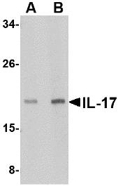
Left: Western blot analysis of IL-17 in A-20 lysate with IL-17 antibody at (A) 2 and (B) 4 µg/ml.
Source:IL-17 antibody was raised against a 16 amino acid peptide from near the amino terminus of human IL-17A.
Purification: Affinity chromatography purified via peptide column
Clonality and Clone: This is a polyclonal antibody.
Host: IL-17 antibody was raised in rabbit.
Please use anti-rabbit secondary antibodies.
Application: IL-17 antibody can be used for the detection of IL-17 by Western blot at 2 – 4 µg/ml.
Tested Application(s): E, WB
Buffer: Antibody is supplied in PBS containing 0.02% sodium azide.
Blocking Peptide:Cat.No. 4877P - IL-17 Peptide
Long-Term Storage: IL-17 antibody can be stored at 4ºC, stable for one year. As with all antibodies care should be taken to avoid repeated freeze thaw cycles. Antibodies should not be exposed to prolonged high temperatures.
Positive Control:
1. Cat. No. 1288 - A20 Cell Lysate
Species Reactivity: H, M
GI Number: 4504651
Accession Number: NP_002181
Short Description: (NT) Interleukin-17A
References
1. Miossec P. Are T cells in rheumatoid synovium aggressors or bystanders? Curr. Opin. Rheumatol. 2000; 12:181-5.
2. Paunovic V, Carroll HP, Vandenbroeck K, et al. Crossed signals: the role of interleukin (IL)-12, -17, -23, and -27 in autoimmunity. Rheumatol. 2008; 47:771-6.
3. Steinman L. A brief history of TH17, the first major revision in the TH1/TH2 hypothesis of T cell-mediated tissue damage. Nat. Med. 2007; 13:139-145.
IL-17 Antibody
Catalog#:4887
Interleukin 17 (IL-17) is a family of pro-inflammatory cytokines produced by activated T cells and is thought to have a major role in the initiation and perpetuation of rheumatoid arthritis. IL-17 regulates the activities of NF-kB and mitogen-activated protein kinases such as ERK and JNK. In addition, IL-17 stimulates the expression of IL-6 and cyclooxygenase-2 and enhances the production of nitric oxide. IL-17-producing T helper cells (TH-17 cells) have been the subject of much attention due to the importance of IL-17 in the pathogenesis of autoimmune inflammation. Because of its role in autoimmune diseases, it is thought that targeting the production and action of IL-17 would be beneficial therapeutically in these diseases.
Additional Names: IL-17 (IN), Interleukin-17, IL-17A, CTLA8
Description
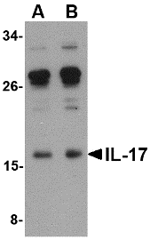
Left: Western blot analysis of IL-17 in A-20 lysate with IL-17 antibody at (A) 0.5 and (B) 1 µg/ml.
Source:IL-17 antibody was raised against a 19 amino acid peptide from near the center of human IL-17A.
Purification: Affinity chromatography purified via peptide column
Clonality and Clone: This is a polyclonal antibody.
Host: IL-17 antibody was raised in rabbit.
Please use anti-rabbit secondary antibodies.
Application: IL-17 antibody can be used for the detection of IL-17 by Western blot at 0.5 – 1 µg/ml.
Tested Application(s): E, WB
Buffer: Antibody is supplied in PBS containing 0.02% sodium azide.
Blocking Peptide:Cat.No. 4887P - IL-17 Peptide
Long-Term Storage: IL-17 antibody can be stored at 4ºC, stable for one year. As with all antibodies care should be taken to avoid repeated freeze thaw cycles. Antibodies should not be exposed to prolonged high temperatures.
Positive Control:
1.Cat. No. 1288 - A20 Cell Lysate
Species Reactivity: H, M
GI Number: 4504651
Accession Number: NP_002181
Short Description: (IN) Interleukin-17A
References
1. Miossec P. Are T cells in rheumatoid synovium aggressors or bystanders? Curr. Opin. Rheumatol. 2000; 12:181-5.
2. Paunovic V, Carroll HP, Vandenbroeck K, et al. Crossed signals: the role of interleukin (IL)-12, -17, -23, and -27 in autoimmunity. Rheumatol. 2008; 47:771-6.
3. Steinman L. A brief history of TH17, the first major revision in the TH1/TH2 hypothesis of T cell-mediated tissue damage. Nat. Med. 2007; 13:139-145.
IKK gamma Antibody
Catalog#:2335
Nuclear factor kappa B (NF-kappaB) is a ubiquitous transcription factor and an essential mediator of gene expression during activation of immune and inflammatory responses. NF-kappaB mediates the expression of a great variety of genes in response to extracellular stimuli. NF-kappaB is associated with IkB proteins in the cell cytoplasm, which inhibit NF-kappaB activity. The IkB kinase (IKKalpha and IKKbeta) phosphorylates IkB and mediates NF-kappaB activation. A novel molecule in the IKK complex was recently identified and termed IKKgamma/NEMO/FIP3/IKKAP1 (1-5). IKKg interacts with IKKbeta and is required for the activation of IKK complex and NF-kappaB by LPS, PMA, TNF, and IL-1 stimulation (1-4). FIP3 was also shown to bind to RIP and NIK and to mediate TNF-induced NF-kappaB activation (3).
Additional Names: IKK gamma (CT),IKKg / NEMO, NEMO, FIP3

Description
Left: Western blot analysis of IKK gamma in HeLa whole cell lysate with IKK gamma antibody at 1 µg/ml.
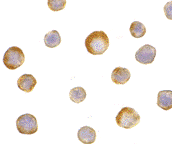
Below: Immunocytochemistry of IKK gamma in HeLa cells with IKK gamma antibody at 5 µg/ml.
Other Product Images
 Source:IKK gamma antibody was raised against a peptide corresponding to amino acids 400 to 416 of human IKK gamma, which are identical to those of mouse homologue.
Purification: Affinity chromatography purified via peptide column
Clonality and Clone: This is a polyclonal antibody.
Host: IKK gamma antibody was raised in rabbit.
Please use anti-rabbit secondary antibodies.
Immunogen: Human IKK gamma Peptide (Cat. No. 2335P)
Application: IKK gamma antibody can be used for detection of IKK gamma by Western blot at 0.5 to 1 µg/ml.A 52 kDa band should be detected. IKK gamma has no cross response to IKK alpha or IKK beta.
Tested Application(s): E, WB, ICC
Buffer: Antibody is supplied in PBS containing 0.02% sodium azide.
Blocking Peptide:Cat. No. 2335P - IKK gamma Peptide
Long-Term Storage: IKK gamma antibody can be stored at 4ºC, stable for one year. As with all antibodies care should be taken to avoid repeated freeze thaw cycles. Antibodies should not be exposed to prolonged high temperatures.
Positive Control:
1.Cat. No. 1201 - HeLa Cell Lysate
Species Reactivity: H, M, R
GI Number: 3641279
Accession Number: AF074382
Short Description: (CT) IkB Kinase gamma
References
1. Rothwarf DM, Zandi E, Natoli G, Karin M. IKK-gamma is an essential regulatory subunit of the IkappaB kinase complex. Nature 1998;395(6699):297-300
2. Yamaoka S, Courtois G, Bessia C, Whiteside ST, Weil R, Agou F, Kirk HE, Kay RJ, Israel A. Complementation cloning of NEMO, a component of the IkappaB kinase complex essential for NF-kappaB activation. Cell. 1998;93(7):1231-40.
3. Li Y, Kang J, Friedman J, Tarassishin L, Ye J, Kovalenko A, Wallach D, Horwitz MS. Identification of a cell protein (FIP-3) as a modulator of NF-kappaB activity and as a target of an adenovirus inhibitor of tumor necrosis factor alpha-induced apoptosis. Proc Natl Acad Sci U S A 1999;96(3):1042-7
4. Mercurio F, Murray BW, Shevchenko A, Bennett BL, Young DB, Li JW, Pascual G, Motiwala A, Zhu H, Mann M, Manning AM. IkappaB kinase (IKK)-associated protein 1, a common component of the heterogeneous IKK complex. Mol Cell Biol. 1999;19(2):1526-38.
Wednesday, June 22, 2011
Clone Antibody
Bio-Synthesis, with 20 years of experience in developing polyclonal antibodies, is a leading global provider of custom antibody production services. Our facility generates high-affinity custom antibodies with industry-leading titer guarantees and comprehensive antibody production packages that simplify the ordering process and present cost-effective methods to isolate epitope-specific antibodies. Visit www.biosyn.com and see why partner with Bio-Synthesis for your custom antibodies needs.
Antibodies
From Antigen selection to antibody purification, BSI has optimized each step of the antibody production process to provide the best antibodies available.
Custom peptide antibody production
Keyhole Limpet Hemocyanin (KLH), Bovine Serum Albumin (BSA), Rabbit Serum Albumin (RSA), Ovalbumin, Thyroglobulin, or Human Gamma Globulin (HGG).Amino Acid Analysis of Conjugates also available.
Isotypes
Antibodies can come in different varieties known as isotypes or classes. In placental mammals there are five antibody isotypes known as IgA, IgD, IgE,IgG and IgM.The antibody isotype of a B cell changes during the cell's development and activation.

For more information visit: www.biosyn.com
IKK epsilon Antibody
Catalog#:2329
Nuclear factor kappa B (NF-kappaB) is a ubiquitous transcription factor and an essential mediator of gene expression during activation of immune and inflammatory responses. NF-kappaB mediates the expression of a great variety of genes in response to extracellular stimuli. NF-kappaB is associated with IkB proteins in the cell cytoplasm, which inhibit NF-kappaB activity. IkB is phosphorylated by IkB kinase (IKK) complex that contains IKKalpha, IKKbeta, and IKKgamma. A novel molecule in the IKK complex was recently identified and designated IKKepsilon/IKK-i (1,2). IKK epsilon is required for the activation of NF-kappaB by PMA and T cell receptors but not by TNFalpha and IL-1 (1). IKKepsilon/IKK-i message is expressed in a variety of tissues and is inducible by TNFalpha, IL-1, and LPS (2).
Additional Names: IKK epsilon (CT), IKKi, IKKe

Description
Left: Western blot analysis of IKK epsilon in Jurkat whole cell lysate with IKK epsilon/IKK-i antibody at 1 µg/ml.
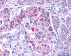
Below: Immunohistochemistry of IKK epsilon in human pancreas tissue with IKK epsilon antibody at 10 µg/ml.
Other Product Images
 Source:IKK epsilon antibody was raised against a peptide corresponding to amino acids 701 to 716 of human IKK epsilon/IKK-i.
Purification: Affinity chromatography purified via peptide column
Clonality and Clone: This is a polyclonal antibody.
Host: IKK epsilon antibody was raised in rabbit.
Please use anti-rabbit secondary antibodies.
Immunogen: Human IKK epsilon Peptide (Cat. No. 2329P)
Application: IKK epsilon antibody can be used for detection of IKK epsilon by Western blot at 0.5 to 1 µg/ml.An 80 kDa band should be detected. It has no cross response to IKKa, IKKb, or IKKy.
Tested Application(s): E, WB, IHC
Buffer: Antibody is supplied in PBS containing 0.02% sodium azide.
Blocking Peptide:Cat. No. 2329P - IKK epsilon Peptide
Long-Term Storage: IKK epsilon antibody can be stored at 4ºC, stable for one year. As with all antibodies care should be taken to avoid repeated freeze thaw cycles. Antibodies should not be exposed to prolonged high temperatures.
Positive Control:
1. Cat. No. 1205 - Jurkat Cell Lysate
2. Cat. No. 1307 - Human Pancreas Tissue Lysate
Species Reactivity: H
GI Number: 7288877
Accession Number: AF241789
Short Description: (CT) IkB Kinase epsilon
References
1. Peters RT, Liao SM, Maniatis T. IKK epsilonpsilon is part of a novel PMA-inducible IkappaB kinase complex. Mol Cell 2000;5(3):513-22
2. Shimada T, Kawai T, Takeda K, Matsumoto M, Inoue J, Tatsumi Y, Kanamaru A, Akira S. IKK-i, a novel lipopolysaccharide-inducible kinase that is related to IkappaB kinases. Int Immunol 1999;11(8):1357-62 (WD010

Description
Left: Western blot analysis of IKK epsilon in Jurkat whole cell lysate with IKK epsilon/IKK-i antibody at 1 µg/ml.
Below: Immunohistochemistry of IKK epsilon in human pancreas tissue with IKK epsilon antibody at 10 µg/ml.
Other Product Images
 Source:IKK epsilon antibody was raised against a peptide corresponding to amino acids 701 to 716 of human IKK epsilon/IKK-i.
Purification: Affinity chromatography purified via peptide column
Clonality and Clone: This is a polyclonal antibody.
Host: IKK epsilon antibody was raised in rabbit.
Please use anti-rabbit secondary antibodies.
Immunogen: Human IKK epsilon Peptide (Cat. No. 2329P)
Application: IKK epsilon antibody can be used for detection of IKK epsilon by Western blot at 0.5 to 1 µg/ml.An 80 kDa band should be detected. It has no cross response to IKKa, IKKb, or IKKy.
Tested Application(s): E, WB, IHC
Buffer: Antibody is supplied in PBS containing 0.02% sodium azide.
Blocking Peptide:Cat. No. 2329P - IKK epsilon Peptide
Long-Term Storage: IKK epsilon antibody can be stored at 4ºC, stable for one year. As with all antibodies care should be taken to avoid repeated freeze thaw cycles. Antibodies should not be exposed to prolonged high temperatures.
Positive Control:
1. Cat. No. 1205 - Jurkat Cell Lysate
2. Cat. No. 1307 - Human Pancreas Tissue Lysate
Species Reactivity: H
GI Number: 7288877
Accession Number: AF241789
Short Description: (CT) IkB Kinase epsilon
References
1. Peters RT, Liao SM, Maniatis T. IKK epsilonpsilon is part of a novel PMA-inducible IkappaB kinase complex. Mol Cell 2000;5(3):513-22
2. Shimada T, Kawai T, Takeda K, Matsumoto M, Inoue J, Tatsumi Y, Kanamaru A, Akira S. IKK-i, a novel lipopolysaccharide-inducible kinase that is related to IkappaB kinases. Int Immunol 1999;11(8):1357-62 (WD010
IKK beta Antibody
Catalog#:2121
Nuclear factor kappa B (NF-kappaB) is a ubiquitous transcription factor and an essential mediator of gene expression during activation of immune and inflammatory responses. NF-kappaB mediates the expression of a great variety of genes in response to extracellular stimuli including IL-1, TNFalpha, and bacteria product LPS. NF-kappaB is associated with IkappaB proteins in the cell cytoplasm, which inhibit NF-kappaB activity. The long-sought IkappaB kinase (IKK), which phosphorylates IkappaB, and mediates IkappaB degradation and NF-kappaB activation, was recently identified by several laboratories (1-5). IKK is a serine protein kinase, and the IKK complex contains alpha and beta subunits (IKKalpha and IKKbeta). IKKalpha and IKKbeta interact with each other and both are essential for NF-kappaB activation. IKKbeta phosphorylates both IkappaB-alpha and IkappaB-beta. IKKbeta is expressed in variety of human tissues.
Additional Names: IKK beta (C3), IKK beta, I-kappa-B-kinase beta, I-kappa-B-kinase 2
Description
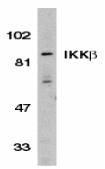
Left: Western blot analysis of IKK beta in Jurkat whole cell lysate with IKK beta antibody (C3) at 1:500 dilution.
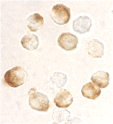
Below: Immunocytochemistry staining of HeLa cells using IKK beta antibody at 10 µg/ml.
Other Product Images
 Source:IKK beta antibody was raised against a peptide corresponding to amino acids near the carboxy-terminus of human IKK beta (Genbank accession NoO14920), which differs from corresponding murine sequence by one amino acid.
Purification: Affinity chromatography purified via peptide column
Clonality and Clone: This is a polyclonal antibody.
Host: IKK beta antibody was raised in rabbit.
Please use anti-rabbit secondary antibodies
Immunogen: Human IKK beta (C-Terminus 3) Peptide (Cat. No. 2121P)
Application: IKK beta antibody can be used for detection of IKK beta by Western blot at 1 – 2 µg/ml.A 87 kDa band should be detected. This polyclonal antibody has no cross response to IKKa or IKKy.
Tested Application(s): E, WB, ICC
Buffer: Antibody is supplied in PBS containing 0.02% sodium azide.
Blocking Peptide:Cat. No. 2121P - IKK beta Peptide
Long-Term Storage: IKK beta antibody can be stored at 4ºC, stable for one year. As with all antibodies care should be taken to avoid repeated freeze thaw cycles. Antibodies should not be exposed to prolonged high temperatures.
Positive Control:
1. Cat. No. 1205 - Jurkat Cell Lysate
2. Cat. No. 1201 - HeLa Whole Cell Lysate
Species Reactivity: H
GI Number: 14285497
Accession Number: O14920
Short Description: (C3) Ikappa B kinase beta
References
1. DiDonato JA, Hayakawa M, Rothwarf DM, Zandi E, Karin M. A cytokine-responsive IkappaB kinase that activates the transcription factor NF-kappaB. Nature 1997;388:548-54
2. Regnier CH, Song HY, Gao X, Goeddel DV, Cao Z, Rothe M. Identification and characterization of an IkappaB kinase. Cell 1997;90:373-83
3. Zandi E, Rothwarf DM, Delhase M, Hayakawa M, Karin M. The IkappaB kinase complex (IKK) contains two kinase subunits, IKKalpha and IKKbeta, necessary for IkappaB phosphorylation and NF-kappaB activation. Cell 1997;91:243-52
4. Woronicz JD, Gao X, Cao Z, Rothe M, Goeddel DY. IkappaB kinase-alpha: NF-kappaB activation and complex formation with IkappaB kinase-alpha and NIK. Science 1997;278:866-9

Description
Left: Western blot analysis of IKK beta in Jurkat whole cell lysate with IKK beta antibody (C3) at 1:500 dilution.
Below: Immunocytochemistry staining of HeLa cells using IKK beta antibody at 10 µg/ml.
Other Product Images
 Source:IKK beta antibody was raised against a peptide corresponding to amino acids near the carboxy-terminus of human IKK beta (Genbank accession NoO14920), which differs from corresponding murine sequence by one amino acid.
Purification: Affinity chromatography purified via peptide column
Clonality and Clone: This is a polyclonal antibody.
Host: IKK beta antibody was raised in rabbit.
Please use anti-rabbit secondary antibodies
Immunogen: Human IKK beta (C-Terminus 3) Peptide (Cat. No. 2121P)
Application: IKK beta antibody can be used for detection of IKK beta by Western blot at 1 – 2 µg/ml.A 87 kDa band should be detected. This polyclonal antibody has no cross response to IKKa or IKKy.
Tested Application(s): E, WB, ICC
Buffer: Antibody is supplied in PBS containing 0.02% sodium azide.
Blocking Peptide:Cat. No. 2121P - IKK beta Peptide
Long-Term Storage: IKK beta antibody can be stored at 4ºC, stable for one year. As with all antibodies care should be taken to avoid repeated freeze thaw cycles. Antibodies should not be exposed to prolonged high temperatures.
Positive Control:
1. Cat. No. 1205 - Jurkat Cell Lysate
2. Cat. No. 1201 - HeLa Whole Cell Lysate
Species Reactivity: H
GI Number: 14285497
Accession Number: O14920
Short Description: (C3) Ikappa B kinase beta
References
1. DiDonato JA, Hayakawa M, Rothwarf DM, Zandi E, Karin M. A cytokine-responsive IkappaB kinase that activates the transcription factor NF-kappaB. Nature 1997;388:548-54
2. Regnier CH, Song HY, Gao X, Goeddel DV, Cao Z, Rothe M. Identification and characterization of an IkappaB kinase. Cell 1997;90:373-83
3. Zandi E, Rothwarf DM, Delhase M, Hayakawa M, Karin M. The IkappaB kinase complex (IKK) contains two kinase subunits, IKKalpha and IKKbeta, necessary for IkappaB phosphorylation and NF-kappaB activation. Cell 1997;91:243-52
4. Woronicz JD, Gao X, Cao Z, Rothe M, Goeddel DY. IkappaB kinase-alpha: NF-kappaB activation and complex formation with IkappaB kinase-alpha and NIK. Science 1997;278:866-9
IKK alpha Antibody
Catalog#:2117
Nuclear factor kappa B (NF-kappaB) is a ubiquitous transcription factor and an essential mediator of gene expression during activation of immune and inflammatory responses. NF-kappaB mediates the expression of a great variety of genes in response to extracellular stimuli including IL-1, TNFalpha and bacteria product LPS. NF-kappaB is associated with IkB proteins in the cell cytoplasm, which inhibit NF-kappaB activity. The long-sought IΚB kinase (IKK), which phosphorylates IkappaB, and mediates IkappaB degradation and NF-kappaB activation, was recently identified by several laboratories (1-5). IKK is a serine protein kinase, and the IKK complex contains alpha and beta subunits (IKKalpha and IKKΒ). IKKalpha and IKKbeta interact with each other and both are essential for the NF-kappaB activation. IKKalpha specifically phosphorylates IkappaB-alpha. IKKalpha is expressed in a variety of human tissues.
Additional Names: IKK alpha (C3), IKK-1, IKKa
Description
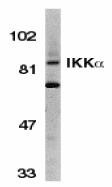
Left: Western blot analysis of IKK alpha in Jurkat whole cell lysate with IKK alpha antibody (C3) at 1:500 dilution.
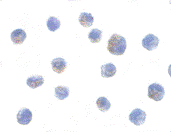
Below: Immunocytochemistry of IKK alpha in Jurkat cells with IKK alpha antibody at 10 µg/ml.
Other Product Images
 Source:IKK alpha antibody was raised against a peptide corresponding to amino acids 658 to 674 of human IKK alpha, which differs from corresponding murine sequence by one amino acid.
Purification: Affinity chromatography purified via peptide column
Clonality and Clone: This is a polyclonal antibody.
Host: IKK alpha antibody was raised in rabbit.
Please use anti-rabbit secondary antibodies.
Immunogen: Human IKK alpha (C-Terminus 3) Peptide (Cat. No. 2117P)
Application: IKK alpha antibody can be used for detection of IKK alpha by Western blot at 1:500 to 1:1000 dilution. An 85 kDa band should be detected. This polyclonal antibody has no cross response to IKKb or IKKy.
Tested Application(s): E, WB, ICC
Buffer: Antibody is supplied in PBS containing 0.02% sodium azide.
Blocking Peptide:Cat. No. 2117P - IKK alpha Peptide
Long-Term Storage: IKK alpha antibody can be stored at 4ºC, stable for one year. As with all antibodies care should be taken to avoid repeated freeze thaw cycles. Antibodies should not be exposed to prolonged high temperatures.
Positive Control:
1. Cat. No. 1205 - Jurkat Cell Lysate
Species Reactivity: H
GI Number: 2327068
Accession Number: AF009225
Short Description: (C3) Ikappa B kinase alpha
References
1. DiDonato JA, Hayakawa M, Rothwarf DM, Zandi E, Karin M. A cytokine-responsive IkappaB kinase that activates the transcription factor NF-kappaB. Nature 1997;388:548-54
2. Regnier CH, Song HY, Gao X, Goeddel DV, Cao Z, Rothe M. Identification and characterization of an IkappaB kinase. Cell 1997;90:373-83
3. Zandi E, Rothwarf DM, Delhase M, Hayakawa M, Karin M. The IkappaB kinase complex (IKK) contains two kinase subunits, IKKalpha and IKKbeta, necessary for IkappaB phosphorylation and NF-kappaB activation. Cell 1997;91:243-52
4. Woronicz JD, Gao X, Cao Z, Rothe M, Goeddel DY. IkappaB kinase-alpha: NF-kappaB activation and complex formation with IkappaB kinase-alpha and NIK. Science 1997;278:866-9

Description
Left: Western blot analysis of IKK alpha in Jurkat whole cell lysate with IKK alpha antibody (C3) at 1:500 dilution.
Below: Immunocytochemistry of IKK alpha in Jurkat cells with IKK alpha antibody at 10 µg/ml.
Other Product Images
 Source:IKK alpha antibody was raised against a peptide corresponding to amino acids 658 to 674 of human IKK alpha, which differs from corresponding murine sequence by one amino acid.
Purification: Affinity chromatography purified via peptide column
Clonality and Clone: This is a polyclonal antibody.
Host: IKK alpha antibody was raised in rabbit.
Please use anti-rabbit secondary antibodies.
Immunogen: Human IKK alpha (C-Terminus 3) Peptide (Cat. No. 2117P)
Application: IKK alpha antibody can be used for detection of IKK alpha by Western blot at 1:500 to 1:1000 dilution. An 85 kDa band should be detected. This polyclonal antibody has no cross response to IKKb or IKKy.
Tested Application(s): E, WB, ICC
Buffer: Antibody is supplied in PBS containing 0.02% sodium azide.
Blocking Peptide:Cat. No. 2117P - IKK alpha Peptide
Long-Term Storage: IKK alpha antibody can be stored at 4ºC, stable for one year. As with all antibodies care should be taken to avoid repeated freeze thaw cycles. Antibodies should not be exposed to prolonged high temperatures.
Positive Control:
1. Cat. No. 1205 - Jurkat Cell Lysate
Species Reactivity: H
GI Number: 2327068
Accession Number: AF009225
Short Description: (C3) Ikappa B kinase alpha
References
1. DiDonato JA, Hayakawa M, Rothwarf DM, Zandi E, Karin M. A cytokine-responsive IkappaB kinase that activates the transcription factor NF-kappaB. Nature 1997;388:548-54
2. Regnier CH, Song HY, Gao X, Goeddel DV, Cao Z, Rothe M. Identification and characterization of an IkappaB kinase. Cell 1997;90:373-83
3. Zandi E, Rothwarf DM, Delhase M, Hayakawa M, Karin M. The IkappaB kinase complex (IKK) contains two kinase subunits, IKKalpha and IKKbeta, necessary for IkappaB phosphorylation and NF-kappaB activation. Cell 1997;91:243-52
4. Woronicz JD, Gao X, Cao Z, Rothe M, Goeddel DY. IkappaB kinase-alpha: NF-kappaB activation and complex formation with IkappaB kinase-alpha and NIK. Science 1997;278:866-9
Tuesday, June 21, 2011
DNA Test For Free
At Biosynthesis DNA Diagnostics is a multi-species molecular genetics laboratory specializing in human and animal DNA testing for individuals, law enforcement, attorneys, breed associations, private investigators and state regulatory agencies.
DNA Familial Relationship Tests
DNA testing can definitively identify distant biological relationships. Our laboratory routinely performs DNA tests in cases of questioned grandparent age, siblingship, twin zygosity, maternity and more.
DNA Identification and Verification
The DNA Identity Testing Center also provides genetic profiling via our SAFEDNATM ID card. Your SAFEDNA ID card will provide positive identification and may be used to ensure child safety, prevent fraud and protect asset.
DNA-Binding Proteins
Structural proteins that bind DNA are well-understood examples of non-specific DNA-protein interactions. Within chromosomes, DNA is held in complexes with structural proteins. These proteins organize the DNA into a compact structure called chromatin.
For more information visit: www.biosyn.com
IKK alpha Antibody
Catalog#:2115
Nuclear factor kappa B (NF-kappaB) is a ubiquitous transcription factor and an essential mediator of gene expression during activation of immune and inflammatory responses. NF-kappaB mediates the expression of a great variety of genes in response to extracellular stimuli including IL-1, TNFalpha and bacteria product LPS. NF-kappaB is associated with IkB proteins in the cell cytoplasm, which inhibit NF-kappaB activity. The long-sought IkappaB kinase (IKK), which phosphorylates IkappaB, and mediates IkappaB degradation and NF-kappaB activation, was recently identified by several laboratories (1-5). IKK is a serine protein kinase, and the IKK complex contains alpha and beta subunits (IKKalpha and IKKΒ). IKKalpha and IKKb interact with each other and both are essential for the NF-kappaB activation. IKKalpha specifically phosphorylates IkappaB-Α.IKKalpha is expressed in a variety of human tissues.
Additional Names: IKK alpha (C2), Inhibitor of nuclear factor kappa-B kinase alpha subunit, IkappaB kinase alpha, NFkappaB inhibitor kinase alpha, IKKa

Description
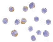
Left: Western blot analysis of IKK alpha in HeLa whole cell lysate with IKK alpha antibody at 1:500 dilution.
Below: Immunocytochemistry of IKK alpha in HeLa cells with IKK alpha antibody at 10 µg/ml.
Other Product Images
 Source:IKK alpha antibody was raised against a peptide corresponding to amino acids 699 to 715 of human IKK alpha, which differs from corresponding murine sequence by one amino acid
Purification: Affinity chromatography purified via peptide column
Clonality and Clone: This is a polyclonal antibody.
Host: IKK alpha antibody was raised in rabbit.
Please use anti-rabbit secondary antibodies.
Immunogen: Human IKK alpha (C-Terminus 2) Peptide (Cat. No. 2115P)
Application: IKK alpha antibody can be used for detection of IKK alpha by Western blot at 1:500 to 1:1000 dilution. A 85 kDa band should be detected. This polyclonal antibody has no cross response to IKKb or IKKy.
Tested Application(s): E, WB, ICC
Buffer: Antibody is supplied in PBS containing 0.02% sodium azide.
Blocking Peptide:Cat. No. 2115P - IKK alpha Peptide
Long-Term Storage: IKK alpha antibody can be stored at 4ºC, stable for one year. As with all antibodies care should be taken to avoid repeated freeze thaw cycles. Antibodies should not be exposed to prolonged high temperatures.
Positive Control:
1. Cat. No. 1201 - HeLa Cell Lysate
Species Reactivity: H
GI Number: 2327068
Accession Number: AF009225
Short Description: (C2) Ikappa B kinase alpha
References
1. DiDonato JA, Hayakawa M, Rothwarf DM, Zandi E, Karin M. A cytokine-responsive IkappaB kinase that activates the transcription factor NF-kappaB. Nature 1997;388:548-54
2. Regnier CH, Song HY, Gao X, Goeddel DV, Cao Z, Rothe M. Identification and characterization of an IkappaB kinase. Cell 1997;90:373-83
3. Zandi E, Rothwarf DM, Delhase M, Hayakawa M, Karin M. The IkappaB kinase complex (IKK) contains two kinase subunits, IKKalpha and IKKalpha, necessary for IkappaB phosphorylation and NF-kappaB activation. Cell 1997;91:243-52
4. Woronicz JD, Gao X, Cao Z, Rothe M, Goeddel DY. IkappaB kinase-beta: NF-kappaB activation and complex formation with IkappaB kinase-alpha and NIK. Science 1997;278:866-9

Description
Left: Western blot analysis of IKK alpha in HeLa whole cell lysate with IKK alpha antibody at 1:500 dilution.
Below: Immunocytochemistry of IKK alpha in HeLa cells with IKK alpha antibody at 10 µg/ml.
Other Product Images
 Source:IKK alpha antibody was raised against a peptide corresponding to amino acids 699 to 715 of human IKK alpha, which differs from corresponding murine sequence by one amino acid
Purification: Affinity chromatography purified via peptide column
Clonality and Clone: This is a polyclonal antibody.
Host: IKK alpha antibody was raised in rabbit.
Please use anti-rabbit secondary antibodies.
Immunogen: Human IKK alpha (C-Terminus 2) Peptide (Cat. No. 2115P)
Application: IKK alpha antibody can be used for detection of IKK alpha by Western blot at 1:500 to 1:1000 dilution. A 85 kDa band should be detected. This polyclonal antibody has no cross response to IKKb or IKKy.
Tested Application(s): E, WB, ICC
Buffer: Antibody is supplied in PBS containing 0.02% sodium azide.
Blocking Peptide:Cat. No. 2115P - IKK alpha Peptide
Long-Term Storage: IKK alpha antibody can be stored at 4ºC, stable for one year. As with all antibodies care should be taken to avoid repeated freeze thaw cycles. Antibodies should not be exposed to prolonged high temperatures.
Positive Control:
1. Cat. No. 1201 - HeLa Cell Lysate
Species Reactivity: H
GI Number: 2327068
Accession Number: AF009225
Short Description: (C2) Ikappa B kinase alpha
References
1. DiDonato JA, Hayakawa M, Rothwarf DM, Zandi E, Karin M. A cytokine-responsive IkappaB kinase that activates the transcription factor NF-kappaB. Nature 1997;388:548-54
2. Regnier CH, Song HY, Gao X, Goeddel DV, Cao Z, Rothe M. Identification and characterization of an IkappaB kinase. Cell 1997;90:373-83
3. Zandi E, Rothwarf DM, Delhase M, Hayakawa M, Karin M. The IkappaB kinase complex (IKK) contains two kinase subunits, IKKalpha and IKKalpha, necessary for IkappaB phosphorylation and NF-kappaB activation. Cell 1997;91:243-52
4. Woronicz JD, Gao X, Cao Z, Rothe M, Goeddel DY. IkappaB kinase-beta: NF-kappaB activation and complex formation with IkappaB kinase-alpha and NIK. Science 1997;278:866-9
IKK alpha Antibody
Catalog#:2025
Nuclear factor kappa B (NF-kappaB) is a ubiquitous transcription factor and an essential mediator of gene expression during activation of immune and inflammatory responses. NF-kappaB mediates the expression of a great variety of genes in response to extracellular stimuli including IL-1, TNFa, and bacteria product LPS. NF-kappaB is associated with IkappaB proteins in the cell cytoplasm, which inhibit NF-kappaB activity. The long-sought IkappaB kinase (IKK), which phosphorylates IkappaB, and mediates IΚB degradation and NF-kappaB activation, was recently identified by several laboratories (1-5). IKK is a serine protein kinase, and the IKK complex contains alpha and beta subunits (IKKalpha and IKKbeta). IKKa and IKKbeta interact with each other and both are essential for the NF-kappaB activation. IKKalpha specifically phosphorylates IkB-alpha. IKKalpha is expressed in variety of human tissues.
Additional Names: IKK alpha (C1), IKKa, IKK-1

Description
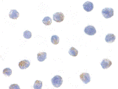
Left: Western blot analysis of IKK alpha in HeLa whole cell lysate with IKK alpha antibody at 1:1000 dilution.
Below: Immunocytochemistry of IKK alpha in Jurkat cells with IKK alpha antibody at 1µg/ml.
Other Product Images
 Source:IKK alpha antibody was raised against a peptide corresponding to amino acids 716 to 734 of human IKK alpha, which differs from corresponding murine sequence by four amino acids.
Purification: Affinity chromatography purified via peptide column
Clonality and Clone: This is a polyclonal antibody.
Host: IKK alpha antibody was raised in rabbit.
Immunogen: Human IKK alpha (C-Terminus 1) Peptide (Cat. No. 2025P)
Application: IKK alpha can be used for detection of IKK alpha by Western blot at 1:1000 to 1:2000 dilution. An 85 kDa band should be detected. antibody has no cross response to IKKb or IKKg.
Tested Application(s): E, WB, ICC
Buffer: Antibody is supplied in PBS containing 0.02% sodium azide.
Blocking Peptide:Cat. No. 2025P - IKK alpha Peptide
Long-Term Storage: IKK alpha antibody can be stored at 4ºC, stable for one year. As with all antibodies care should be taken to avoid repeated freeze thaw cycles. Antibodies should not be exposed to prolonged high temperatures.
Positive Control:
1. Cat. No. 1201 - HeLa Cell Lysate
2. Cat. No. 1205 Jurkat Whole Cell Lysate
Species Reactivity: H
GI Number: 2327068
Accession Number: AF009225
Short Description: (C1) Ikappa B kinase alpha
References
1. DiDonato JA, Hayakawa M, Rothwarf DM, Zandi E, Karin M. A cytokine-responsive IkappaB kinase that activates the transcription factor NF-kappaB. Nature 1997;388:548-54
2. Regnier CH, Song HY, Gao X, Goeddel DV, Cao Z, Rothe M. Identification and characterization of an IkappaB kinase. Cell 1997;90:373-83
3. Zandi E, Rothwarf DM, Delhase M, Hayakawa M, Karin M. The IkappaB kinase complex (IKK) contains two kinase subunits, IKKalpha and IKKbeta, necessary for IkappaB phosphorylation and NF-kappaB activation. Cell 1997;91:243-52
4. Woronicz JD, Gao X, Cao Z, Rothe M, Goeddel DY. IkappaB kinase-beta: NF-kappaB activation and complex formation with IkappaB kinase-alpha and NIK. Science 1997;278:866-9

Description
Left: Western blot analysis of IKK alpha in HeLa whole cell lysate with IKK alpha antibody at 1:1000 dilution.
Below: Immunocytochemistry of IKK alpha in Jurkat cells with IKK alpha antibody at 1µg/ml.
Other Product Images
 Source:IKK alpha antibody was raised against a peptide corresponding to amino acids 716 to 734 of human IKK alpha, which differs from corresponding murine sequence by four amino acids.
Purification: Affinity chromatography purified via peptide column
Clonality and Clone: This is a polyclonal antibody.
Host: IKK alpha antibody was raised in rabbit.
Immunogen: Human IKK alpha (C-Terminus 1) Peptide (Cat. No. 2025P)
Application: IKK alpha can be used for detection of IKK alpha by Western blot at 1:1000 to 1:2000 dilution. An 85 kDa band should be detected. antibody has no cross response to IKKb or IKKg.
Tested Application(s): E, WB, ICC
Buffer: Antibody is supplied in PBS containing 0.02% sodium azide.
Blocking Peptide:Cat. No. 2025P - IKK alpha Peptide
Long-Term Storage: IKK alpha antibody can be stored at 4ºC, stable for one year. As with all antibodies care should be taken to avoid repeated freeze thaw cycles. Antibodies should not be exposed to prolonged high temperatures.
Positive Control:
1. Cat. No. 1201 - HeLa Cell Lysate
2. Cat. No. 1205 Jurkat Whole Cell Lysate
Species Reactivity: H
GI Number: 2327068
Accession Number: AF009225
Short Description: (C1) Ikappa B kinase alpha
References
1. DiDonato JA, Hayakawa M, Rothwarf DM, Zandi E, Karin M. A cytokine-responsive IkappaB kinase that activates the transcription factor NF-kappaB. Nature 1997;388:548-54
2. Regnier CH, Song HY, Gao X, Goeddel DV, Cao Z, Rothe M. Identification and characterization of an IkappaB kinase. Cell 1997;90:373-83
3. Zandi E, Rothwarf DM, Delhase M, Hayakawa M, Karin M. The IkappaB kinase complex (IKK) contains two kinase subunits, IKKalpha and IKKbeta, necessary for IkappaB phosphorylation and NF-kappaB activation. Cell 1997;91:243-52
4. Woronicz JD, Gao X, Cao Z, Rothe M, Goeddel DY. IkappaB kinase-beta: NF-kappaB activation and complex formation with IkappaB kinase-alpha and NIK. Science 1997;278:866-9
IKAP Antibody
Catalog#:2337
IKAP was initially identified as a scaffold protein of the IkappaB kinase complex that could bind to IKKalpha, IKKbeta, NF-kappaB, and the NF-kappaB-inducing kinase (NIK), although later evidence has cast doubt on this. More recent reports show that mutations in IKAP such as a frameshift leading to a truncated protein or a missense mutation that leads to defective phosphorylation are responsible for the autosomal recessive genetic disease familial dysautonomia (FD). Reports indicating that it forms part of the RNA polymerase II transcription elongation complex suggest that this disease may be due to compromised transcription elongation. More recently, it was shown that IKAP associates with c-Jun N-terminal kinase (JNK) and could specifically enhance JNK activation induced by the upstream JNK activators MEKK1 and ASK1, indicating another possible cause for FD. At least two isoforms of IKAP are known two exist.
Additional Names: IKAP, IKK complex-associated protein, IKBKAP
Description
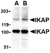
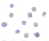
Left: Western blot analysis of IKAP in A-20 cell lysate with IKAP antibody at in (A) 0.5, and (B) 1 µg/ml.
Below: Immunocytochemistry of IKAP in A-20 cells with IKAP antibody at 1 µg/ml.
Other Product Images
 Source:IKAP antibody was raised against a 16 amino acid peptide from near the carboxy terminus of human IKAP.
Purification: Affinity chromatography purified via peptide column
Clonality and Clone: This is a polyclonal antibody.
Host: IKAP antibody was raised in rabbit.
Please use anti-rabbit secondary antibodies.
Application: IKAP antibody can be used for detection of IKAP by Western blot at 0.5 to 1 µg/ml.
Tested Application(s): E, WB, ICC
Buffer: Antibody is supplied in PBS containing 0.02% sodium azide.
Blocking Peptide:Cat.No. 2337P - IKAP Peptide
Long-Term Storage: IKAP antibody can be stored at 4ºC, stable for one year. As with all antibodies care should be taken to avoid repeated freeze thaw cycles. Antibodies should not be exposed to prolonged high temperatures.
Positive Control:
1. Cat. No. 1288 - A20 Cell Lysate
Species Reactivity: H, M
GI Number: 3757822
Accession Number: AAC64258
Short Description: part of the RNA polymerase II transcription elongation complex
References
1. Cohen L, Henzel WJ, and Baeuerle PA. IKAP is a scaffold protein of the IkB kinase complex. Nature 1998; 395:292-6.
2. Krappmann D, Hatada EN, Tegethoff S, et al. The I kappa B kinase (IKK) complex is tripartite and contains IKK gamma but not IKAP as a regular component. J. Biol. Chem. 2000; 275:29779-87.
3. Anderson SL, Coli R, Daly IW, et al. Familial dysautonomia is caused by mutations of the IKAP gene. Am. J. Hum. Genet. 2001; 68:753-8.
4. Hawkes NA, Otero G, Winkler GS, et al. Purification and characterization of the human elongator complex. J. Biol. Chem. 2002; 277:3047-52.

Description
Left: Western blot analysis of IKAP in A-20 cell lysate with IKAP antibody at in (A) 0.5, and (B) 1 µg/ml.
Below: Immunocytochemistry of IKAP in A-20 cells with IKAP antibody at 1 µg/ml.
Other Product Images
 Source:IKAP antibody was raised against a 16 amino acid peptide from near the carboxy terminus of human IKAP.
Purification: Affinity chromatography purified via peptide column
Clonality and Clone: This is a polyclonal antibody.
Host: IKAP antibody was raised in rabbit.
Please use anti-rabbit secondary antibodies.
Application: IKAP antibody can be used for detection of IKAP by Western blot at 0.5 to 1 µg/ml.
Tested Application(s): E, WB, ICC
Buffer: Antibody is supplied in PBS containing 0.02% sodium azide.
Blocking Peptide:Cat.No. 2337P - IKAP Peptide
Long-Term Storage: IKAP antibody can be stored at 4ºC, stable for one year. As with all antibodies care should be taken to avoid repeated freeze thaw cycles. Antibodies should not be exposed to prolonged high temperatures.
Positive Control:
1. Cat. No. 1288 - A20 Cell Lysate
Species Reactivity: H, M
GI Number: 3757822
Accession Number: AAC64258
Short Description: part of the RNA polymerase II transcription elongation complex
References
1. Cohen L, Henzel WJ, and Baeuerle PA. IKAP is a scaffold protein of the IkB kinase complex. Nature 1998; 395:292-6.
2. Krappmann D, Hatada EN, Tegethoff S, et al. The I kappa B kinase (IKK) complex is tripartite and contains IKK gamma but not IKAP as a regular component. J. Biol. Chem. 2000; 275:29779-87.
3. Anderson SL, Coli R, Daly IW, et al. Familial dysautonomia is caused by mutations of the IKAP gene. Am. J. Hum. Genet. 2001; 68:753-8.
4. Hawkes NA, Otero G, Winkler GS, et al. Purification and characterization of the human elongator complex. J. Biol. Chem. 2002; 277:3047-52.
Monday, June 20, 2011
Amino Acid Serum
Bio-Synthesis, with 20 years of experience in developing polyclonal antibodies, is a leading global provider of custom antibody production services. Our facility generates high-affinity custom antibodies with industry-leading titer guarantees and comprehensive antibody production packages that simplify the ordering process and present cost-effective methods to isolate epitope-specific antibodies. Visit www.biosyn.com and see why partner with Bio-Synthesis for your custom antibodies needs.
Amino Acid
In chemistry, an amino acid is a molecule containing both amine and carboxyl functional groups. In biochemistry, this term refers to alpha-amino acids with the general formula H2NCHRCOOH, where R is an organic subsistent. In the alpha amino acids, the amino and carboxylate groups are attached to the same carbon, which is called the α–carbon. The various alpha amino acids differ in which side chain (R group) is attached to their alpha carbon.
An Essential Amino Acid
An essential amino acid or indispensable amino acid is an amino acid that cannot be synthesized de novo by the organism and therefore must be supplied in the diet.Nine amino acids are generally regarded as essential for humans: phenylalanine, valine, threonine, tryptophan, isoleucine, methionine, histidine, leucine, and lysine. Arginine is required by infants and growing kids. They are called essential not because they are more important to life than the others, but because the body does not synthesize them, making it essential to include them in one's diet in order to obtain them.
Amino acids and DNA
DNA is a sequence of nucleotides. There are four nucleotides: adenine, thymine, cytosine, guanine. The exact sequence of these determines the code of each gene. When DNA is transcribed (the first step in expression of the gene), RNA is synthesized using this code. The RNA is a complementary copy of one strand of the DNA. The RNA leaves the nucleus and in the cytoplasm it is translated into a protein.
For more information visit: www.biosyn.com
Hrk Antibody
Catalog#:3771
Apoptosis plays a major role in normal organism development, tissue homeostasis, and removal of damaged cells. Hrk, a pro-apoptotic member of the Bcl-2 homology domain-3 (BH3)-only group of the Bcl-2 family of proteins, was also identified as novel protein induced during programmed neuronal death. It lacks significant homology to other Bcl-2 family members except for an 8-amino acid region that is similar to the BH3 motif of Bik. Hrk regulates apoptosis through interaction with the anti-apoptotic proteins Bcl-2 and Bcl-XL via this domain. It does not interact with the pro-apoptotic proteins Bax, Bak, or Bcl-XS. Hrk localizes to mitochondrial membranes in a pattern similar to that previously reported for Bcl-2 and Bcl-XL. Despite its predicted molecular weight, Hrk often migrates at 12-15 kDa.
Additional Names: Hrk, Harakiri, Neuronal death proteinDP
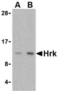
Description
Left: Western blot analysis of Hrk in mouse pancreas tissue lysate with Hrk antibody at (A) 2.5 and (B) 5 µg/ml.
Source:HRK antibody was raised against a 15 amino acid peptide from near the center of human Hrk.
Purification: Affinity chromatography purified via peptide column
Clonality and Clone: This is a polyclonal antibody.
Host: Hrk antibody was raised in rabbit.
Please use anti-rabbit secondary antibodies.
Application: Hrk antibody can be used for the detection of Hrk by Western blot at 2.5 - 5 µg/ml.
Tested Application(s): E, WB
Buffer: Antibody is supplied in PBS containing 0.02% sodium azide.
Blocking Peptide:Cat.No. 3771P - Hrk Peptide
Long-Term Storage: Hrk antibody can be stored at 4ºC, stable for one year. As with all antibodies care should be taken to avoid repeated freeze thaw cycles. Antibodies should not be exposed to prolonged high temperatures.
Positive Control:
1. Cat. No. 1414 - Mouse Pancreas Tissue Lysate
Species Reactivity: H, M
GI Number: 4504493
Accession Number: NP_003797
Short Description: a Bcl-2 family member involved durring programmed neuronal death
References
1. Lockshin RA, Osborne B, and Zakeri Z. Cell death in the third millennium. Cell Death Differ. 2000; 7:2-7.
2. Imaizumi K, Tsuda M, Imai Y, et al. Molecular cloning of a novel polypeptide, DP5, induced during programmed neuronal death. J. Biol. Chem. 1997; 272:18842-8.
3. Inohara N, Ding L, Chen S, et al. harakiri, a novel regulator of cell death, encodes a protein that activates apoptosis and interacts selectively with survival-promoting proteins Bcl-2 and Bcl-XL. EMBO J. 1997; 16:1686-94.
GDF6 Antibody
Catalog#:4691
Growth differentiation factors (GDFs) are members of the transforming growth factor (TGF) superfamily that is involved in embryonic development and adult tissue homeostasis. Both GDF6 and GDF7 are closely related to GDF5 which has been shown to induce activation of plasminogen activator, thereby inducing angiogenesis. It is predominantly expressed in long bones during fetal embryonic development and is involved in bone formation. In Xenopus, GDF6 is expressed at the edge of the neural plate and within the anterior neural plate including the eye fields. GDF6 is required for normal formation of some bones and joints in the limbs, skull, and axial skeleton. It may regulate patterning of the ectoderm by interacting with bone morphogenetic proteins (BMPs), and control eye development. Mutations in this gene result in colobomata, which are congenital abnormalities in ocular development, and in Klippel-Feil syndrome (KFS), a congenital disorder of spinal segmentation.
Additional Names: GDF6 (CT), Growth Differentiation Factor 6, GDF16, GDF-6, CDMP2
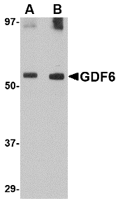
Description
Left: Western blot analysis of GDF6 in SK-N-SH lysate with GDF6 antibody at (A) 0.5 and (B) 1 µg/ml.
Source:GDF6 antibody was raised against a 17 amino acid peptide near the carboxy terminus of the human GDF6.
Purification: Affinity chromatography purified via peptide column
Clonality and Clone: This is a polyclonal antibody.
Host: GDF6 antibody was raised in rabbit.
Please use anti-rabbit secondary antibodies
Application: GDF6 antibody can be used for detection of GDF6 by Western blot at 1 µg/ml.
Tested Application(s): E, WB
Buffer: Antibody is supplied in PBS containing 0.02% sodium azide.
Blocking Peptide:Cat.No. 4691P - GDF6 Peptide
Long-Term Storage: GDF6 antibody can be stored at 4ºC, stable for one year. As with all antibodies care should be taken to avoid repeated freeze thaw cycles. Antibodies should not be exposed to prolonged high temperatures.
Positive Control:
1.Cat. No. 1220 - SK-N-SH Cell Lysate
Species Reactivity: H, M, R
GI Number: 48475062
Accession Number: NP_001001557
Short Description: (CT) Growth Differentiation Factor 6
References
1. Massague J. 1990. The transforming growth factor-beta family. Ann. Rev. Cell Biol. 6:597-641.
2. McPherron AC, Lawler AM, and Lee SJ. Regulation of skeletal muscle mass in mice by a new TGF-beta superfamily member. Nature 1997; 387:83-90.
3. Hanel ML and Hensey C. Eye and neural defects associated with loss of GDF6. BMC Dev. Biol. 2006; 6:43.
4. Settle SH Jr., Rountree RB, Sinha A, et al. Multiple joint and skeletal patterning defects caused by single and double mutations in the mouse Gdf6 and Gdf5 genes. Dev. Biol. 2003; 254:116-130.
Subscribe to:
Posts (Atom)
