Thursday, July 28, 2011
ACV and Related Compounds Products
Definition
Acyclovir (ACV) is the key intermediate in the biosynthesis of all penicillins and cephalosporins by eukaryotic and prokaryotic microorganisms.
Discovery
Gnann J W et al., in 1983 studied the mechanism of action, pharmacokinetics and clinical applications of ACV . Tadepalli S et al., in 1986 developed a simple and sensitive enzyme-linked immunosorbent assay for the detection and quantitation of ACV in human plasma and urine. ACV immobilized on a solid phase and free acyclovir in the sample solution were allowed to compete for a limited amount of anti-acyclovir monoclonal antibody. The specific antibody bound to the immobilized ACV was detected by the use of alkaline phosphatase conjugated anti-mouse imtnunoglobulin . Balzarini J et al., 2002 designed a number of acyclovir and ganciclovir derivatives that are endowed with intrinsic strong fluorescent characteristics by synthesizing tricyclic ACV and GCV derivatives containing an additional aromatic entity attached to the purine system .
Structural Characteristics
ACV also referred to as acycloguanosine, demonstrates strong and selective activity against herpes simplex and varicellazoster viruses. ACV structure is [9-(2-hydroxyethoxy)methylguanine]. Tadepalli S et al., in 1986 synthesized 1-O-octadecyl-sn-glycero-3-phospho-acyclovir (ODG-P-ACV), 1-O-hexadecylpropanediol-3-phospho-acyclovir (HDP-P-ACV), and 1-O-octadecyl-sn-glycero-3-phospho-azidothymidine (ODG-P-AZT), and evaluated their antiviral activity in human hepatoma cells that constitutively produce HBV. Results from specificity studies showed that there was a high degree of cross-reactivity with ACV and ACV succinate, minor cross-reactivity with the primnary metabolite of ACV (9-carboxymethoxymethylguanine [CMMG]), and significant cross-reactivity with 8-hydroxy-9- (2-hydroxyethoxy)methylguanine (8-OH-ACV), a minor metabolite. 9-(2-14ydroxy-1-hydroxymethylethoxy)methylguanine (BW B759U), structurally similar to ACV, exhibited low but measurable cross-reactivity.
Mode of Action
The mechanism of action of ACV and its bioavailability and pharmacokinetics in humans have been established for both intravenous and oral formulations of the drug. ACV triphosphate inhibit the DNA polymerase of human hepatitis B virus (HBV) by 50% at submicromolar concentrations, but no effects of ACV treatment have been noted on the clinical manifestations of hepatitis B. However, HDP-P-ACV and ODG-P-ACV inhibited viral replication by 50% at 0.5 and 6.8 μM, respectively. Based on the EC50, HDP-P-ACV, ODG-P-ACV, and ODG-P-AZT were > 200, > 14.7, and > 48 times more active than their free nucleosides in reducing HBV replication in 2.2.15 cells. To evaluate the biochemical basis for the increased antiviral activity, authors studied the uptake and metabolism of 1-O-octadecyl-sn-glycero-3-phospho-[3H]acyclovir (ODG-P-[3H]ACV) in HepG2 cells. Cellular uptake of ODG-P-[3H]ACV was found to be substantially greater than that of [3H]ACV, and cellular levels of ACV-mono-, -di-, and -triphosphate were much higher with ODG-P-ACV. ODG-P-[3H]ACV was well absorbed orally. Based on urinary recovery of tritium after oral or parenteral administration of the radio labeled compounds, oral absorption of ODG-P-ACV in mice was 100% versus 37% for ACV. ODG-P-ACV plasma area under the curve was more than 7-fold greater than that of ACV. Lipid prodrugs of this type is useful orally in treating viral diseases 4,5,6.
Functions
ACV is an antiviral drug, which selectively inhibits replication of members of the herpes group of DNA viruses with low cell toxicity. This drug is used clinically in treating a variety of herpesvirus infections.
Prodrug of ACV, Valaciclovir (VACV), a prodrug of ACV is usually preferred in the oral treatment of viral infections, mainly herpes simplex virus (HSV).
A number of tricyclic ACV and ganciclovir (GCV) derivatives substituted with bulky lipophilic groups have been identified as potent and highly selective cytostatic agents against herpes simplex virus type (HSV-1)-thymidine kinase (TK) gene-transduced human osteosarcoma and murine mammary carcinoma tumor cells.
Effect in cell culture, the tricyclic ACV derivatives were also endowed with a very pronounced bystander effect in cell culture, albeit at relatively high drug concentrations.
ACV acts against cytomegalovirus (CMV) in general and the latter against CMV retinitis mainly via phosphorylation and inhibition of the viral DNA polymerase .
For more information visit: www.biosyn.com
Wednesday, July 27, 2011
Introduction to Swine Flu
The origin. It is well known that most higher organisms carry the genomes of certain viruses that normally lie dormant in a quiescent state until they become activated by different factors and causing a number of problems. However in most cases, after infecting their hosts, viruses are cleared by an immunological response mounted by the host or eventually they kill their host this way putting an end to their replication cycle. Yet in either case, the newly produced virus can infect other hosts and start the reproductive cycle once more.
Swine Flu Transmission and Vaccine
Transmission. At this point, the infected human can get sick and transmit the virus via aerosolized micro drops of saliva produced during coughing, and he is also capable of passing the virus by direct contact with non-infected people. About vaccines against it. Currently there are not vaccines against the new swine flu virus strain and producing a vaccine can take 6-9 months. Vaccines against the influenza virus are strain specific, however because the virus has a high rate of mutations in those regions responsible for initiating the infection process there is a need to develop new vaccines every year.
Tamiflu
This drug manufactured by Roche appears to be effective against the swine flu; influenza virus initiates the infection process by latching to sialoglycoproteins present of the cell surface via their neuraminidase. Inhibition of the viral neuraminidase interferes with the infection process contributing to reduce symptoms and complications.
Influenza Peptide Virus
The influenza virus is an enveloped RNA virus and its genome is split between 7 to 8 RNA fragments, each encoding for one to two genes. This large number of RNA fragments allows their mixing or reassorment if more than one virus strain is infecting a cell, to yield new viral strains containing genetic information from different viral strains.
Immunity and Viral Peptide
Although the protective immunity stimulated by the vaccines is humoral and mediated by neutralizing antibodies there is significant body of evidence pointing to the crucial role of T cell immunity. In effect, the increased susceptibility of young children and older people correlates well with a low Th1 immunity.
For more information visit: www.biosyn.com
SIRNA Synthesis
Small interfering RNA (siRNA), sometimes known as short interfering RNA or silencing RNA, are a class of 20-25 nucleotide-long double-stranded RNA molecules that play a variety of roles in biology. Most notably, siRNA is involved in the RNA interference (RNAi) pathway where the siRNA interferes with the expression of a specific gene. In addition to their role in the RNAi pathway, siRNAs also act in RNAi-related pathways, e.g. as an antiviral mechanism or in shaping the chromatin structure of a genome; the complexity of these pathways is only now being elucidated.
siRNAs
siRNAs were first discovered by David Baulcombe's group in Norwich, England, as part of post-transcriptional gene silencing (PTGS) in plants, and published their findings in Science in a paper titled "A species of small antisense RNA in posttranscriptional gene silencing in plants". Shortly thereafter, in 2001, synthetic siRNAs were then shown to be able to induce RNAi in mammalian cells by Thomas Tuschl and colleagues in a paper published in Nature. This discovery led to a surge in interest in harnessing RNAi for biomedical research and drug development.
RNA Interference
RNA Interference Home has everything you need for your RNAi and siRNA research. You will find information here and links on RNAi protocols, an RNAi Forum, and RNAi bioinformatic software for design of RNAi and SiRNA inhibitory molecules.
For more information visit: www.biosyn.com
IRE1p Antibody
Catalog#:3655
Accumulation of malfolded proteins in the endoplasmic reticulum (ER) activates the unfolded protein response (UPR) and the upregulation of the ER molecular chaperones GRP78 and GRP 94 (1,2). These proteins are normally bound to ER transmembrane proteins such as IRE1p and ATF6 (3,4) but ER stress causes their dissociation. This allows IRE1p, a serine-threonine protein kinase to transduce the unfolded protein signal from the ER to the nucleus. IRE1p also has an endoribonuclease activity that is required to splice X-box binding protein (XBP1) mRNA converting it to a potent UPR transcriptional activation (5). Depletion of IRE1p through the expression of a dominant negative form of IRE1p has no effect on transfected cells, but cell death via apoptosis occurs under stress conditions that cause unfolded proteins to accumulate in the ER (6). Two alternatively spliced transcript variants encoding different isoforms have been found for this gene.
Additional Names: IRE1p (CT), Endoplasmic reticulum-to-nucleus signaling 1, ERN1
Description
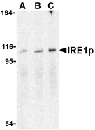
Left: Western blot analysis of IRE1p in A-20 cell lysate with IRE1p antibody at (A) 0.5, (B) 1 and (C) 2 µg/ml.
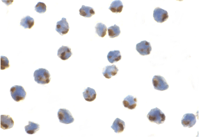
Below: Immunocytochemistry of IRE1p in A-20 cells with IRE1p antibody at 1 µg/ml.
Other Product Images
 Source:IRE1p antibody was raised against a 16 amino acid peptide from near the carboxy terminus of human IRE1p.
Purification: Affinity chromatography purified via peptide column
Clonality and Clone: This is a polyclonal antibody.
Host: IRE1p antibody was raised in rabbit.
Please use anti-rabbit secondary antibodies.
Immunogen: Human IRE1p (C-Terminus) Peptide (Cat. No. 3655P)
Application: IRE1p antibody can be used for the detection of IRE1p by Western blot at 1 – 2 µg/ml.
Tested Application(s): E, WB, ICC
Buffer: Antibody is supplied in PBS containing 0.02% sodium azide.
Blocking Peptide:Cat. No. 3655P - IRE1p Peptide
Long-Term Storage: IRE1p antibody can be stored at 4ºC, stable for one year. As with all antibodies care should be taken to avoid repeated freeze thaw cycles. Antibodies should not be exposed to prolonged high temperatures.
Positive Control:
1. Cat. No. 1288 - A-20 Cell Lysate
Species Reactivity: H, M, R
GI Number: 193806335
Accession Number: O75460
Short Description: (CT) Endoplasmic reticulum-to-nucleus signaling 1
References
1. Little E, Ramakrishnan M, Roy B, et al. The glucose-regulated proteins (GRP78 and GRP94): functions, gene regulation, and applications. Crit. Rev. Eukaryot. Gene Expr. 1994; 4:1-18.
2. Lee AS. The ER chaperone and signaling regulator GRP78/BiP as a monitor of endoplasmic reticulum stress. Methods 2005; 35:373-81.
3. Bertolotti A, Zhang Y, Hendershot LM, et al. Dynamic interaction of BiP and ER stress transducers in the unfolded-protein response. Nat. Cell Biol. 2000; 2:326-32.
4. Shen J, Chen X, Hendershot L, et al. ER stress regulation of ATF6 localization by dissociation of BiP/GRP78 binding and unmasking of Golgi localization signals. Dev. Cell 2002; 3:99-111.
Description
Left: Western blot analysis of IRE1p in A-20 cell lysate with IRE1p antibody at (A) 0.5, (B) 1 and (C) 2 µg/ml.
Below: Immunocytochemistry of IRE1p in A-20 cells with IRE1p antibody at 1 µg/ml.
Other Product Images
Source:IRE1p antibody was raised against a 16 amino acid peptide from near the carboxy terminus of human IRE1p.Purification: Affinity chromatography purified via peptide column
Clonality and Clone: This is a polyclonal antibody.
Host: IRE1p antibody was raised in rabbit.
Please use anti-rabbit secondary antibodies.
Immunogen: Human IRE1p (C-Terminus) Peptide (Cat. No. 3655P)
Application: IRE1p antibody can be used for the detection of IRE1p by Western blot at 1 – 2 µg/ml.
Tested Application(s): E, WB, ICC
Buffer: Antibody is supplied in PBS containing 0.02% sodium azide.
Blocking Peptide:Cat. No. 3655P - IRE1p Peptide
Long-Term Storage: IRE1p antibody can be stored at 4ºC, stable for one year. As with all antibodies care should be taken to avoid repeated freeze thaw cycles. Antibodies should not be exposed to prolonged high temperatures.
Positive Control:
1. Cat. No. 1288 - A-20 Cell Lysate
Species Reactivity: H, M, R
GI Number: 193806335
Accession Number: O75460
Short Description: (CT) Endoplasmic reticulum-to-nucleus signaling 1
References
1. Little E, Ramakrishnan M, Roy B, et al. The glucose-regulated proteins (GRP78 and GRP94): functions, gene regulation, and applications. Crit. Rev. Eukaryot. Gene Expr. 1994; 4:1-18.
2. Lee AS. The ER chaperone and signaling regulator GRP78/BiP as a monitor of endoplasmic reticulum stress. Methods 2005; 35:373-81.
3. Bertolotti A, Zhang Y, Hendershot LM, et al. Dynamic interaction of BiP and ER stress transducers in the unfolded-protein response. Nat. Cell Biol. 2000; 2:326-32.
4. Shen J, Chen X, Hendershot L, et al. ER stress regulation of ATF6 localization by dissociation of BiP/GRP78 binding and unmasking of Golgi localization signals. Dev. Cell 2002; 3:99-111.
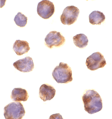
IRAK-M Antibody
Catalog#:2355
Interleukin-1 (IL-1) and lipopolysaccharide (LPS) induces cellular response through IL-1 receptor (IL-1R) and Toll like receptors (TLR). IL-1 receptor-associated kinase (IRAK and IRAK2) mediates the activation of NF-betaB by IL-1/Toll receptors (1,2), which is a pivotal transcription factor mediating inflammatory and immune response. A novel member in the IRAK/Pelle family was recently identified and designated IRAK-M (3). IRAKs associate with IL-1/Toll receptors after IL-1 or LPS stimulation and the dominant negative mutants of IRAKs inhibit IL-1 or LPS induced NF-betaB activation. Members in IRAK/Pelle family play a central role in IL-1R/TLR mediated inflammatory responses to cytokine IL-1 and LPS.
Additional Names: IRAK-M
Description
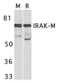
Left: Western blot analysis of IRAK-M in mouse spleen (M) and rat liver (R) tissue lysates with IRAK-M antibody at 1 µg/ml.
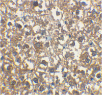
Below: Immunohistochemical staining of rat liver cells using IRAK-M antibody at 2 µg/ml.
Other Product Images
 Source:IRAK-M antibody was raised against a peptide corresponding to amino acids from the carboxy terminus of human IRAK-M .
Purification: Affinity chromatography purified via peptide column
Clonality and Clone: This is a polyclonal antibody.
Host: IRAK-M antibody was raised in rabbit.
Please use anti-rabbit secondary antibodies.
Immunogen: Human IRAK-M Peptide (Cat. No. 2355P)
Application: IRAK-M antibody can be used for detection of IRAK-M by Western blot at 0.5 to 1 µg/ml.A 68 kDa major band can be detected. IRAK-M antibody can also detect IRAK-M by immunohistochemistry at 2 µg/ml.IRAK-M antibody has no cross response to IRAK or IRAK2.
Tested Application(s): E, WB, IHC
Buffer: Antibody is supplied in PBS containing 0.02% sodium azide.
Blocking Peptide:Cat. No. 2355P - IRAK-M Peptide
Long-Term Storage: IRAK-M antibody can be stored at 4ºC, stable for one year. As with all antibodies care should be taken to avoid repeated freeze thaw cycles. Antibodies should not be exposed to prolonged high temperatures.
Positive Control:
1. Cat. No. 1406 - Mouse Spleen Tissue Lysate
2. Cat. No. 1465 - Rat Kidney Lysate
Species Reactivity: H, M, R
GI Number: 5225377
Accession Number: AF113136
Short Description: A novel IL-1R Associated Kinase
References
1. Cao Z; Henzel WJ; Gao X. IRAK: a kinase associated with the interleukin-1 receptor. Science 1996;271:1128-31.
2. Muzio M, Ni J, Feng P, Dixit VM. IRAK (Pelle) family member IRAK-2 and MyD88 as proximal mediators of IL-1 signaling. Science 1997;278:1612-5
3. Wesche H, Gao X, Li X, Kirschning CJ, Stark GR, Cao Z. IRAK-M is a novel member of the Pelle/interleukin-1 receptor-associated kinase (IRAK) family. J Biol Chem. 1999;274(27):19403-10.
Description
Left: Western blot analysis of IRAK-M in mouse spleen (M) and rat liver (R) tissue lysates with IRAK-M antibody at 1 µg/ml.
Below: Immunohistochemical staining of rat liver cells using IRAK-M antibody at 2 µg/ml.
Other Product Images
Source:IRAK-M antibody was raised against a peptide corresponding to amino acids from the carboxy terminus of human IRAK-M .Purification: Affinity chromatography purified via peptide column
Clonality and Clone: This is a polyclonal antibody.
Host: IRAK-M antibody was raised in rabbit.
Please use anti-rabbit secondary antibodies.
Immunogen: Human IRAK-M Peptide (Cat. No. 2355P)
Application: IRAK-M antibody can be used for detection of IRAK-M by Western blot at 0.5 to 1 µg/ml.A 68 kDa major band can be detected. IRAK-M antibody can also detect IRAK-M by immunohistochemistry at 2 µg/ml.IRAK-M antibody has no cross response to IRAK or IRAK2.
Tested Application(s): E, WB, IHC
Buffer: Antibody is supplied in PBS containing 0.02% sodium azide.
Blocking Peptide:Cat. No. 2355P - IRAK-M Peptide
Long-Term Storage: IRAK-M antibody can be stored at 4ºC, stable for one year. As with all antibodies care should be taken to avoid repeated freeze thaw cycles. Antibodies should not be exposed to prolonged high temperatures.
Positive Control:
1. Cat. No. 1406 - Mouse Spleen Tissue Lysate
2. Cat. No. 1465 - Rat Kidney Lysate
Species Reactivity: H, M, R
GI Number: 5225377
Accession Number: AF113136
Short Description: A novel IL-1R Associated Kinase
References
1. Cao Z; Henzel WJ; Gao X. IRAK: a kinase associated with the interleukin-1 receptor. Science 1996;271:1128-31.
2. Muzio M, Ni J, Feng P, Dixit VM. IRAK (Pelle) family member IRAK-2 and MyD88 as proximal mediators of IL-1 signaling. Science 1997;278:1612-5
3. Wesche H, Gao X, Li X, Kirschning CJ, Stark GR, Cao Z. IRAK-M is a novel member of the Pelle/interleukin-1 receptor-associated kinase (IRAK) family. J Biol Chem. 1999;274(27):19403-10.
IRAK-4 Antibody
Catalog#:3125
Interleukin-1 (IL-1) and lipopolysaccharide (LPS) induces cellular responses through IL-1 receptor (IL-1R) and Toll-like receptors (TLR). IL-1R-associated kinases (IRAK, IRAK2, and IRAK-M) regulate the activation of NF-kappaB and MAP kinase (MAPK) by IL-1R/TLR (1-3). A novel member in the IRAK/Pelle family was recently identified and designated IRAK-4 (4,5). Overexpression of IRAK-4 activates NF-kB and MAPK pathways. IRAK-4 interacts with and phosphorylates IRAK-1. IRAK-4-deficient animals are completely resistant to the challenge with LPS. Animals and humans lacking IRAK-4 are impaired in their responses to viral and bacterial challenges (5,6). Members in IRAK/Pelle family play a central role in IL-1R/TLR mediated inflammatory responses and in innate immunity.
Additional Names: IRAK-4
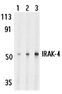
Description
Left: Western blot analysis of IRAK-4 in HeLa cell lysate with IRAK-4 antibody at 1 (lane 1), 2 (lane 2), and 4 (lane 3) µg/ml, respectively.
Below: Immunocytochemistry of IRAK-4 in K562 cells with IRAK-4 antibody at 10 µg/ml.
Other Product Images
 Source:
IRAK-4 antibody was raised against a synthetic peptide corresponding to amino acids at the carboxy terminus of human IRAK-4.
Purification: Affinity chromatography purified via peptide column
Clonality and Clone: This is a polyclonal antibody.
Host: IRAK-4 antibody was raised in rabbit.
Please use anti-rabbit secondary antibodies.
Immunogen: Human IRAK4 Peptide (Cat. No. 3125P)
Application: IRAK-4 antibody can be used for the detection of IRAK-4 by Western blot at 1 to 2 µg/ml.A 50 kDa band can be detected. Anti-IRAK-4 has no cross response to other IRAKs.
Tested Application(s): E, WB, ICC
Buffer: Antibody is supplied in PBS containing 0.02% sodium azide.
Blocking Peptide:Cat. No. 3125P - IRAK-4 Peptide
Long-Term Storage: IRAK-4 antibody can be stored at 4ºC, stable for one year. As with all antibodies care should be taken to avoid repeated freeze thaw cycles. Antibodies should not be exposed to prolonged high temperatures.
Positive Control:
1. Cat. No. 1204 - K562 Cell Lysate
Species Reactivity: H
GI Number: 20219010
Accession Number: AAM15772
Short Description: A member in IRAK/Pelle family
References
1. Cao Z; Henzel WJ; Gao X. IRAK: a kinase associated with the interleukin-1 receptor. Science 1996;271:1128-31.
2. Muzio M, Ni J, Feng P, Dixit VM. IRAK (Pelle) family member IRAK-2 and MyD88 as proximal mediators of IL-1 signaling. Science 1997;278:1612-5
3. Wesche H, Gao X, Li X, Kirschning CJ, Stark GR, Cao Z. IRAK-M is a novel member of the Pelle/interleukin-1 receptor-associated kinase (IRAK) family. J Biol Chem. 1999;274(27):19403-10.
4. Li S, Strelow A, Fontana EJ, Wesche H. IRAK-4: a novel member of the IRAK family with the properties of an IRAK-kinase. Proc Natl Acad Sci USA . 2002;99(8):5567-72.
Tuesday, July 19, 2011
DNA Structure Animation
For over 20 years, BioSynthesis has provided custom oligonucleotide production services worldwide, including researcher at university, biotechnology and pharmaceutical institutions. We offer wide variety of oligo modifications and labelings from small discovery research scale to multi-gram ASR/cGMP for clinical diagnostic production. As always, quality is guaranteed!
DNA Structure
DNA has a compact, highly organized structure that is reminiscent of a spiral staircase. There are several structural characteristics that are readily identifiable in DNA.
DNA Synthesis
At bio-Synthesis any synthesis of DNA that happens outside the s phase of the cell cycle. The term is to describe the replication of dna during the nucleotide excision repair of dna damage and thus distinct from the semi-conservative replication of dna which is confined to the s-phase of the eukaryotic cell cycle.
DNA Replication
DNA replication is the process of copying a double-stranded DNA molecule to form two double-stranded molecules. The process of DNA replication is a fundamental process used by all living organisms as it is the basis for biological inheritance.
For more Information visit: www.biosyn.com
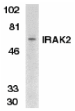
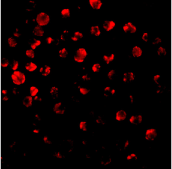
IRAK-2 Antibody
Catalog#:2213
The pro-inflammatory cytokine IL-1 induces cellular response through two subunits of its receptor, IL-1 receptor I (IL-1RI) and IL-1 receptor accessory protein (IL-1RAcP). IL-1 receptor-associated kinase (IRAK) mediates activation of NF-kappaB , which is a pivotal transcription factor mediating inflammatory and immune response. A novel member in the IRAK/Pelle family was recently identified and designated IRAK2 (2). Both IRAK and IRAK2 recruit to the subunits of the IL-1R complex after IL-1 binding and lead to NF-kappaB activation. IRAKs also associate with Toll-like receptor (TLR) and the dominant negative mutants of IRAKs inhibit LPS-induced NF-kappaB activation (3,4). Members in IRAK/Pelle family play a central role in IL-1R and TLR mediated inflammatory responses to cytokine IL-1 and LPS. IRAK2 is expressed in a variety of human tissues.
Additional Names: IRAK-2 (CT), IRAK-2
Description
Left: Western blot analysis of IRAK2 in K562 whole cell lysate with IRAK2 antibody at 1:500 dilution.
Below: Immunofluorescence of IRAK2 in HeLa cells with IRAK antibody at 10 µg/ml.
Other Product Images
 Source:IRAK2 antibody was raised against a peptide corresponding to amino acids near the carboxy terminus of human IRAK2.
Purification: Affinity chromatography purified via peptide column
Clonality and Clone: This is a polyclonal antibody.
Host: IRAK-2 antibody was raised in rabbit.
Please use anti-rabbit secondary antibodies.
Immunogen: Human IRAK2 (C-Terminus) Peptide (Cat. No. 2213P)
Application: IRAK-2 antibody can be used for detection of IRAK2 by Western blot at 1:500 to 1:1000 dilution. A 65 kDa band can be detected. Anti-IRAK2 has no cross response to IRAK.
Tested Application(s): E, WB, IF
Buffer: Antibody is supplied in PBS containing 0.02% sodium azide.
Blocking Peptide:Cat. No. 2213P - IRAK-2 Peptide
Long-Term Storage: IRAK-2 antibody can be stored at 4ºC, stable for one year. As with all antibodies care should be taken to avoid repeated freeze thaw cycles. Antibodies should not be exposed to prolonged high temperatures.
Positive Control:
1.Cat. No. 1204 - K562 Cell Lysate
2. Cat. No. 1201 - HeLa Cell Lysate
Species Reactivity: H
GI Number: 37725373
Accession Number: AAO24761
Short Description: (CT) IL-1R Associated Kinase
References
1. Cao Z; Henzel WJ; Gao X. IRAK: a kinase associated with the interleukin-1 receptor. Science 1996;271:1128-31.
2. Muzio M, Ni J, Feng P, Dixit VM. IRAK (Pelle) family member IRAK-2 and MyD88 as proximal mediators of IL-1 signaling. Science 1997;278:1612-5
3. Zhang FX, Kirschning CJ, Mancinelli R, Xu XP, Jin Y, Faure E, Mantovani A, Rothe M, Muzio M, Arditi M. Bacterial lipopolysaccharide activates nuclear factor-kappaB through interleukin-1 signaling mediators in cultured human dermal endothelial cells and mononuclear phagocytes. J Biol Chem 1999;274:7611-4
4. Yang RB, Mark MR, Gurney AL , Godowski PJ. Signaling events induced by lipopolysaccharide-activated toll-like receptor 2. J Immunol 1999;163:639-43 (RD0300)
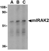
IRAK2 Antibody
Catalog#:3597
The pro-inflammatory cytokine IL-1 induces cellular response through two subunits of its receptor, IL-1 receptor I (IL-1RI) and IL-1 receptor accessory protein (IL-1RAcP). IL-1 receptor-associated kinase (IRAK) mediates activation of NF-kappaB, which is a pivotal transcription factor mediating inflammatory and immune response. A novel member in the IRAK/Pelle family has been identified and designated IRAK2 (1). Both IRAK and IRAK2 recruit to the subunits of the IL-1R complex after IL-1 binding and lead to NF-kappaB activation. IRAKs also associate with Toll like receptor (TLR) and the dominant negative mutants of IRAKs inhibit LPS-induced NF-?B activation (2,3). Members in IRAK/Pelle family play a central role in IL-1R and TLR mediated inflammatory response. Unlike human IRAK2, murine IRAK2 exists as four alternately spliced isoforms (IRAK2a-d), with two isoforms (IRAK2c and d) acting in an inhibitory fashion (4). IRAK2 is expressed in a variety of tissues.
Additional Names: IRAK2 (C2), mIRAK2
Description
Left: Western blot analysis of IRAK2 in A-20 whole cell lysate with IRAK2 antibody (C2) at (A) 0.5, (B) 1, and (C) 2 µg/ml.
Source:RAK2 antibody was raised against a peptide corresponding to 16 amino acids near the carboxy-terminus of mouse IRAK2a which is common to all four isoforms. Anti-IRAK2 is mouse specific.
Purification: Affinity chromatography purified via peptide column
Clonality and Clone: This is a polyclonal antibody.
Host: IRAK2 antibody was raised in rabbit.
Please use anti-rabbit secondary antibodies.
Immunogen: Human IRAK2 (C-Terminus 2) Peptide (Cat. No. 3597P)
Application: IRAK2 antibody can be used for detection of mouse IRAK2 by Western blot at 0.5 - 1 µg/ml.Anti-IRAK2 has no cross response to IRAK.
Tested Application(s): E, WB
Buffer: Antibody is supplied in PBS containing 0.02% sodium azide.
Blocking Peptide:Cat. No. 3597P - IRAK2 Peptide
Long-Term Storage: IRAK2 antibody can be stored at 4ºC, stable for one year. As with all antibodies care should be taken to avoid repeated freeze thaw cycles. Antibodies should not be exposed to prolonged high temperatures.
Positive Control:
1. Cat. No. 1288 - A-20 Cell Lysate
Species Reactivity: M
GI Number: 37725373
Accession Number: AAO24761
Short Description: (C2) mouse IL-1R associated kinase
References
1. Muzio M, Ni J, Feng P, et al. IRAK (Pelle) family member IRAK-2 and MyD88 as proximal mediators of IL-1 signaling. Science 1997; 278:1612-5.
2. Zhang FX, Kirschning CJ, Mancinelli R, et al. Bacterial lipopolysaccharide activates nuclear factor-kappaB through interleukin-1 signaling mediators in cultured human dermal endothelial cells and mononuclear phagocytes. J. Biol. Chem. 1999; 274:7611-4.
3. Yang RB, Mark MR, Gurney AL, et al. Signaling events induced by lipopolysaccharide-activated toll-like receptor 2. J. Immunol. 1999; 163:639-43.
4. Hardy MP and O’Neill LAJ. The murine IRAK2 gene encodes four alternately spliced isoforms, two of which are inhibitory. J. Biol. Chem. 2004; 279:27699-708.
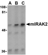
IRAK2 Antibody
Catalog#:3595
The pro-inflammatory cytokine IL-1 induces cellular response through two subunits of its receptor, IL-1 receptor I (IL-1RI) and IL-1 receptor accessory protein (IL-1RAcP). IL-1 receptor-associated kinase (IRAK) mediates activation of NF-kappaB, which is a pivotal transcription factor mediating inflammatory and immune response. A novel member in the IRAK/Pelle family has been identified and designated IRAK2 (1). Both IRAK and IRAK2 recruit to the subunits of the IL-1R complex after IL-1 binding and lead to NF-kappaB activation. IRAKs also associate with Toll like receptor (TLR) and the dominant negative mutants of IRAKs inhibit LPS-induced NF-?B activation (2,3). Members in IRAK/Pelle family play a central role in IL-1R and TLR mediated inflammatory response. Unlike human IRAK2, murine IRAK2 exists as four alternately spliced isoforms (IRAK2a-d), with two isoforms (IRAK2c and d) acting in an inhibitory fashion (4). IRAK2 is expressed in a variety of tissues.
Additional Names: IRAK2 (CT), mIRAK2
 Description
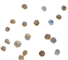
Left: Western blot analysis of IRAK2 in RAW264.7 whole cell lysate with mIRAK2 antibody at (A) 0.5, (B) 1, and (C) 2 µg/ml.
Below: Immunocytochemistry of IRAK2 in A-20 cells with IRAK2 antibody at 1 µg/ml.
Other Product Images
 Source:IRAK2 antibody was raised against a peptide corresponding to 17 amino acids near the carboxy-terminus of mouse IRAK2a which is common to all four isoforms. mIRAK2 antibody will also recognize human IRAK2.
Purification: Affinity chromatography purified via peptide column
Clonality and Clone: This is a polyclonal antibody.
Host: IRAK2 antibody was raised in rabbit.
Please use anti-mouse secondary antibodies.
Immunogen: Human IRAK2 (C-Terminus) Peptide (Cat. No. 3595P)
Application: IRAK2 antibody can be used for detection of mouse IRAK2 by Western blot at 0.5 - 1 µg/ml.A 65 kDa band should be detected. Anti-IRAK2 has no cross response to IRAK. Anti-IRAK2 will also recognize human IRAK2.
Tested Application(s): E, WB, ICC
Buffer: Antibody is supplied in PBS containing 0.02% sodium azide.
Blocking Peptide:Cat. No. 3595P - IRAK2 Peptide
Long-Term Storage: IRAK2 antibody can be stored at 4ºC, stable for one year. As with all antibodies care should be taken to avoid repeated freeze thaw cycles. Antibodies should not be exposed to prolonged high temperatures.
Positive Control:
1. Cat. No. 1283 - RAW264.7 Cell Lysate
2. Cat. No. 1288 - A-20 Cell Lysate
Species Reactivity: H, M
GI Number: 37725373
Accession Number: AAO24761
Short Description: (CT) mouse IL-1R associated kinase
References
1. Muzio M, Ni J, Feng P, et al. IRAK (Pelle) family member IRAK-2 and MyD88 as proximal mediators of IL-1 signaling. Science 1997; 278:1612-5.
2. Zhang FX, Kirschning CJ, Mancinelli R, et al. Bacterial lipopolysaccharide activates nuclear factor-kappaB through interleukin-1 signaling mediators in cultured human dermal endothelial cells and mononuclear phagocytes. J. Biol. Chem. 1999; 274:7611-4.
3. Yang RB, Mark MR, Gurney AL, et al. Signaling events induced by lipopolysaccharide-activated toll-like receptor 2. J. Immunol. 1999; 163:639-43.
4. Hardy MP and O’Neill LAJ. The murine IRAK2 gene encodes four alternately spliced isoforms, two of which are inhibitory. J. Biol. Chem. 2004; 279:27699-708.
Wednesday, July 13, 2011
Extinct Biological Hazard Organisms
Biosynthesis DNA Testing (Ancestry/DNA Identity Profile) provides you with a simple and objective description of your deep ancestral origins as well as a certificate of your unique genetic fingerprint. The test gives you an estimated percentage of ancestry from the four major historical population groups.
Biological Hazard
A biological hazard or biohazard is an organism, or substance derived from an organism, that poses a threat to (primarily) human health. This can include medical waste or samples of a microorganism, virus or toxin (from a biological source) that can impact human health. It can also include substances harmful to animals. The term and its associated symbol is generally used as a warning, so that those potentially exposed to the substances will know to take precautions. There is also a biohazard HCS/WHMIS logo which utilizes the same symbol.
Biological Agents
Biological agents include prions, viruses, microorganisms (bacteria and fungi) and some unicellular and multicellular eukaryotes (for example parasites) and their associated toxins. They have the ability to adversely affect human health in a variety of ways, ranging from allergic reactions that are usually relatively mild, to serious medical conditions, even death. These organisms are ubiquitous in the natural environment; they are found in water, soil, plants, and animals.
For more information visit: www.biosyn.com
IRAK Monoclonal Antibody
Catalog#:PM-5317
Nuclear factor kappa B (NF-kappaB) is a ubiquitous transcription factor and an essential mediator of gene expression during activation of immune and inflammatory responses. NF-kappaB mediates the expression of a great variety of genes in response to extracellular stimuli including IL-1, TNFalpha and LPS. A serine/threonine protein kinase associated with IL-1 receptor (IRAK) and its homologue mouse pelle-like protein kinase (mPLK) were identified recently. IRAK is associated with the IL-1 receptor subunits IL-1RI and IL-1RAcP after IL-1 binding and serves as a signaling molecule to mediate IL-1 response. IRAK mediates a signaling cascade leading to NF-kB activation by members in IL-1 family including IL-1 and a novel cytokine IL-18 (also termed IGIF).
Additional Names: IRAK (8F1A7),IL-1-Receptor Associated Kinase
Description
Left: Western blot analysis of IRAK in mouse liver lysate with IRAK antibody at (A) 1 and (B) 2 µg/ml.
Source:Mouse monoclonal IRAK antibody was raised against a recombinant protein corresponding to amino acids 618 - 712 of human IRAK.
Purification: Immunoaffinity chromotography purified IgG
Clonality and Clone: This is a monoclonal antibody. (Clone 8F1A7)
Host: IRAK monoclonal antibody was raised in mouse.
Please use anti-mouse secondary antibodies.
Immunogen: Recombinant protein corresponding to amino acids 618 - 712 of human IRAK.
Application: IRAK antibody can be used for Western blot at 1 - 2 µg/ml.
Tested Application(s): E, WB
Buffer: Antibody is supplied in PBS containing 0.02% sodium azide.
Blocking Peptide:
Long-Term Storage: IRAK monoclonal antibody can be stored at 4ºC, stable for one year.
Positive Control:
1. Cat. No. 95-112 - IRAK Recombinant Protein
Species Reactivity: H, M
Short Description: IL-1-Receptor Associated Kinase
References
1. Cao Z,; Henzel WJ, and Gao X. IRAK: a kinase associated with the interleukin-1 receptor. Science 1996; 271:1128-31.
2. Trofimova M, Sprenkle AB, Green M; et al. Developmental and tissue-specific expression of mouse pelle-like protein kinase. J. Bio. Chem. 1996; 271: 17609-12.
3. Jianing Huang, Xiong Gao, Shyun Li, et al. Recruitment of IRAK to the interleukin 1 receptor complex requires interleukin-1 receptor accessory protein. Proc. Natl. Acad. Sci. USA 1997; 94:12829-32.
4. Robinson D, Shibuya K, Mui A, et al. IGIF does not drive Th1 development but synergizes with IL-12 for interferon-gamma production and activates IRAK and NF-kB. Immunity 1997; 7:571-81.
IRAK Monoclonal Antibody
Catalog#:PM-5315
Nuclear factor kappa B (NF-kappaB) is a ubiquitous transcription factor and an essential mediator of gene expression during activation of immune and inflammatory responses. NF-kappaB mediates the expression of a great variety of genes in response to extracellular stimuli including IL-1, TNFalpha and LPS. A serine/threonine protein kinase associated with IL-1 receptor (IRAK) and its homologue mouse pelle-like protein kinase (mPLK) were identified recently. IRAK is associated with the IL-1 receptor subunits IL-1RI and IL-1RAcP after IL-1 binding and serves as a signaling molecule to mediate IL-1 response. IRAK mediates a signaling cascade leading to NF-kB activation by members in IL-1 family including IL-1 and a novel cytokine IL-18 (also termed IGIF).
Additional Names: IRAK (1F12C1),IL-1-Receptor Associated Kinase
Description
Left: Western blot analysis of IRAK in HeLa lysates with IRAK antibody at (A) 1 and (B) 2 µg/ml.
Source:Mouse monoclonal IRAK antibody was raised against a recombinant protein corresponding to amino acids 618 - 712 of human IRAK.
Purification: Immunoaffinity chromotography purified IgG
Clonality and Clone: This is a monoclonal antibody. (Clone 1F12C1)
Host: IRAK monoclonal antibody was raised in mouse.
Please use anti-mouse secondary antibodies.
Immunogen: Recombinant protein corresponding to amino acids 618 - 712 of human IRAK.
Application: RAK antibody can be used for Western blot at 1 µg/ml.
Tested Application(s): E, WB
Buffer: Antibody is supplied in PBS containing 0.02% sodium azide.
Blocking Peptide:
Long-Term Storage: IRAK monoclonal antibody can be stored at 4ºC, stable for one year.
Positive Control:
1. Cat. No. 95-112 - IRAK Recombinant Protein
Species Reactivity: H, M
Short Description: IL-1-Receptor Associated Kinase
References
1. Cao Z,; Henzel WJ, and Gao X. IRAK: a kinase associated with the interleukin-1 receptor. Science 1996; 271:1128-31.
2. Trofimova M, Sprenkle AB, Green M; et al. Developmental and tissue-specific expression of mouse pelle-like protein kinase. J. Bio. Chem. 1996; 271: 17609-12.
3. Jianing Huang, Xiong Gao, Shyun Li, et al. Recruitment of IRAK to the interleukin 1 receptor complex requires interleukin-1 receptor accessory protein. Proc. Natl. Acad. Sci. USA 1997; 94:12829-32.
4. Robinson D, Shibuya K, Mui A, et al. IGIF does not drive Th1 development but synergizes with IL-12 for interferon-gamma production and activates IRAK and NF-kB. Immunity 1997; 7:571-81.
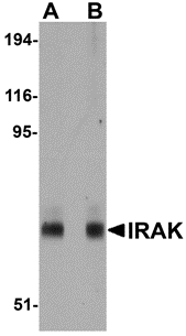
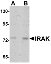
IRAK Antibody
Catalog#:1007
Nuclear factor kappa B (NF-kappaB) is a ubiquitous transcription factor and an essential mediator of gene expression during activation of immune and inflammatory responses. NF-kappaB mediates the expression of a great variety of genes in response to extracellular stimuli including IL-1, TNFalpha and LPS. A serine/threonine protein kinase associated with IL-1 receptor (IRAK) and its homologue mouse pelle-like protein kinase (mPLK) were identified recently. IRAK is associated with the IL-1 receptor subunits IL-1RI and IL-1RAcP after IL-1 binding and serves as a signaling molecule to mediate IL-1 response. IRAK mediates a signaling cascade leading to NF-kappaB activation by members in IL-1 family including IL-1 and a novel cytokine IL-18 (also termed IGIF).
Additional Names: IRAK (CT), IL-1-Receptor Associated Kinase
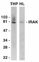
Description
Left: Western blot analysis of IRAK in THP-1 (THP) and HeLa (HL) whole cell lysates with IRAK antibody at 1:2000 dilution.
Below: Immunocytochemistry of IRAK in HeLa cells with IRAK antibody at 10 µg/ml.
Other Product Images
 Source:IRAK antibody was raised against a peptide corresponding to amino acids near the carboxy terminus of human IRAK.
Purification: Antibody is DEAE purified
Clonality and Clone: This is a polyclonal antibody.
Host: IRAK antibody was raised in rabbit.
Please use anti-rabbit secondary antibodies.
Immunogen: Human IRAK (C-Terminus) Peptide (Cat. No. 1007P)
Application: IRAK antibody can be used for Western blot at 1:1000 dilution and for immunoprecipitation with 2 to 4 ug per sample. An 80 kDa band should be detected in the lysate from non-activated cells. It has no cross response to IRAK2.
Tested Application(s): E, WB, ICC, IP
Buffer: Antibody is supplied in PBS containing 0.02% sodium azide.
Blocking Peptide:Cat. No. 1007P - IRAK Peptide
Long-Term Storage: IRAK antibody can be stored at 4ºC, stable for one year. As with all antibodies care should be taken to avoid repeated freeze thaw cycles. Antibodies should not be exposed to prolonged high temperatures.
Positive Control:
1. Cat. No. 1201 - HeLa Cell Lysate
2. Cat. No. 1208 - THP-1 Whole Cell Lysate
Species Reactivity: H, M, R
GI Number: 8928535
Accession Number: P51617
Short Description: (CT) IL-1R associated kinase
References
1. Cao Z; Henzel WJ; Gao X. IRAK: a kinase associated with the interleukin-1 receptor. Science 1996;271:1128-31.
2. Trofimova M; Sprenkle AB; Green M; Sturgill TW; Goebl MG; Harrington MA. Developmental and tissue-specific expression of mouse pelle-like protein kinase. J Bio Chem 1996; 271: 17609-1
3. Jianing Huang, Xiong Gao, Shyun Li, and Zhaodan Cao. Recruitment of IRAK to the interleukin 1 receptor complex requires interleukin-1 receptor accessory protein. Proc Natl Acad Sci USA 1997;94:12829-12832
4. Robinson D, Shibuya K, Mui A, Zonin F, Murphy E, Sana T, Hartley SB, Menon S, Kastelein R, Bazan F, O'Garra A. IGIF does not drive Th1 development but synergizes with IL-12 for interferon-gamma production and activates IRAK and NF-kappaB. Immunity 1997;7:571-581
Wednesday, July 6, 2011
DNA Sequence
BIO-SYNTHESIS is second to none in its ability to meet our customer's needs, often providing modified DNA with wide variety of selections. BSI offer BNA/DNA chimera synthesis at competitive prices and quick delivery. Bio-Synthesis continued to provide quality DNA products and services for the research community, but it has also become a world leader in providing custom peptide products and services.
DNA Sequence
A DNA sequence or genetic sequence is a succession of letters representing the primary structure of a real or hypothetical DNA molecule or strand, with the capacity to carry information. The possible letters are A, C, G, and T, representing the four nucleotide subunits of a DNA strand - adenine, cytosine, guanine, thymine bases covalently linked to phospho-backbone. In the typical case, the sequences are printed abutting one another without gaps, as in the sequence AAAGTCTGAC, going from 5' to 3' from left to right. A succession of any number of nucleotides greater than four is liable to be called a sequence.
DNA
The concept was to mimic an oligonucleotide binding to double stranded DNA via Hoogsteen base pairing in the major groove. Thus the nucleobases of DNA were retained, but the deoxyribose phosphodiester backbone of DNA was replaced by a pseudo-peptide backbone that according to computer model building was homomorphous with the DNA backbone.
Deoxyribonucleic Acid
Deoxyribonucleic acid (DNA) is a nucleic acid that contains the genetic instructions used in the development and functioning of all known living organisms and some viruses. The main role of DNA molecules is the long-term storage of information.
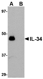
Catalog#:4781
Interleukin-34 (IL-34) was identified through a functional screening of a library of secreted proteins after transfection of an embryonic kidney cell line with recombinant cDNAs. Flow cytometry and monocyte viability assays demonstrated increased survival and/or proliferation of monocytes induced by IL-34 and specific binding of IL-34 to CD14+ monocytes. IL-34 is a second ligand to the colony stimulating factor (CSF)-1 receptor in addition to CSF1. Like CSF1, IL-34 stimulates phosphorylation of ERK1 and ERK2 in human monocytes and induces differentiation of bone marrow cells into macrophages. Despite its predicted molecular weight, IL-34 will often run at higher molecular weight in SDS-PAGE.
Additional Names: IL-34 (IN), Interleukin-34
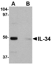
Description
Left: Western blot analysis of IL-34 in human brain tissue lysate with IL-34 antibody at 0.25 µg/ml in (A) the absence and (B) the presence of blocking peptide.
Source:IL-34 antibody was raised against a 13 amino acid peptide from near the center of human IL-34.
Purification: Affinity chromatography purified via peptide column
Clonality and Clone: This is a polyclonal antibody.
Host: IL-34 antibody was raised in rabbit.
Please use anti-rabbit secondary antibodies.
Application: IL-34 antibody can be used for the detection of IL-34 by Western blot at 0.25 – 0.5 µg/ml.
Tested Application(s): E, WB
Buffer: Antibody is supplied in PBS containing 0.02% sodium azide.
Blocking Peptide:Cat.No. 4781P - IL-34 Peptide
Long-Term Storage: IL-34 antibody can be stored at 4ºC, stable for one year. As with all antibodies care should be taken to avoid repeated freeze thaw cycles. Antibodies should not be exposed to prolonged high temperatures.
Positive Control:
1.Cat. No. 1303 - Human Brain Tissue Lysate
Species Reactivity: H, M, R
GI Number: 187610438
Accession Number: ACD13474
Short Description: (IN) Interleukin 34
References
1. Lin H, Lee E, Hestir K, et al. Discovery of a cytokine and its receptor by functional screening of the extracellular proteome. Science 2008; 320:807-11.
Catalog#:4779
Interleukin-34 (IL-34) was identified through a functional screening of a library of secreted proteins after transfection of an embryonic kidney cell line with recombinant cDNAs. Flow cytometry and monocyte viability assays demonstrated increased survival and/or proliferation of monocytes induced by IL-34 and specific binding of IL-34 to CD14+ monocytes. IL-34 is a second ligand to the colony stimulating factor (CSF)-1 receptor in addition to CSF1. Like CSF1, IL-34 stimulates phosphorylation of ERK1 and ERK2 in human monocytes and induces differentiation of bone marrow cells into macrophages. Despite its predicted molecular weight, IL-34 will often run at higher molecular weight in SDS-PAGE.
Additional Names: IL-34 (NT), Interleukin-34
 Description
Left: Western blot analysis of IL-34 in human brain tissue lysate with IL-34 antibody at 0.25 µg/ml in (A) the absence and (B) the presence of blocking peptide.
Source:IL-34 antibody was raised against a 16 amino acid peptide from near the amino terminus of human IL-34.
Purification: Affinity chromatography purified via peptide column
Clonality and Clone: This is a polyclonal antibody.
Host: IL-34 antibody was raised in rabbit.
Please use anti-rabbit secondary antibodies.
Application: IL-34 antibody can be used for the detection of IL-34 by Western blot at 0.25 – 0.5 µg/ml.
Tested Application(s): E, WB
Buffer: Antibody is supplied in PBS containing 0.02% sodium azide.
Blocking Peptide:Cat.No. 4779P - IL-34 Peptide
Long-Term Storage: IL-34 antibody can be stored at 4ºC, stable for one year. As with all antibodies care should be taken to avoid repeated freeze thaw cycles. Antibodies should not be exposed to prolonged high temperatures.
Positive Control:
1. Cat. No. 1303 - Human Brain Tissue Lysate
Species Reactivity: H, M, R
GI Number: 187610438
Accession Number: ACD13474
Short Description: (NT) Interleukin 34
References
1. Lin H, Lee E, Hestir K, et al. Discovery of a cytokine and its receptor by functional screening of the extracellular proteome. Science 2008; 320:807-11.
DescriptionLeft: Western blot analysis of IL-34 in human brain tissue lysate with IL-34 antibody at 0.25 µg/ml in (A) the absence and (B) the presence of blocking peptide.
Source:IL-34 antibody was raised against a 16 amino acid peptide from near the amino terminus of human IL-34.
Purification: Affinity chromatography purified via peptide column
Clonality and Clone: This is a polyclonal antibody.
Host: IL-34 antibody was raised in rabbit.
Please use anti-rabbit secondary antibodies.
Application: IL-34 antibody can be used for the detection of IL-34 by Western blot at 0.25 – 0.5 µg/ml.
Tested Application(s): E, WB
Buffer: Antibody is supplied in PBS containing 0.02% sodium azide.
Blocking Peptide:Cat.No. 4779P - IL-34 Peptide
Long-Term Storage: IL-34 antibody can be stored at 4ºC, stable for one year. As with all antibodies care should be taken to avoid repeated freeze thaw cycles. Antibodies should not be exposed to prolonged high temperatures.
Positive Control:
1. Cat. No. 1303 - Human Brain Tissue Lysate
Species Reactivity: H, M, R
GI Number: 187610438
Accession Number: ACD13474
Short Description: (NT) Interleukin 34
References
1. Lin H, Lee E, Hestir K, et al. Discovery of a cytokine and its receptor by functional screening of the extracellular proteome. Science 2008; 320:807-11.
Catalog#:3751
Interleukin-32 (IL-32) was initially identified as a transcript (NK4) that is selectively expressed in lymphocytes and NK cells and whose expression is increased following activation by IL-2 . It was later re-isolated from an IL-18-treated lung carcinoma cell line and re-named IL-32 (2). IL-32 is unusual in that it does not share sequence homology with known cytokine families and is highly expressed in immune tissues, existing in at least four differentially spliced isoforms. Because treatment of human monocytic and mouse macrophage cells with IL-32 induces several proinflammatory cytokines such as TNF-alpha, IL-8 and MIP-2, and because it is also induced in human peripheral lymphocyte cells after mitogen stimulation and in epithelial cells by IFNgamma, it has been suggested that IL-32 may play a role in autoimmune and inflammatory diseases such as rheumatoid arthritis (3).
Additional Names: IL-32 (IN), Interleukin 32
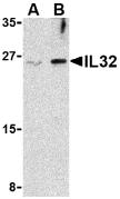
Description
Left: Western blot analysis of IL-32 in Jurkat cell lysate with IL-32 antibody at (A) 2.5 and (B) 5 µg/ml.
Source:IL-32 antibody was raised against a 16 amino acid peptide from near the center of human IL-32.
Purification: Affinity chromatography purified via peptide column
Clonality and Clone: This is a polyclonal antibody.
Host: IL-32 antibody was raised in rabbit.
Please use anti-rabbit secondary antibodies.
Immunogen: Human IL-32 (Intermediate Domain) Peptide (Cat. No. 3751P)
Application: IL-32 antibody can be used for the detection of IL-32 by Western blot at 2.5 – 5 µg/ml.This antibody detects the largest isoform of IL-32.
Tested Application(s): E, WB
Buffer: Antibody is supplied in PBS containing 0.02% sodium azide.
Blocking Peptide:Cat. No. 3751P - IL-32 Peptide
Long-Term Storage: IL-32 antibody can be stored at 4ºC, stable for one year. As with all antibodies care should be taken to avoid repeated freeze thaw cycles. Antibodies should not be exposed to prolonged high temperatures.
Positive Control:
1. Cat. No. 1205 - Jurkat Cell Lysate
Species Reactivity: H, M
GI Number: 14424787
Accession Number: AAH09401
Short Description: (IN) Interleukin 32
References
1. Dahl CA, Schall RP, He HL, et al. Identification of a novel gene expressed in activated natural killer cells and T cells. J. Immunol. 1992; 148:597-603.
2. Kim S-H, Han S-Y, Azam T, et al. Interleukin-32: a cytokine and inducer of TNF-a. Immunity 2005; 22:131-42.
3. Cagnard N, Letourneur F, Essabbani A, et al. Interleukin-32, CCL2, PF4F1 and GFD10 are the only cytokine/chemokine genes differentially expressed by in vitro cultured rheumatoid and osteoarthritis fibroblast-like synoviocytes. Eur. Cyto. Network 2005; 16:289-92.
Subscribe to:
Posts (Atom)