PIG-Y Antibody
Catalog# :4945
Glycosylphosphatidylinositol (GPI) lipid anchoring is an important post-translational modification of proteins that takes place in the endoplasmic reticulum. The synthesis of GPI is initiated by GPI-N-acetylglucosaminyltransferase (GPI-GnT), a complex of proteins including PIG-A, PIG-H, PIG-C, GPI1, and DPM2. PIG-Y, the mammalian homolog to yeast Eri1p, is also thought to be involved in the biosynthesis of GPI. The PIG-Y gene encodes two proteins, one of which arises from leaky scanning of the mRNA. This antibody only detects isoform 1 of PIG-Y. Despite its predicted molecular weight, PIG-Y often migrates at 28-30 kDa in SDS-PAGE.
Additional Names : PIG-Y (CT), Phosphatidylinositol glycan anchor biosynthesis class Y, PreY, PIGY
 Description
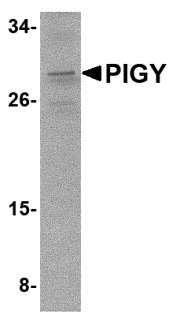
Left: Western blot analysis of PIG-Y in A-20 cell lysate with PIG-Y antibody at 2 µg/ml.
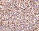
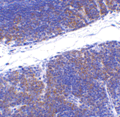
Below: Immunohistochemistry of PIGY in human spleen tissue with PIGY antibody at 2.5 µg/ml.
Other Product Images

Source : PIG-Y antibody was raised against a 16 amino acid peptide near the carboxy terminus of human PIG-Y.
Purification : Affinity chromatography purified via peptide column
Clonality and Clone : This is a polyclonal antibody.
Host : PIG-Y antibody was raised in rabbit. Please use anti-rabbit secondary antibodies.
Application : PIG-Y antibody can be used for detection of PIG-Y by Western blot at 1 – 2 µg/ml.
Tested Application(s) : E, WB, IHC
Buffer : Antibody is supplied in PBS containing 0.02% sodium azide.
Blocking Peptide :Cat.No. 4945P - PIG-Y Peptide
Long-Term Storage : PIG-Y antibody can be stored at 4ºC, stable for one year. As with all antibodies care should be taken to avoid repeated freeze thaw cycles. Antibodies should not be exposed to prolonged high temperatures.
Positive Control :
1. Cat. No. 1288 - A20 Cell Lysate
Species Reactivity : H, M
GI Number : 14249680
Accession Number : NP_116295
Short Description : (CT) Phosphatidylinositol glycan anchor biosynthesis class Y
References
1. Eisenhaber B, Maurer-Stroh S, Novatchkova M, et al. Enzymes and auxiliary factors for GPI lipid anchor biosynthesis and post-translational transfer to proteins. Bioessays 2003; 25:367-85.
2. Watanabe R, Murakami Y, Marmor MD, et al. Initial enzyme for glycosylphosphatidylinositol biosynthesis requires PIG-P and is regulated by DPM2. EMBO J. 2000; 19:4402-11.
3. Murakami Y, Siripanyaphinoyo U, Hong Y, et al. The initial enzyme for glycosylphosphatidylinositol biosynthesis requires PIG-Y, a seventh component. Mol. Biol. Cell 2005; 16:5236-46.
DC-SIGN Antibody
Catalog# : 2349
Dendritic cells (DCs) that control immune responses were recently found to capture and transport HIV from the mucosal area to remote lymph nodes (1), where DCs hand over HIV to CD4+ T lymphocytes. DCs also amplify the amount of virus and extend the duration of viral infectivity. Multiple strains of HIV-1, HIV-2 and SIV bind to DCs via DC-SIGN (2). ICAM-3 is the natural ligand for DC-SIGN (3). A DC-SIGN homologue (termed DC-SIGNR, L-SIGN, and DC-SIGN2) was identified recently (4-8). DC-SIGN forms a novel gene family with DC-SIGNR and many alternatively spliced isoforms of DC-SIGN and DC-SIGNR (8). The expression of DC-SIGN was found in mucosal tissues including placenta, small intestine, and rectum.
Additional Names : DC-SIGN (ED), DC-SIGN
 Description
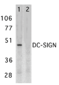
Left: Western blot analysis of DC-SIGN expression in human placenta tissue lysate in the absence (lane 1) and presence (lane 2) of blocking peptide with DC-SIGN antibody at 2 µg /ml.
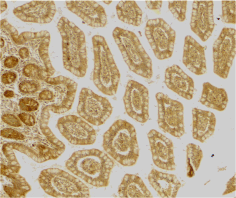
Below: Immunohistochemistry of DC-SIGN in human small intestine tissue with DC-SIGN antibody at 10 µg/ml.
Other Product Images
 Source : DC-SIGN antibody was raised against a synthetic peptide corresponding to amino acids near the center of human DC-SIGN .
Purification : Affinity chromatography purified via peptide column
Clonality and Clone : This is a polyclonal antibody.
Host : DC-SIGN antibody was raised in rabbit. Please use anti-rabbit secondary antibodies.
Immunogen : Human DC-SIGN (Extracellular Domain) Peptide (Cat. No. 2349P)
Application : DC-SIGN antibody can be used for detection of DC-SIGN by Western blot at 1 – 2 µg/ml.A band at approximately 44 kDa can be detected.
Tested Application(s) : E, WB, IHC
Buffer : Antibody is supplied in PBS containing 0.02% sodium azide.
Blocking Peptide : Cat. No. 2349P - DC-SIGN Peptide
Long-Term Storage : DC-SIGN antibody can be stored at 4ºC, stable for one year. As with all antibodies care should be taken to avoid repeated freeze thaw cycles. Antibodies should not be exposed to prolonged high temperatures.
Positive Control
1. Cat. No. 1309 - Human Placenta Lysate
2. Cat. No. 1308 - Human Small Intestine Tissue Lysate
Species Reactivity :H
GI Number : 46396012
Accession Number : Q9NNX6
Short Description : (ED) A novel HIV biding protein
References
1. Geijtenbeek TB, Kwon DS, Torensma R, et al. DC-SIGN, a dendritic cell-specific HIV-1-binding protein that enhances trans-infection of T cells. Cell. 2000;100:587-97.
2. Pohlmann S, Baribaud F, Lee B, et al. RW. DC-SIGN interactions with human immunodeficiency virus type 1 and 2 and simian immunodeficiency virus. J Virol. 2001;75(10):4664-72.
3. Geijtenbeek TB, Torensma R, van Vliet SJ, et al. Identification of DC-SIGN, a novel dendritic cell-specific ICAM-3 receptor that supports primary immune responses. Cell. 2000;100(5):575-85.
4. Soilleux EJ, Barten R, Trowsdale J. DC-SIGN; a related gene, DC-SIGNR; and CD23 form a cluster on 19p13. J Immunol. 2000;165(6):2937-42.
TNFRSF14 Antibody
Catalog# : 3775
Tumor necrosis factor receptor (TNFR) superfamily members are defined by cysteine-rich domains in their extracellular regions that bind TNF-related ligands that share a common structural homology in their extracellular domain. TNFRSF14 was initially identified as the Herpesvirus entry mediator and upon binding to the herpes simplex virus (HSV) envelope glycoprotein D or either of its natural ligands LIGHT and lymphotoxin alpha (LT), activates the transcription factors NF-kappaB and AP-1. Activation of this signal transduction pathway in T cells stimulates T cell proliferation and cytokine production, leading to inflammation and enhanced CTL-mediated tumor immunity, suggesting that these proteins may be useful as potential targets for controlling cellular immune responses.
Additional Names : TNFRSF14 (NT), Tumor necrosis factor receptor superfamily 14, Herpesvirus
 Description
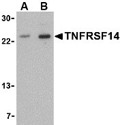
Left: Western blot analysis of TNFRSF14 in mouse thymus tissue lysate with TNFRSF14 antibody at (A) 1 and (B) 2 µg/ml.
Below: Immunohistochemistry of TNFRSF14 in mouse thymus tissue with TNFRSF14 antibody at 1 µg/ml.
Other Product Images
 Source : TNFRSF14 antibody was raised against a 15 amino acid peptide from near the amino terminus of human TNFRSF14.
Purification : Affinity chromatography purified via peptide column
Clonality and Clone : This is a polyclonal antibody.
Host : TNFRSF14 antibody was raised in rabbit. Please use anti-rabbit secondary antibodies.
Application : TNFRSF14 antibody can be used for the detection of TNFRSF14 by Western blot at 1 - 2 µg/ml.
Tested Application(s) : E, WB, IHC
Buffer : Antibody is supplied in PBS containing 0.02% sodium azide.
Blocking Peptide : Cat.No. 3775P - TNFRSF14 Peptide
Long-Term Storage : TNFRSF14 antibody can be stored at 4ºC, stable for one year. As with all antibodies care should be taken to avoid repeated freeze thaw cycles. Antibodies should not be exposed to prolonged high temperatures.
Positive Control
1. Cat. No. 1409 - Mouse Thymus Tissue Lysate
Species Reactivity :H, M
GI Number : 13878821
Accession Number : Q92956
Short Description : (NT) Herpesvirus entry mediator
References
1. Montgomery RI, Warner MS, Lum BJ, et al. Herpes simplex virus-1 entry into cells mediated by a novel member of the TNF/NGF receptor family. Cell 1996; 87:427-36.
2. Montgomery RI, Warner MS, Lum BJ, et al. Herpes simplex virus-1 entry into cells mediated by a novel member of the TNF/NGF receptor family. Cell 1996; 87:427-36.
3. Marsters SA, Ayres TM, Skubatch M, et al. Herpesvirus entry mediator, a member of the tumor necrosis factor receptor (TNFR) family, interacts with members of the TNFR-associated factor family and activates the transcription factors NF-kappaB and AP-1. J. Biol. Chem. 1997; 272:14029-32.
4. Mauri DN, Ebner R, Montgomery RI, et al. LIGHT, a new member of the TNF superfamily, and lymphotoxin alpha are ligands for herpesvirus entry mediator. Immunity 1998; 8:21-30.
GFR alpha 2 Antibody
Catalog# : 1135
Members of the glial cell line-derived neurotrophic factor (GDNF) family, including GDNF and neurturin (NTN), play key roles in the control of vertebrate neuron survival and differentiation. Physiological responses to NTN require the presence of a novel glycosylphosphadidylinositol linked protein NTNRalpha, which is a cell surface receptor for NTN. The cDNAs encoding NTNRa from human, rat, chicken, and mouse have been cloned recently. NTNRalpha was also termed GDNFRbeta, Ret ligand 2 (RETL2) or TGFbeta-related neurotrophic factor receptor 2 (TrnR2) and nominated as GFRalpha-2 recently. GFRalpha-2 binds NTN and mediates activation of RET receptor tyrosine kinase by both NTN and GDNF. Thus, NTN, GFRalpha-2, and the Ret PTK form a complex to transduce NTN signal and to mediate NTN function.
Additional Names : GFR alpha 2 (IN), GFRa-2, GDNFRb, GDNFRbeta
 Description
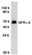
Left: Western blot analysis of GFR alpha 2 in HeLa total cell lysate with GFR alpha 2 antibody at 1:1000 dilution.
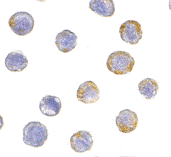
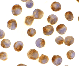
Below: Immunocytochemistry of GFR alpha 2 in HeLa cells with GFR alpha 2 antibody at 5 µg/ml.
Other Product Images
 Source : GFR alpha 2 antibody was raised against a peptide corresponding to amino acids 377 to 391 of human GFR alpha 2. The amino acid sequences of immunogenic peptide are identical to those of mouse and rat.
Purification : Antibody is DEAE purified
Clonality and Clone : This is a polyclonal antibody.
Host : GFR alpha 2 antibody was raised in rabbit. Please use anti-rabbit secondary antibodies.
Immunogen : Human GFR alpha 2 Peptide (Cat. No. 1135P)
Application : GFR alpha 2 antibody can be used for detection of GFR alpha 2 by Western blot at 1:1000 to 1:2000 dilution. A 52 kDa band should be detected.
Tested Application(s) : E, WB, ICC
Buffer : Antibody is supplied in PBS containing 0.02% sodium azide.
Blocking Peptide : Cat. No. 1135P - GFR alpha 2 Peptide
Long-Term Storage : GFR alpha 2 antibody can be stored at 4ºC, stable for one year. As with all antibodies care should be taken to avoid repeated freeze thaw cycles. Antibodies should not be exposed to prolonged high temperatures.
Positive Control
1. Cat. No. 1201 - HeLa Cell Lysate
Species Reactivity :H, M, R
GI Number : 2145080
Accession Number : AAC52036
Short Description : (IN) Neurturin receptor
References
1. Kotzbauer PT, Lampe PA, Heuckeroth RO, Golden JP, Creedon DJ, Johnson EM Jr, lbrandt J. Neurturin, a relative glial-cell-line-derived neurotrophic factor. Nature 1996;384:467-470
2. Klein RD, Sherman D, Ho WH, Stone D, Bennett GL, Moffat B, Vandlen R, Simmons L, Gu Q, Hongo JA, Devaux B, Poulsen K, Armanini M, Nozaki C, Asai N, Goddard A, Phillips H, Henderson CE, Takahashi M, Rosenthal A. A GPI-linked protein that interacts with Ret to form a candidate neurturin receptor. Nature 1997;387:717-721
3. Buj-Bello A, Adu J, Pinon LG, Horton A, Thompson J, Rosenthal A, Chinchetru M, Buchman VL, Davies AM. Neurturin responsiveness requires a GPI-linked receptor and the Ret receptor tyrosine kinase. Nature 1997;387:721-724
4. Baloh RH, Tansey MG, Golden JP, Creedon DJ, Heuckeroth RO, Keck CL, Zimonjic DB, Popescu NC, Johnson EM Jr, Milbrandt J. TrnR2, a novel receptor that mediates neurturin and GDNF signaling through Ret. Neuron 1997;18:793-802
IL-21 Antibody
Catalog# : 2465
A novel cytokine was recently identified in human and mouse and designated IL-21 , which has significant homology to IL-2, IL-4, and IL-15. The receptor for IL-21 (IL-21R, also termed NILR for novel Interleukin receptor) is a new member of the class I cytokine receptor family (1,2). IL-21R forms a complex with the common cytokine receptor g chain, gc, and mediates IL-21 signaling (3,4). IL-21 and its receptor activate JAK-STAT signaling pathway. IL-21 is expressed in activated T cells, and HL-60 and THP-1 cell lines. IL-21 plays a role in the proliferation and maturation of NK, B and T cell populations.
Additional Names : IL-21 (IN), IL-21
 Description
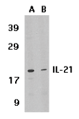
Left: Western blot analysis of IL-21 expression in HL-60 cell lysate in the absence (lane A) or presence of blocking peptide (lane B) with IL-21 antibody at 1 µg /ml.
Below: Immunocytochemistry of IL-21 in K562 cells with IL-21 antibody at 2 µg/ml.
Other Product Images
 Source : IL-21 antibody was raised against a synthetic peptide corresponding to 15 amino acids near the center of human IL-21 precursor .
Purification : Affinity chromatography purified via peptide column
Clonality and Clone : This is a polyclonal antibody.
Host : IL-21 antibody was raised in rabbit. Please use anti-rabbit secondary antibodies.
Immunogen : Human IL-21 (Intermediate Domain) Peptide (Cat. No. 2465P)
Application : IL-21 antibody can be used for detection of IL-21 by Western blot at 0.5 to 1 µg/ml.An approximately 18 kDa band can be detected.
Tested Application(s) : E, WB, ICC
Buffer : Antibody is supplied in PBS containing 0.02% sodium azide.
Blocking Peptide : Cat. No. 2465P - IL-21 Peptide
Long-Term Storage : IL-21 antibody can be stored at 4ºC, stable for one year. As with all antibodies care should be taken to avoid repeated freeze thaw cycles. Antibodies should not be exposed to prolonged high temperatures.
Positive Control
1. Cat. No. 1209 - HL60 Cell Lysate
2. Cat. No. 1208 - THP-1 Whole Cell Lysate
Species Reactivity :H
GI Number : 11141875
Accession Number : NP_068575
Short Description : (IN) Interleukin 21, related to IL2 and IL15
References
1. Parrish-Novak J, Dillon SR, Nelson A, et al. Interleukin 21 and its receptor are involved in NK cell expansion and regulation of lymphocyte function. Nature. 2000;408(6808):57-63.
2. Ozaki K, Kikly K, Michalovich D, et al. Cloning of a type I cytokine receptor most related to the IL-2 receptor beta chain. Proc Natl Acad Sci USA . 2000;97(21):11439-44.
3. Asao H, Okuyama C, Kumaki S, et al. the common gamma-chain is an indispensable subunit of the IL-21 receptor complex. J Immunol. 2001;167(1):1-5.
erbB-2 Antibody
Catalog# : 5153
The ErbB family consists of four closely related tyrosine kinase receptors that act as potent mediators of normal cell growth and development. Aberrant expression or function of one or more of these receptors can play a major role in the development and evolution of cancer. ErbB-2, also known as HER2, has been implicated in the evolution of both breast and gastric cancers, and is evident in other cancer types such as ovarian and salivary gland tumors. ErbB-2 possesses an active tyrosine kinase domain, but no direct ligand has been identified yet. ErbB-2 is the preferred binding partner to the other members of the ErbB family and is thought to act primarily through the Ras-MAPK, PI3k-PKB/Akt, and PLC-PKC signaling pathways. Numerous anti-cancer strategies have been employed against erbB-2, such as antibody-based therapies to prevent ligand binding or receptor activation through dimerization, antibody-dependent cell mediated cytotoxicity, in addition to direct kinase inhibition to prevent molecular activation/downstream signaling.
Additional Names : erbB-2, v-erb-b2 erythroblastic leukemia viral oncogene homolog 2, NEU, NGL, HER2, HER-2, TKR1, CD340
 Description
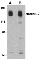
Left: Western blot analysis of erbB-2 in rat liver tissue lysate with erbB-2 antibody at (A) 1 and (B) 2 µg/ml.
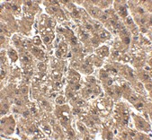
Below: Immunohistochemistry of erbB-2 in human liver tissue with erbB-2 antibody at 2.5 μg/ml.
Other Product Images
 Source : erbB-2 antibody was raised against a 19 amino acid peptide near the carboxy terminus of human erbB-2.
Purification : Affinity chromatography purified via peptide column
Clonality and Clone : This is a polyclonal antibody.
Host : erbB-2 antibody was raised in chicken. Please use anti-rabbit secondary antibodies.
Application : erbB-2 antibody can be used for detection of erbB-2 by Western blot at 1 – 2 µg/ml.
Tested Application(s) : E, WB
Buffer : Antibody is supplied in PBS containing 0.02% sodium azide.
Blocking Peptide : Cat.No. 5153P - erbB-2 Peptide
Long-Term Storage : erbB-2 antibody can be stored at 4ºC, stable for one year. As with all antibodies care should be taken to avoid repeated freeze thaw cycles. Antibodies should not be exposed to prolonged high temperatures.
Positive Control
1. Cat. No. 1464 - Rat Liver Tissue Lysate
Species Reactivity :H, M, R
GI Number : 54792096
Accession Number : NP_004439
Short Description : v-erb-b2 erythroblastic leukemia viral oncogene homolog 2
References
1. Baselga J and Swain SM. Novel anticancer targets: revisiting ERBB2 and discovering ERBB3. Nature Rev. 2009; 9:463-475.
2. Slamon DJ, Clark GM, Wong SG, et al. Human breast cancer: correlation of relapse and survival with amplification of the HER-2/neu oncogene. Science 1987; 235:177-82.
3. Vermeij J, Teugels E, Bourgain C, et al. Genomic activation of the EGFR and HER2-neu genes in a significant proportion of invasive epithelial ovarian cancers. BMC Cancer 2008; 8:3.
4. Marmor MD, Skaria KB, and Yarden Y. Signal transduction and oncogenesis by ErbB/HER receptors. Int. J. Rad. Oncol. Biol. Phys. 2004; 58:903-13.

 Description