Showing posts with label Custom Antibody. Show all posts

Tuesday, May 31, 2011

Aak1 Antibody


Catalog#:4831

AP2-associated protein kinase 1 (Aak1) is a member of the Ark1/Prk1 subfamily of Ser/Thr protein kinases that are thought to regulate endocytosis by phosphorylating the accessory endocytic components. Aak1 interacts with and phosphorylates the mu2 subunit of the AP-2 complex, which promotes binding of the AP-2 to tyrosine based (Yxxf) internalization motif-containing receptors and subsequent receptor endocytosis. At least two isoforms of Aak1 are known to exist; the longer isoform contains an extended carboxy-terminus that contains an additional clathrin-binding domain. Overexpression of this long isoform or Aak1 depletion by RNA interference impairs transferrin recycling from the early/sorting endosome, suggesting that Aak1 functions at multiple steps of the endosomal pathway by regulating transferrin internalization and its recycling back to the plasma membrane.

Additional Names:
Aak1 (CT), AP2-associated protein kinase 1
Description

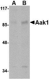
Left: Western blot analysis of Aak1 in A-20 lysate with Aak1 antibody at (A) 1 and (B) 2 µg/m


Source:
Aak1 antibody was raised against a 18 amino acid peptide near the carboxy terminus of the human Aak1.
Purification: Affinity chromatography purified via peptide column
Clonality and Clone: This is a polyclonal antibody.
Host: Aak1 antibody was raised in rabbit.
Please use anti-rabbit secondary antibodies.
Application: Aak1 antibody can be used for detection of Aak1 by Western blot at 1 – 2 µg/ml.
Tested Application(s): E, WB
Buffer: Antibody is supplied in PBS containing 0.02% sodium azide.
Blocking Peptide:Cat.No. 4831P - Aak1 Peptide
Long-Term Storage: Aak1 antibody can be stored at 4ºC, stable for one year. As with all antibodies care should be taken to avoid repeated freeze thaw cycles. Antibodies should not be exposed to prolonged high temperatures.
Positive Control:
1. Cat. No. 1288 - A20 Cell Lysate
Species Reactivity: H, M, R
GI Number: 148277037
Accession Number: NP_055726
Short Description: (CT) AP2-associated protein kinase 1
References

1. Connor SD and Schmid SL. Identification of an adaptor-associated kinase, AAK1, as a regulator of clathrin-mediated endocytosis. J. Cell Biol. 2002; 156:921-9.
2. Smythe E and Ayscough KR. The Ark1/Prk1 family of protein kinases. Regulators of endocytosis and the actin skeleton. EMBO Rep. 2003; 4:246-51.
3. Ricotta D, Connor SD, Schmid SL, et al. Phosphorylation of the AP2 m2 subunit by AAK1 mediates high affinity binding to membrane protein sorting signals. J. Cell Biol. 2002; 156:791-5.
4. Connor SD and Henderson DM. A novel AAK1 splice variant functions at multiple steps of the endocytic pathway. Mol. Biol. Cell 2007; 18:2698-706.

Sunday, May 8, 2011

SAPAP4 Antibody

SAPAP4 Antibody

Catalog# :4633

SAP90/PSD-95-associated protein 4 (SAPAP4, also known as DLGAP4) is a member of a protein family whose members specifically interact with PSD-95/SAP90, a membrane-associated guanylate kinase localized at postsynaptic density (PSD) in neuronal cells. Like the other SAPAP proteins, SAPAP4 is thought to be an adaptor protein that also interacts with different synaptic scaffolding proteins, cytoskeletal and signaling components, such as focal adhesion kinase (FAK) and proline-rich tyrosine kinase 2 (PYK2). SAPAP4 mRNA is targeted to cell bodies in a similar manner to SAPAP1 and -2, whereas SAPAP3 mRNA is detected mainly in cell bodies. At least three isoforms of SAPAP2 are known to exist. This SAPAP4 antibody will not cross-react with other SAPAP proteins.

Additional Names : SAPAP4, SAP90/PSD-95-associated protein 4, Disks large-associated protein 4, DLGAP4, DAP4

Description
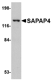
Left: Western blot analysis of SAPAP4 in SK-N-SH cell lysate with SAPAP4 antibody at 1 µg/ml.


Source :SAPAP4 antibody was raised against a 13 amino acid peptide from near the center of human SAPAP4.
Purification : Affinity chromatography purified via peptide column
Clonality and Clone : This is a polyclonal antibody.
Host : SAPAP4 antibody was raised in rabbit.
Please use anti-rabbit secondary antibodies.
Application : SAPAP4 antibody can be used for detection of SAPAP4 by Western blot at 0.5 – 1 µg/ml.
Tested Application(s) : E, WB
Buffer : Antibody is supplied in PBS containing 0.02% sodium azide.
Blocking Peptide :Cat.No. 4633P - SAPAP4 Peptide
Long-Term Storage : SAPAP4 antibody can be stored at 4ºC, stable for one year. As with all antibodies care should be taken to avoid repeated freeze thaw cycles. Antibodies should not be exposed to prolonged high temperatures.
Positive Control :
1. Cat. No. 1220 - SK-N-SH Cell Lysate
Species Reactivity : H
GI Number : 34335253
Accession Number : NP_055717
Short Description : SAP90/PSD-95-associated protein 4
References
1. SAPAPs. A family of PSD-95/SAP90-associated proteins localized at postsynaptic density. J. Biol. Chem. 1997; 272:11943-51.
2. Ranta S, Zhang Y, Ross B, et al. Positional cloning and characterization of the human DLGAP2 gene and its exclusion in progressive epilepsy with mental retardation. Eur. J. Hum. Genet. 2000; 8:381-4.
3. Kindler S, Rehbein M, Classen B, et al. Distinct spatiotemporal expression of SAPAP transcripts in the developing rat brain: a novel dendritically localized mRNA. Brain Res. Mol. Brain Res. 2004; 126:14-21.
4. Bongiorno-Borbone L, Kadare G, Benfenati F, et al. FAK and PYK2 interact with SAP/PSD-95-associated protein-3. Biochem. Biophys. Res. Commun. 2005; 337:641-6.

Thursday, May 5, 2011

Custom Organic Synthesis


With over two decades of combined experience and expertise in range of chemical synthesis, BSI has been a one-stop synthesis solution station for its clients worldwide. Our team of Ph.D organic chemists have expertise in a wide variety of chemistries from intermediates, reference compounds, and starting materials, to derivatives of lead compounds.

Custom Chemical Synthesis

Chemical synthesis is an essential part of drug development. However, the number of technical and managerial tasks involved in the process – many of which prove to be "one-off" operations - can serve to hamper the overall effectiveness of the research team. Outsourcing the activity to a specialist custom synthesis house is an efficient, cost-effective means of ensuring the smooth and timely passage of new drug compounds through the pipeline.

Laboratory Custom Synthesis Projects

Under laboratory custom synthesis projects, our customer should provide literatures, methods, or their own synthesis route describing all in details. The more information provided, the shorter the project timelines and the lower the cost. Our chemists will evaluate synthesis feasibility prior to the initiation of the projects, and provided an estimate of cost and delivery time.

Research and Development Process Optimization

Whether is an expansion of existing scalable process or to patent development, BSI can develop new and proprietary route by using our in-house staff with state-of-art technology including design of experiments, real-time reaction profiles and total process development. BSI can assist clients in transferring their research laboratory projects to pilot productions.
SAPAP3 Antibody

Catalog# :4411

SAP90/PSD-95-associated protein 3 (SAPAP3, also known as DLGAP3) is a member of a protein family whose members specifically interact with PSD-95/SAP90, a membrane-associated guanylate kinase localized at postsynaptic density (PSD) in neuronal cells. Like the other SAPAP proteins, SAPAP3 is thought to be an adaptor protein that also interacts with different synaptic scaffolding proteins, cytoskeletal and signaling components, such as focal adhesion kinase (FAK) and proline-rich tyrosine kinase 2 (PYK2). Both SAPAP3 protein and mRNA are targeted to dendrites, whereas SAPAP1, -2, and -4 mRNAs are detected mainly in cell bodies. Recent experiments have suggested that SAPAP3 may be involved in obsessive-compulsive disorder (OCD), as mice lacking SAPAP3 exhibited OCD-like symptoms which could be relieved by lentiviral-mediated selective expression of SAPAP3 in the striatum of SAPAP3-mutant mice. At least two isoforms are known to exist.

Additional Names : SAPAP3, SAP90/PSD-95-associated protein 3, Disks large-associated protein 3, DLGAP3
Description
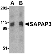
Left:Western blot analysis of SAPAP3 in rat brain tissue lysate with SAPAP3 antibody at (A) 1 (B) 2 µg/ml.

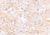
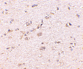
Below:Immunohistochemical staining of human brain tissue using SAPAP3 antibody at 2.5 µg/ml.

Other Product Images
Source :SAPAP3 antibody was raised against a 18 amino acid peptide from near the carboxy terminus of human SAPAP3.
Purification : Affinity chromatography purified via peptide column
Clonality and Clone : This is a polyclonal antibody.
Host : SAPAP3 antibody was raised in rabbit.
Please use anti-rabbit secondary antibodies.
Application : SAPAP3 antibody can be used for detection of SAPAP3 by Western blot at 1 – 2 µg/ml.
Tested Application(s) : E, WB, IHC
Buffer : Antibody is supplied in PBS containing 0.02% sodium azide.
Blocking Peptide :Cat.No. 4411P - SAPAP3 Peptide
Long-Term Storage : SAPAP3 antibody can be stored at 4ºC, stable for one year. As with all antibodies care should be taken to avoid repeated freeze thaw cycles. Antibodies should not be exposed to prolonged high temperatures.
Positive Control :
1. Cat. No. 1463 - Rat Brain Tissue Lysate
Species Reactivity : H, M, R
GI Number : 71153509
Accession Number : O95886
Short Description : SAP90/PSD-95-associated protein 3
References
1. SAPAPs. A family of PSD-95/SAP90-associated proteins localized at postsynaptic density. J. Biol. Chem. 1997; 272:11943-51.
2. Kindler S, Rehbein M, Classen B, et al. Distinct spatiotemporal expression of SAPAP transcripts in the developing rat brain: a novel dendritically localized mRNA. Brain Res. Mol. Brain Res. 2004; 126:14-21.
3. Bongiorno-Borbone L, Kadare G, Benfenati F, et al. FAK and PYK2 interact with SAP/PSD-95-associated protein-3. Biochem. Biophys. Res. Commun. 2005; 337:641-6.
4. Welch JM, Wang D, and Feng G. Differential mRNA expression and protein localization of the SAP90/PSD-95-associated proteins (SAPAPs) in the nervous system of the mouse. J. Comp. Neurol. 2004; 472:24-39.

SAPAP2 Antibody

SAPAP2 Antibody

Catalog# :4625

SAP90/PSD-95-associated protein 2 (SAPAP2, also known as DLGAP2) is a member of a protein family whose members specifically interact with PSD-95/SAP90, a membrane-associated guanylate kinase localized at postsynaptic density (PSD) in neuronal cells. Like the other SAPAP proteins, SAPAP2 is thought to be an adaptor protein that also interacts with different synaptic scaffolding proteins, cytoskeletal and signaling components, such as focal adhesion kinase (FAK) and proline-rich tyrosine kinase 2 (PYK2). SAPAP2 mRNA is targeted to cell bodies in a similar manner to SAPAP1 and -4, whereas SAPAP3 mRNA is detected mainly in cell bodies. At least three isoforms of SAPAP2 are known to exist. This SAPAP2 antibody will not cross-react with other SAPAP proteins.

Additional Names : SAPAP2,SAP90/PSD-95-associated protein 2, Disks large-associated protein 2, DLGAP2, DAP-2

Description
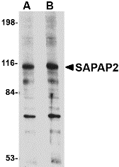
b>Left: Western blot analysis of SAPAP2 in L1210 cell lysate with SAPAP2 antibody at (A) 0.5 and (B) 1 µg/ml.

Below:Immunohistochemistry of SAPAP2 in human brain with SAPAP2 antibody at 5 µg/ml.

Other Product Images

Source :SAPAP2 antibody was raised against a 16 amino acid peptide from near the center of human SAPAP2.
Purification : Affinity chromatography purified via peptide column
Clonality and Clone : This is a polyclonal antibody.
Host : SAPAP2 antibody was raised in rabbit.
Please use anti-rabbit secondary antibodies.
Application : SAPAP2 antibody can be used for detection of SAPAP2 by Western blot at 0.5 – 1 µg/ml.
Tested Application(s) : E, WB, IHC
Buffer : Antibody is supplied in PBS containing 0.02% sodium azide.
Blocking Peptide :Cat.No. 4625P - SAPAP2 Peptide
Long-Term Storage : SAPAP2 antibody can be stored at 4ºC, stable for one year. As with all antibodies care should be taken to avoid repeated freeze thaw cycles. Antibodies should not be exposed to prolonged high temperatures.
Positive Control :
1. Cat. No. 1284 - L1210 Cell Lysate
Species Reactivity : H, M, R
GI Number : 160395585
Accession Number : Q9P1A6
Short Description : SAP90/PSD-95-associated protein 2
References
1. SAPAPs. A family of PSD-95/SAP90-associated proteins localized at postsynaptic density. J. Biol. Chem. 1997; 272:11943-51.
2. Ranta S, Zhang Y, Ross B, et al. Positional cloning and characterization of the human DLGAP2 gene and its exclusion in progressive epilepsy with mental retardation. Eur. J. Hum. Genet. 2000; 8:381-4.
3. Kindler S, Rehbein M, Classen B, et al. Distinct spatiotemporal expression of SAPAP transcripts in the developing rat brain: a novel dendritically localized mRNA. Brain Res. Mol. Brain Res. 2004; 126:14-21.
4. Bongiorno-Borbone L, Kadare G, Benfenati F, et al. FAK and PYK2 interact with SAP/PSD-95-associated protein-3. Biochem. Biophys. Res. Commun. 2005; 337:641-6.

SAPAP1 Antibody

SAPAP1 Antibody

Catalog# :4623

SAP90/PSD-95-associated protein 1 (SAPAP1, also known as DLGAP1 or GKAP) is a member of a protein family whose members specifically interact with PSD-95/SAP90, a membrane-associated guanylate kinase localized at postsynaptic density (PSD) in neuronal cells. Like the other SAPAP proteins, SAPAP1 is thought to be an adaptor protein that also interacts with different synaptic scaffolding proteins, cytoskeletal and signaling components, such as focal adhesion kinase (FAK) and proline-rich tyrosine kinase 2 (PYK2). SAPAP1 mRNA is targeted to cell bodies in a similar manner to SAPAP2 and -4, whereas SAPAP3 mRNA is detected mainly in cell bodies. SAPAP1 protein however, is targeted to the synapse and is not reliant on the synaptic localization of PSD-95 or the synaptic scaffolding molecule (S-SCAM). At least three isoforms of SAPAP1 are known to exist. This SAPAP1 antibody will not cross-react with other SAPAP proteins.

Additional Names : SAPAP1, SAP90/PSD-95-associated protein 1, Disks large-associated protein 1, DLGAP1, DAP-1, GKAP

Description
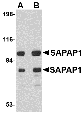
Left: Western blot analysis of SAPAP1 in rat brain tissue lysate with SAPAP1 antibody at (A) 0.5 and (B) 1 µg/ml.

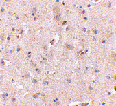
Below:Immunohistochemistry of SAPAP1 in human brain with SAPAP1 antibody at 5 µg/ml.

Other Product Images
Source :SAPAP1 antibody was raised against a 18 amino acid peptide from near the center of human SAPAP1.
Purification : Affinity chromatography purified via peptide column
Clonality and Clone : This is a polyclonal antibody.
Host : SAPAP1 antibody was raised in rabbit.
Please use anti-rabbit secondary antibodies
Application : SAPAP1 antibody can be used for detection of SAPAP1 by Western blot at 0.5 – 1 µg/ml.
Tested Application(s) : E, WB, IHC
Buffer : Antibody is supplied in PBS containing 0.02% sodium azide.
Blocking Peptide :Cat.No. 4623P - SAPAP1 Peptide
Long-Term Storage : SAPAP1 antibody can be stored at 4ºC, stable for one year. As with all antibodies care should be taken to avoid repeated freeze thaw cycles. Antibodies should not be exposed to prolonged high temperatures.
Positive Control :
1. Cat. No. 1303 - Human Brain Tissue Lysate
Species Reactivity : H, M, R
GI Number : 4758822
Accession Number : NP_004737
Short Description : SAP90/PSD-95-associated protein 1
References
1. SAPAPs. A family of PSD-95/SAP90-associated proteins localized at postsynaptic density. J. Biol. Chem. 1997; 272:11943-51.
2. Kindler S, Rehbein M, Classen B, et al. Distinct spatiotemporal expression of SAPAP transcripts in the developing rat brain: a novel dendritically localized mRNA. Brain Res. Mol. Brain Res. 2004; 126:14-21.
3. Bongiorno-Borbone L, Kadare G, Benfenati F, et al. FAK and PYK2 interact with SAP/PSD-95-associated protein-3. Biochem. Biophys. Res. Commun. 2005; 337:641-6.
4. Welch JM, Wang D, and Feng G. Differential mRNA expression and protein localization of the SAP90/PSD-95-associated proteins (SAPAPs) in the nervous system of the mouse. J. Comp. Neurol. 2004; 472:24-39.

Rim3 Antibody

Rim3 Antibody

Catalog# :4471

Rab3-interacting molecules (RIMs) are synaptic proteins are synaptic proteins necessary for neural transmission and plasticity. While both Rim1 and Rim 2 are thought to be effector proteins for Rab3, binding to Rab3 on synaptic vesicles in a GTP-dependent manner, less is known of Rim3. Expression of Rim3 in PC12 cells induced a significant increase in calcium-triggered exocytosis, with no appreciable change in the baseline release, suggesting that it plays a role in the regulation of exocytosis. Rim3 protein localizes primarily to neuronal dendrites and the postsynaptic densities, as opposed to Rim1 which is found in presynapse locations, indicating that Rim3 may contribute to synapse transmission and plasticity. This antibody is predicted to have no cross-reactivity to other Rim proteins.

Additional Names : Rim3 (NT), Rab3-interacting molecule 3, regulating synaptic membrane exocytosis 3, RIM3 gamma, RIMS3, NIM3
Description
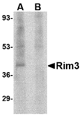
Left: Western blot analysis of Rim3 in human brain tissue lysate with Rim3 antibody at 1 µg/ml in the (A) absence and (B) presence of blocking peptide.

Below:Immunohistochemistry of Rim3 in human brain tissue with Rim3 antibody at 2.5 µg/ml.

Other Product Images

Source :Rim3 antibody was raised against a 17 amino acid peptide from near the amino terminus of human Rim3.
Purification : Affinity chromatography purified via peptide column
Clonality and Clone : This is a polyclonal antibody.
Host : Rim3 antibody was raised in rabbit.
Application : Rim3 antibody can be used for detection of Rim3 by Western blot at 0. 5 – 1 µg/ml.
Tested Application(s) : E, WB, IHC
Buffer : Antibody is supplied in PBS containing 0.02% sodium azide.
Blocking Peptide :Cat.No. 4471P - Rim3 Peptide
Long-Term Storage : Rim3 antibody can be stored at 4ºC, stable for one year. As with all antibodies care should be taken to avoid repeated freeze thaw cycles. Antibodies should not be exposed to prolonged high temperatures.
Positive Control :
1. Cat. No. 1303 - Human Brain Tissue Lysate
Species Reactivity : H, M
GI Number : 48476344
Accession Number : NP_055562
Short Description : (NT) Rab3-interacting molecule 3
References
1. Wang Y, Sugita S, and Sudhof TC. The RIM/NIM family of neuronal C2 domain proteins: interactions with Rab3 and a new class of Src homology 3 domain proteins. J. Biol. Chem. 2000; 275:20033-44.
2. Liang F, Zhang B, Tang J, et al. RIM3gamma is a postsynaptic protein in the rat central nervous system. J. Comp. Neurol. 2007; 503:501-10.

Wednesday, May 4, 2011

Presenilin1 Antibody


Catalog# :4203

Presenilin1 was initially identified a marker of susceptibility to early-onset Alzheimer’s disease. In addition to PEN2, nicastrin and APH-1, Presenilin1 forms the g-secretase protein complex, a membrane-bound aspartyl protease that can cleave certain proteins at peptide bonds buried within the hydrophobic environment of the lipid bilayer. This cleavage is responsible for a key step in signaling from several cell-surface receptors and is thought to be required for the generation of the neurotoxic amyloid peptides that are central to the pathogenesis of Alzheimer’s disease. Like the tumor necrosis factor-a-converting enzyme (TACE) and the b-site cleavage enzyme (BACE) protease families, g-secretase will cleave the amyloid precursor protein (APP), but within the intramembrane region of APP, resulting in either the non-toxic p3 (from the a and g cleavage site) or the toxic Ab amyloid peptide (from the b and g cleavage site). It is thought that accumulation of the Ab peptide is the precursor to Alzheimer’s disease. Multiple isoforms of presenilin1 are known to exist. This antibody has no cross-reactivity to presenilin2.

Additional Names : Presenilin1, PSEN1, PS1, Gamma-secretase subunit presenilin-1

Description

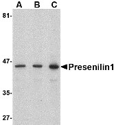
Left:Western blot analysis of presenilin1 in human brain lysate with presenilin1 antibody at (A) 0.5, (B) 1, and (C) 2 µg/ml.

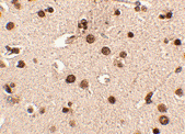
Below:Immunohistochemistry of Presenilin1 in human brain tissue with Presenilin1 antibody at 2.5 µg/ml.

Other Product Images

Source :Presenilin1 antibody was raised against a 23 amino acid peptide from near the carboxy terminus of human presenilin1.
Purification : Affinity chromatography purified via peptide column
Clonality and Clone : This is a polyclonal antibody.
Host : Presenilin1 antibody was raised in rabbit.
Please use anti-rabbit secondary antibodies.
Application : Presenilin1 antibody can be used for detection of presenilin1 by Western blot at 0.5 – 1 µg/ml.
Tested Application(s) : E, WB, IHC
Buffer : Antibody is supplied in PBS containing 0.02% sodium azide.
Blocking Peptide :Cat.No. 4203P - Presenilin1 Peptide
Long-Term Storage : Presenilin1 antibody can be stored at 4ºC, stable for one year. As with all antibodies care should be taken to avoid repeated freeze thaw cycles. Antibodies should not be exposed to prolonged high temperatures.
Positive Control :
1.Cat. No. 1303 - Human Brain Tissue Lysate
Species Reactivity : H, M, R
GI Number : 4506163
Accession Number : NP_000012
Short Description : a gamma-secretase subunit
References
1. Sherrington R, Rogaev EI, Liang Y, et al. Cloning of a gene bearing missense mutations in early-onset familial Alzheimer’s disease. Nature 1995; 375:754-60.
2. Weihofen A and Martoglio B. Intramembrane-cleaving proteases: controlled liberation of proteins and bioactive peptides. Trends Cell Biol. 2003; 13:71-8.
3. Periz G and Fortini ME. Functional reconstitution of g-secretase through coordinated expression of presenilin, nicastrin, aph-1, and pen-2. J. Neurosci. Res. 2004; 77:309-22.
4. Selkoe DJ. The cell biology of b-amyloid precursor protein and presenilin in Alzheimer’s disease. Trends Cell Biol. 1998; 8:447-53.

Monday, April 25, 2011

LRFN1 Antibody

LRFN1 Antibody

Catalog# :5065

LRFN1 is one of a family of five transmembrane glycoproteins that are highly expressed in neuronal tissues. LRFN proteins share leucine-rich repeat (LRR)-immunoglobulin-like (Ig)-fibronectin type III (Fn)-transmembrane domain structure with other members of the LRR-Ig-Fn protein superfamily such as the Slitrk family of proteins. Expression of LRFN1, -3, and -4 mRNA was detected in embryonic neuronal cells, while Lrfn2 and Lrfn5 expression was primarily restricted to more mature cells. LRFN1, -2, and -4 bound to PDZ domains of postsynaptic PSD95, re-distributing PSD95 to the cell periphery. It has been suggested that the Lrfn proteins play a role in the developing and/or mature vertebrate nervous system. At least two isoforms of LRFN1 are known to exist. This antibody is predicted to not cross-react with other members of the LRFN family.

Additional Names : LRFN1, Leucine-rich repeat and fibronectin typeIII domain-containing protein 1, synaptic adhesion-like molecule 2
Description
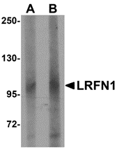
Left: Western blot analysis of LRFN1 in human brain lysate with LRFN1 antibody at (A) 1 and (B) 2 µg/ml.

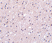
Below:Immunohistochemistry of LRFN1 in mouse brain tissue with LRFN1 antibody at 2.5 µg/ml.

Other Product Images
Source :LRFN1 antibody was raised against an 18 amino acid peptide near the carboxy terminus of human LRFN1.
Purification : Affinity chromatography purified via peptide column
Clonality and Clone : This is a polyclonal antibody.
Host : LRFN1 antibody was raised in rabbit.
Please use anti-rabbit secondary antibodies.
Application : LRFN1 antibody can be used for detection of LRFN1 by Western blot at 1 – 2 µg/ml.
Tested Application(s) : E, WB, IHC
Buffer : Antibody is supplied in PBS containing 0.02% sodium azide.
Blocking Peptide :Cat.No. 5065P - LRFN1 Peptide
Long-Term Storage : LRFN1 antibody can be stored at 4ºC, stable for one year. As with all antibodies care should be taken to avoid repeated freeze thaw cycles. Antibodies should not be exposed to prolonged high temperatures.
Positive Control :
1. Cat. No. 1303 - Human Brain Tissue Lysate
Species Reactivity : H, M, R
GI Number : 189028858
Accession Number : Q9P244
Short Description : Leucine-rich repeat and fibronectin type III domain-containing protein 1
References
1. Morimura N, Inoue T, Katayama K, et al. Comparative analysis of structure, expression and PSD95-binding capacity of Lrfn, a novel family of neuronal transmembrane proteins. Gene 2006; 380:72-83.
2. Kobe B and Kajava AV. The leucine-rich repeat as a protein recognition motif. Curr. Opin. Struct. Biol. 2001; 11:725-32.
3. Aruga J and Mikoshiba K. Identification and characterization of Slitrk, a novel transmembrane protein family controlling neurite outgrowth. Mol. Cell Neurosci. 2003; 24:117-29.

Friday, April 8, 2011

Nucleotide Synthesis


Take advantage of Bio Synthesis's 20 years experience in quality custom synthesis for all your custom DNA nucleotide needs. With state of the art facilities couple with high trained staff, Bio Synthesis offers FAST and RELIABLE delivery of high quality oligonucleotides.

Nucleotides

Nucleotides are organic compounds that consist of three joined structures: a nitrogenous base, a sugar, and a phosphate group. The most common nucleotides can be divided into two groups (purines and pyrimidines) based on the structure of the nitrogenous base. The joined sugar is either ribose or deoxyribose.

Nucleotide Synthesis And Structure

A nucleotide is composed of a ring of nitrogen, carbon and oxygen atoms, a five carbon sugar (together referred to as a nucleoside) and one phosphate group. Nucleotides can be synthesized through a variety of methods both in vitro and in vivo. This can involve salvage synthesis (the re-use of parts of nucleotides in resynthesizing new nucleotides through breakdown and synthesis reactions in order to exchange useful parts), or the use of protecting groups in a laboratory.

Synthesis

Nucleotides can be synthesized through a variety of methods both in vitro and in vivo. This can involve salvage synthesis (the re-use of parts of nucleotides in resynthesizing new nucleotides through breakdown and synthesis reactions in order to exchange useful parts), or the use of protecting groups in a laboratory. In the latter case, a purified nucleoside or nucleobase is protected to create a phosphoramidite, and can be used to obtain analogues not present in nature and/or to create an oligonucleotide.

Wednesday, April 6, 2011

BACE Antibody

BACE Antibody

Catalog# :2253

Accumulation of the amyloid-b (Ab) plaque in the cerebral cortex is a critical event in the pathogenesis of Alzheimer’s disease. Ab peptide is generated by proteolytic cleavage of the b-amyloid protein precursor (APP) at b- and g-sites by two proteases. APP is first cleaved by b-secretase, producing a soluble derivative of the protein and a membrane anchored 99-amino acid carboxy-terminal fragment (C99). The C99 fragment serves as substrate for g-secretase to generate the 4 kDa amyloid-b peptide, which is deposited in the brains of all suffers of Alzheimer’s disease. The long-sought b-secretase was recently identified by several groups independently and designated beta-site APP cleaving enzyme (BACE) and aspartyl protease 2 (Asp2) (1-4). BACE/Asp2 is a novel transmembrane aspartic protease and colocalizes with APP.

Additional Names : BACE (CT), Asp2

Description
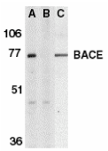
Left:Western blot analysis of BACE in human brain tissue lysate in the absence (A) or presence (B) of blocking peptide (2253P) and in mouse 3T3 cell lysate (C) with BACE antibody at 1 µg/ml.

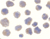
Below:Immunocytochemistry of BACE in 3T3 cells with BACE antibody at 10 µg/ml.

Other Product Images


Source :BACE antibody was raised against a peptide corresponding to 17 amino acids at the carboxy terminus of human BACE.
Purification : Ion exchange chromatography purified
Clonality and Clone : This is a polyclonal antibody.
Host : BACE antibody was raised in rabbit.
Please use anti-rabbit secondary antibodies.
Immunogen : Human BACE (C-Terminus) Peptide (Cat. No. 2253P)
Application : BACE can be used for detection of BACE by Western blot at 0.5 to 1 µg/ml.An approximately 70 kDa band should be detected.
Tested Application(s) : E, WB, ICC
Buffer : Antibody is supplied in PBS containing 0.02% sodium azide.
Blocking Peptide :Cat. No. 2253P - BACE Peptide
Long-Term Storage : BACE antibody can be stored at 4ºC, stable for one year. As with all antibodies care should be taken to avoid repeated freeze thaw cycles. Antibodies should not be exposed to prolonged high temperatures.
Positive Control :
1. Cat. No. 1303 - Human Brain Tissue Lysate
2. Cat. No. 1212 - 3T3 Whole Cell Lysate
Species Reactivity : H, M
GI Number : 6118538
Accession Number : AF190725
Short Description : (CT) Beta-site APP Cleaving Enzyme
References
1. Vassar R, Bennett BD, Babu-Khan S, et al. beta-secretase cleavage of Alzheimer's amyloid precursor protein by the transmembrane aspartic protease BACE. Science 1999;286:735-41
2. Hussain I, Powell D, Howlett DR , et al. Identification of a novel aspartic protease (Asp 2) as beta-secretase. Mol Cell Neurosci 1999;14:419-27
3. Yan R, Bienkowski MJ, Shuck ME, et al. Membrane-anchored aspartyl protease with Alzheimer's disease beta-secretase activity. Nature 1999;402:533-7
4. Sinha S, Anderson JP, Barbour R, et al. Purification and cloning of amyloid precursor protein beta-secretase from human brain. Nature 1999;402:537-40 (WD0500)

APP Antibody

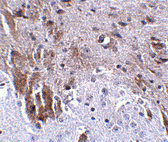
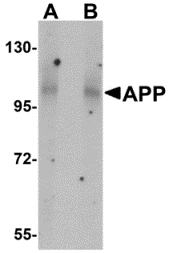
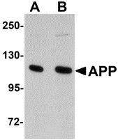
APP Antibody Catalog# :5129 Accumulation of the amyloid-beta peptide (Abeta) in the cerebral cortex is a critical event in the pathogenesis of Alzheimer’s disease. The beta−amyloid protein precursor (APP) is cleaved by one of two beta−secretases (BACE and BACE2), producing a soluble derivative of the protein and a membrane anchored 99-amino acid carboxy-terminal fragment (C99). The C99 fragment serves as substrate for γ−secretase to generate the 4 kDa amyloid-beta peptide (Abeta), which is deposited in the Alzheimer’s disease patients’ brains. Recently, Death Receptor 6 (DR6) was found to interact with an amino-terminal fragment of the Beta-amyloid protein (N-APP) in neurons, activating a caspase 6-dependent apoptotic event leading to axonal degeneration and pruning during development, suggesting that these two proteins are involved in neural development and may possibly play a role in Alzheimer’s disease. Additional Names : APP (NT), Amyloid Precursor Protein, amyloid-beta precursor protein Description Left: Western blot analysis of APP in mouse brain tissue lysate with APP antibody at (A) 1 and (B) 2 µg/ml. Below:Immunohistochemistry of APP in mouse brain tissue with APP antibody at 2.5 μg/ml. Other Product Images

Antibody product image

Source :APP antibody was raised against an 18 amino acid peptide near the amino terminus of human APP.
Purification : Affinity chromatography purified via peptide column
Clonality and Clone : This is a polyclonal antibody.
Host : APP antibody was raised in rabbit.
Please use anti-rabbit secondary antibodies.
Application : APP antibody can be used for detection of APP by Western blot at 1 – 2 µg/ml.
Tested Application(s) : E, WB, IHC
Buffer : Antibody is supplied in PBS containing 0.02% sodium azide.
Blocking Peptide :Cat.No. 5129P - APP Peptide
Long-Term Storage : APP antibody can be stored at 4ºC, stable for one year. As with all antibodies care should be taken to avoid repeated freeze thaw cycles. Antibodies should not be exposed to prolonged high temperatures.
Positive Control :
1. Cat. No. 1403 - Mouse Brain Tissue Lysate
Species Reactivity : H, M, R
GI Number : 112927
Accession Number : P05067
Short Description : (NT) Amyloid precursor protein
References
1. Ponte P, Gonzalez-DeWhitt P, Schilling J, et al. A new A4 amyloid mRNA contains a domain homologous to serine proteinase inhibitors. Nature 1988; 331:525-77.
2. Selkoe DJ. Cell biology of the amyloid beta-protein precursor and the mechanism of Alzheimer's disease. Annu Rev Cell Biol 1994; 10:373-403.
3. Nikolaev A, McLaughlin T, O’Leary DD, et al. APP binds DR6 to trigger axon pruning and neuron death via distinct caspases. Nature 2009; 457:981-9.

APP Antibody

APP Antibody

Catalog# :2136

Accumulation of the amyloid-beta peptide (alphabeta) in the cerebral cortex is a critical event in the pathogenesis of Alzheimer's disease. The b-amyloid protein precursor (APP) is cleaved by beta-secretase, producing a soluble derivative of the protein and a membrane anchored 99-amino acid carboxy-terminal fragment (C99). The C99 fragment serves as substrate for g-secretase to generate the 4 kDa amyloid-beta peptide (alphabeta), which is deposited in the brains of all suffers of Alzheimer’s disease.

Additional Names : APP (CT), APP
Description
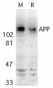
Left: Western blot analysis of APP in mouse (M) and rat (R) brain tissue lysates with APP antibody at 2 µg/ml.

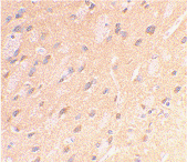
Below: Immunohistochemical staining of rat brain using APP antibody at 2 µg/ml.

Other Product Images

Antibody product image

Source :APP antibody was raised against a peptide corresponding to amino acids 737 to 751 of human amyloid A4 protein precursor (APP) or 85 to 99 of the C99 fragment generated by b-secretase cleavage. The peptide sequences are identical to those of monkey, mouse, rat, chicken, and a variety of other species.
Purification : Affinity chromatography purified via peptide column
Clonality and Clone : This is a polyclonal antibody.
Host : APP antibody was raised in rabbit.
Please use anti-rabbit secondary antibodies.
Immunogen : Human APP (C-Terminus) Peptide (Cat. No. 2136P)
Application : APP antibody can be used for detection of APP and the C99 fragment by Western blot at 1 – 2 µg/ml.
Tested Application(s) : E, WB, IHC
Buffer : Antibody is supplied in PBS containing 0.02% sodium azide.
Blocking Peptide :Cat. No. 2136P - APP Peptide
Long-Term Storage : APP antibody can be stored at 4ºC, stable for one year. As with all antibodies care should be taken to avoid repeated freeze thaw cycles. Antibodies should not be exposed to prolonged high temperatures.
Positive Control :
1. Cat. No. 1403 - Mouse Brain Tissue Lysate
Species Reactivity : H, M, R
GI Number : 28721
Accession Number : CAA30050
Short Description : (CT) Amyloid protein precursor
References
1. Ponte P, Gonzalez-DeWhitt P, Schilling J, Miller J, Hsu D, Greenberg B, Davis K, Wallace W, Lieberburg I, Fuller F. A new A4 amyloid mRNA contains a domain homologous to serine proteinase inhibitors. Nature 1988;331:525-7
2. Selkoe D.J. Cell biology of the amyloid beta-protein precursor and the mechanism of Alzheimer's disease.Annu Rev Cell Biol 1994;10:373-403 (RD1299)

APP Antibody

APP Antibody

Catalog# :2133

Accumulation of the amyloid-beta peptide (alphabeta) in the cerebral cortex is a critical event in the pathogenesis of Alzheimer's disease. The b-amyloid protein precursor (APP) is cleaved by beta-secretase, producing a soluble derivative of the protein and a membrane anchored 99-amino acid carboxy-terminal fragment (C99). The C99 fragment serves as substrate for g-secretase to generate the 4 kDa amyloid-b peptide (alphabeta), which is deposited in the brains of all suffers of Alzheimer's disease.

Additional Names : APP (AbNT), APP(AbNT)
Description
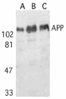
Left: Western blot analysis of APP in human (A), mouse (B), and rat (C) brain tissue lysates with APP antibody at 1 µg/ml.

Below: Immunohistochemistry of APP in human brain (Alzheimer’s disease) tissue with APP antibody at 10 µg/ml.

Other Product Images

Antibody product image

Source :APP antibody was raised against a peptide corresponding to amino acids of human amyloid A4 protein precursor (APP) corresponding to the amino terminus of the 4K Ab peptide generated by b- and g-secretases The peptide sequences are identical to those of rabbit, pig, bovine, guinea pig, and chicken.
Purification : Affinity chromatography purified via peptide column
Clonality and Clone : This is a polyclonal antibody.
Host : APP antibody was raised in rabbit.
Please use anti-rabbit secondary antibodies
Immunogen : Human APP / AbNT / Peptide (Cat. No. 2133P)
Application : APP antibody can be used for detection of APP and Ab peptide by Western blot at 0.5 - 1 µg/ml.
Tested Application(s) : E, WB, IHC
Buffer : Antibody is supplied in PBS containing 0.02% sodium azide.
Blocking Peptide :Cat. No. 2133P - APP (AbNT) Peptide
Long-Term Storage : APP antibody can be stored at 4ºC, stable for one year. As with all antibodies care should be taken to avoid repeated freeze thaw cycles. Antibodies should not be exposed to prolonged high temperatures.
Positive Control :
1. Cat. No. 1403 - Mouse Brain Tissue Lysate
Species Reactivity : H, M, R
GI Number : 28721
Accession Number : CAA30050
Short Description : (AbNT) Amyloid protein precursor
References
1. Ponte P, Gonzalez-DeWhitt P, Schilling J, Miller J, Hsu D, Greenberg B, Davis K, Wallace W, Lieberburg I, Fuller F. A new A4 amyloid mRNA contains a domain homologous to serine proteinase inhibitors. Nature 1988;331:525-7.
2. Selkoe D.J. Cell biology of the amyloid beta-protein precursor and the mechanism of Alzheimer's disease. Annu Rev Cell Biol 1994;10:373-403 (RD1000)

Protein Synthesis RNA


Protein biosynthesis is the process in which cells build proteins. The term is sometimes used to refer only to protein translation but more often it refers to a multi-step process, beginning with amino acid synthesis and transcription which are then used for translation. Protein biosynthesis, although very similar, differs between prokaryotes and eukaryotes.

Amino Acid Synthesis

Amino acids are the monomers which are polymerized to produce proteins. Amino acid synthesis is the set of biochemical processes which build the amino acids from carbon sources like glucose. Not all amino acids may be synthesised by every organism, for example adult humans have to obtain 8 of the 20 amino acids from their diet.

Transcription

Transcription is the process by which an mRNA template, encoding the sequence of the protein in the form of a trinucleotide code, is transcribed from the genome to provide a template for translation. Transcription copies the template from one strand of the DNA double helix, called the template strand.

Ribonucleic Acid or RNA

Ribonucleic acid or RNA is a nucleic acid made from a long chain of nucleotide units. Each nucleotide consists of a nitrogenous base, a ribose sugar, and a phosphate. RNA is very similar to DNA, but differs in a few important structural details: in the cell RNA is usually single stranded, while DNA is usually double stranded. RNA nucleotides contain ribose while DNA contains deoxyribose, and RNA has the nucleotide uracil rather than thymine which is present in DNA.

Tuesday, April 5, 2011

Polyclonal Antibody


Bio-Synthesis, with 20 years of experience in developing polyclonal antibodies, is a leading global provider of custom antibody production services. Our facility generates high-affinity custom antibodies with industry-leading titer guarantees and comprehensive antibody production packages that simplify the ordering process and present cost-effective methods to isolate epitope-specific antibodies.

Polyclonal Antibodies

Polyclonal antibodies (or antisera) are antibodies that are derived from different B cell lines. They are a mixture of immunoglobulin molecules secreted against a specific antigen, each recognising a different epitope.

Antigen Preparation

The size, extent of aggregation and relative nativity of protein antigens can all dramatically affect the quality and quantity of antibody produced. Small polypeptides (<10 ku) and non-protein antigens generally need to be conjugated or cross-linked to larger, immunogenic, carrier proteins to increase immunogenicity and provide T cell epitopes. Generally, the larger the immunogenic protein the better.

Antigen Quantity

Selection of antigen quantity for immunization varies with the properties of the antigen and the adjuvant selected. In general, microgram to milligram quantities of protein in adjuvant are necessary to elicit high titer antibodies. Antigen dosage is generally species, rather than body weight, associated.

Monday, April 4, 2011

APP Antibody

APP Antibody

Catalog# :2136

Accumulation of the amyloid-beta peptide (alphabeta) in the cerebral cortex is a critical event in the pathogenesis of Alzheimer's disease. The b-amyloid protein precursor (APP) is cleaved by beta-secretase, producing a soluble derivative of the protein and a membrane anchored 99-amino acid carboxy-terminal fragment (C99). The C99 fragment serves as substrate for g-secretase to generate the 4 kDa amyloid-beta peptide (alphabeta), which is deposited in the brains of all suffers of Alzheimer’s disease.

Additional Names : APP (CT), APP

Description
Left: Western blot analysis of APP in mouse (M) and rat (R) brain tissue lysates with APP antibody at 2 µg/ml.

Below: Immunohistochemical staining of rat brain using APP antibody at 2 µg/ml.

Other Product Images

Source :APP antibody was raised against a peptide corresponding to amino acids 737 to 751 of human amyloid A4 protein precursor (APP) or 85 to 99 of the C99 fragment generated by b-secretase cleavage. The peptide sequences are identical to those of monkey, mouse, rat, chicken, and a variety of other species .
Purification : Affinity chromatography purified via peptide column
Clonality and Clone : This is a polyclonal antibody.
Host : APP antibody was raised in rabbit.
Please use anti-rabbit secondary antibodies.
Immunogen : Human APP (C-Terminus) Peptide (Cat. No. 2136P)
Application : APP antibody can be used for detection of APP and the C99 fragment by Western blot at 1 – 2 µg/ml.
Tested Application(s) : E, WB, IHC
Buffer : Antibody is supplied in PBS containing 0.02% sodium azide.
Blocking Peptide :Cat. No. 2136P - APP Peptide
Long-Term Storage : APP antibody can be stored at 4ºC, stable for one year. As with all antibodies care should be taken to avoid repeated freeze thaw cycles. Antibodies should not be exposed to prolonged high temperatures.
Positive Control :
1. Cat. No. 1403 - Mouse Brain Tissue Lysate
Species Reactivity : H, M, R
GI Number : 28721
Accession Number : CAA30050
Short Description : (CT) Amyloid protein precursor
References
1. Ponte P, Gonzalez-DeWhitt P, Schilling J, Miller J, Hsu D, Greenberg B, Davis K, Wallace W, Lieberburg I, Fuller F. A new A4 amyloid mRNA contains a domain homologous to serine proteinase inhibitors. Nature 1988;331:525-7
2. Selkoe D.J. Cell biology of the amyloid beta-protein precursor and the mechanism of Alzheimer's disease.Annu Rev Cell Biol 1994;10:373-403 (RD1299)

APP Antibody

APP Antibody

Catalog# :2133

Accumulation of the amyloid-beta peptide (alphabeta) in the cerebral cortex is a critical event in the pathogenesis of Alzheimer's disease. The b-amyloid protein precursor (APP) is cleaved by beta-secretase, producing a soluble derivative of the protein and a membrane anchored 99-amino acid carboxy-terminal fragment (C99). The C99 fragment serves as substrate for g-secretase to generate the 4 kDa amyloid-b peptide (alphabeta), which is deposited in the brains of all suffers of Alzheimer's disease.

Additional Names : APP (AbNT), APP(AbNT)
Description
Left: Western blot analysis of APP in human (A), mouse (B), and rat (C) brain tissue lysates with APP antibody at 1 µg/ml.

Below: Immunohistochemistry of APP in human brain (Alzheimer’s disease) tissue with APP antibody at 10 µg/ml.

Other Product Images

Antibody product image
Source :APP antibody was raised against a peptide corresponding to amino acids of human amyloid A4 protein precursor (APP) corresponding to the amino terminus of the 4K Ab peptide generated by b- and g-secretases The peptide sequences are identical to those of rabbit, pig, bovine, guinea pig, and chicken.
Purification : Affinity chromatography purified via peptide column
Clonality and Clone : This is a polyclonal antibody.
Host : APP antibody was raised in rabbit.
Please use anti-rabbit secondary antibodies
Immunogen : Human APP / AbNT / Peptide (Cat. No. 2133P)
Application : APP antibody can be used for detection of APP and Ab peptide by Western blot at 0.5 - 1 µg/ml.
Tested Application(s) : E, WB, IHC
Buffer : Antibody is supplied in PBS containing 0.02% sodium azide.
Blocking Peptide :Cat. No. 2133P - APP (AbNT) Peptide
Long-Term Storage : APP antibody can be stored at 4ºC, stable for one year. As with all antibodies care should be taken to avoid repeated freeze thaw cycles. Antibodies should not be exposed to prolonged high temperatures.
Positive Control :
1. Cat. No. 1403 - Mouse Brain Tissue Lysate
Species Reactivity : H, M, R
GI Number : 28721
Accession Number : CAA30050
Short Description : (AbNT) Amyloid protein precursor
References
1. Ponte P, Gonzalez-DeWhitt P, Schilling J, Miller J, Hsu D, Greenberg B, Davis K, Wallace W, Lieberburg I, Fuller F. A new A4 amyloid mRNA contains a domain homologous to serine proteinase inhibitors. Nature 1988;331:525-7.
2. Selkoe D.J. Cell biology of the amyloid beta-protein precursor and the mechanism of Alzheimer's disease. Annu Rev Cell Biol 1994;10:373-403 (RD1000)

APP Antibody

APP Antibody

Catalog# :5143

Accumulation of the amyloid-beta peptide (Abeta) in the cerebral cortex is a critical event in the pathogenesis of Alzheimer’s disease. The beta−amyloid protein precursor (APP) is cleaved by one of two beta−secretases (BACE and BACE2), producing a soluble derivative of the protein and a membrane anchored 99-amino acid carboxy-terminal fragment (C99). The C99 fragment serves as substrate for gamma−secretase to generate the 4 kDa amyloid-beta peptide (Abeta), which is deposited in the Alzheimer’s disease patients’ brains. Recently, Death Receptor 6 (DR6) was found to interact with an amino-terminal fragment of the Beta-amyloid protein (N-APP) in neurons, activating a caspase 6-dependent apoptotic event leading to axonal degeneration and pruning during development, suggesting that these two proteins are involved in neural development and may possibly play a role in Alzheimer’s disease.

Additional Names : APP (NT2), Amyloid Precursor Protein, amyloid-beta precursor protein
Description
Left: Western blot analysis of APP in rat brain tissue lysate with APP antibody at (A) 1 and (B) 2 µg/ml.

Below:Immunohistochemistry of APP in mouse brain tissue with APP antibody at 2.5 μg/ml.

Other Product Images
Source :APP antibody was raised against an 18 amino acid peptide near the amino terminus of human APP.
Purification : Affinity chromatography purified via peptide column
Clonality and Clone : This is a polyclonal antibody.
Host : APP antibody was raised in rabbit.
Please use anti-rabbit secondary antibodies.
Application : APP antibody can be used for detection of APP by Western blot at 1 – 2 µg/ml.
Tested Application(s) : E, WB, IHC
Buffer : Antibody is supplied in PBS containing 0.02% sodium azide.
Blocking Peptide :Cat.No. 5143P - APP Peptide
Long-Term Storage : APP antibody can be stored at 4ºC, stable for one year. As with all antibodies care should be taken to avoid repeated freeze thaw cycles. Antibodies should not be exposed to prolonged high temperatures.
Positive Control :
1. Cat. No. 1463 - Rat Brain Tissue Lysate
Species Reactivity : H, M, R
GI Number : 112927
Accession Number : P05067
Short Description : (NT2) Amyloid precursor protein
References
1. Ponte P, Gonzalez-DeWhitt P, Schilling J, et al. A new A4 amyloid mRNA contains a domain homologous to serine proteinase inhibitors. Nature 1988; 331:525-77.
2. Selkoe DJ. Cell biology of the amyloid beta-protein precursor and the mechanism of Alzheimer's disease. Annu Rev Cell Biol 1994; 10:373-403.
3. Nikolaev A, McLaughlin T, O’Leary DD, et al. APP binds DR6 to trigger axon pruning and neuron death via distinct caspases. Nature 2009; 457:981-9.

APH1 Antibody

APH1 Antibody

Catalog# :4003

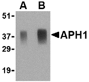
APH1 was initially identified as a component of the Notch pathway in C. elegans. Along with nicastrin, PEN2, and presenilin-1 APH1 is an essential component of the gammasecretase complex which cleave the amyloid precursor protein (APP) at what are known as the gammaand gammasites and can lead to the accumulation of the Amyloid beta peptide Abetacleavage product that is associated with Alzheimer’s disease. APH1 exists in at least three distinct isoforms with APH1a as the principal isoform present in the gammasecretase complex. Mice deficient in this isoform, but not the other two, were lethal at E10.5, with impaired vascular and neural development observed. Despite its predicted molecular weight, APH1 protein often migrates at aberrant locations in SDS-PAGE.

Additional Names : APH1 (IN), Anterior pharynx defective 1, presenilin stabilization factor
Description
Left: Western blot analysis of APH1 in human brain tissue lysate with APH1 antibody at (A) 0.5 and (B) 1 µg/ml.

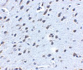
Below:Immunohistochemistry of APH1 in human brain tissue with APH1 antibody at 5 µg/ml.

Other Product Images
Source :APH1 antibody was raised against a 18 amino acid peptide from near the center of human APH1.
Purification : Affinity chromatography purified via peptide column
Clonality and Clone : This is a polyclonal antibody.
Host : APH1 antibody was raised in rabbit.
Please use anti-rabbit secondary antibodies.
Application : APH1 antibody can be used for detection of APH1 by Western blot at 0.5 – 1 µg/ml.
Tested Application(s) : E, WB, IHC
Buffer : Antibody is supplied in PBS containing 0.02% sodium azide.
Blocking Peptide :Cat.No. 4003P - APH1 Peptide
Long-Term Storage : APH1 antibody can be stored at 4ºC, stable for one year. As with all antibodies care should be taken to avoid repeated freeze thaw cycles. Antibodies should not be exposed to prolonged high temperatures.
Positive Control :
1. Cat. No. 1303 - Human Brain Tissue Lysate
Species Reactivity : H, M, R
GI Number : 14250557
Accession Number : AAH08732
Short Description : (IN) a component of the presenilin complex
References
1. Goutte C, Tsunozaki M, Hale VA, et al. APH-1 is a multipass membrane protein essential for the Notch signaling pathway in Caenorhabditis elegans embryos. Proc. Natl. Acad. Sci. USA 2002; 99:775-9.
2. Periz G and Fortini ME. Functional reconstitution of -secretase through coordinated expression of presenilin, nicastrin, aph-1, and pen-2. J. Neurosci. Res. 2004; 77:309-22.
3. Selkoe DJ. The cell biology of-amyloid precursor protein and presenilin in Alzheimer’s disease. Trends Cell Biol. 1998; 8:447-53.
4. Ma G, Li T, Price DL, et al. APH-1a is the principal mammalian aph-1 isoform present in g-secretase complexes during embryonic development. Neuro. Dis. 2005; 25:192-8.