TCCR Antibody
Catalog# : 2483
Upon antigen challenge, T-helper cells differentiate into two functional distinct subsets, Th1 and Th2. Th1 cells produce IL-2, IFN-gamma and lymphotoxin-beta that augment cell mediated immune response while Th2 cells secrete IL-4, IL-5, and IL-10 that enhance humoral immunity. The function of T-helper cells is regulated by cytokines. A novel cytokine receptor was recently identified and cloned (1,2). It is a new member in the type I cytokine receptor family and designated TCCR for T-cell cytokine receptor and WSX-1 (1,2). TCCR deficient mice had impaired Th1 responses to protein antigen challenge, including decreased levels of IFN-gamma and Th1-dependent antibody IgG2a . TCCR is predominately expressed in thymus, spleen, lymph notes and peripheral blood leukocytes.
Additional Names : TCCR (CT), Type I T-cell cytokine receptor, IL-27 receptor alpha, IL-27Ra
 Description
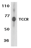
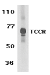
Left: Western blot analysis of TCCR expression in human spleen tissue lysate with TCCR antibody at 1 µg /ml.
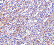
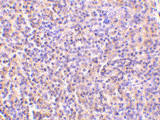
Below: Immunohistochemistry of TCCR in human spleen tissue with TCCR antibody at 5 µg/ml.
Other Product Images
 Source : TCCR antibody was raised against a synthetic peptide corresponding to amino acids near the carboxy terminus of human TCCR precursor This sequence is identical to that of mouse TCCR.
Purification : Affinity chromatography purified via peptide column
Clonality and Clone : This is a polyclonal antibody.
Host : TCCR antibody was raised in rabbit. Please use anti-rabbit secondary antibodies.
Immunogen : Human TCCR (C-Terminus) Peptide (Cat. No. 2483P)
Application : TCCR antibody can be used for detection of TCCR by Western blot at 0.5 to 1 µg/ml.An approximately 70 kDa band can be detected. It is human, mouse, and rat reactive.
Tested Application(s) : E, WB, IHC
Buffer : Antibody is supplied in PBS containing 0.02% sodium azide.
Blocking Peptide : Cat. No. 2483P - TCCR Peptide
Long-Term Storage : TCCR antibody can be stored at 4ºC, stable for one year. As with all antibodies care should be taken to avoid repeated freeze thaw cycles. Antibodies should not be exposed to prolonged high temperatures.
Positive Control
1. Cat. No. 1306 - Human Spleen Tissue Lysate
Species Reactivity :H
GI Number : 4759328
Accession Number : NP_004834
Short Description : (CT) A new T cell cytokine receptor
References
1. Chen Q, Ghilardi N, Wang H, Baker T, Xie MH, Gurney A, Grewal IS and de Sauvage FJ. Development of Th1-type immune responses requires the type I cytokine receptor TCCR Nature 2000;407(6806):916-920
2. Sprecher,C.A., Grant,F.J., Baumgartner,J.W., Presnell,S.R., Schrader,S.K., Yamagiwa,T., Whitmore,T.E., O'Hara,P.J. and Foster,D.F. Cloning and characterization of a novel class I cytokine receptor Biochem. Biophys. Res. Commun. 1998;246(1):82-90
TCCR Antibody
Catalog# : 2481
Upon antigen challenge, T-helper cells differentiate into two functional distinct subsets, Th1 and Th2. Th1 cells produce IL-2, IFN-gamma and lymphotoxin-beta that augment cell mediated immune response while Th2 cells secrete IL-4, IL-5, and IL-10 that enhance humoral immunity. The function of T-helper cells is regulated by cytokines. A novel cytokine receptor was recently identified and cloned (1,2). It is a new member in the type I cytokine receptor family and designated TCCR for T-cell cytokine receptor and WSX-1 (1,2). TCCR deficient mice had impaired Th1 responses to protein antigen challenge, including decreased levels of IFN-gamma and Th1-dependent antibody IgG2a . TCCR is predominately expressed in thymus, spleen, lymph notes and peripheral blood leukocytes.
Additional Names : TCCR (NT), Type I T-cell cytokine receptor, IL-27 receptor alpha, IL-27Ra
 Description
Left: Western blot analysis of TCCR expression in human spleen tissue lysates with TCCR antibody at 1 µg /ml.
Below: Immunohistochemistry of TCCR in human spleen tissue with TCCR antibody at 10 µg/ml.
Other Product Images
 Source : TCCR antibody was raised against a synthetic peptide corresponding to amino acids near the amino terminus of human TCCR precursor.
Purification : Affinity chromatography purified via peptide column
Clonality and Clone : This is a polyclonal antibody.
Host : TCCR antibody was raised in rabbit. Please use anti-rabbit secondary antibodies.
Immunogen : Human TCCR (N-Terminus) Peptide (Cat. No. 2481P)
Application : TCCR antibody can be used for detection of TCCR by Western blot at 0.5 to 1 µg/ml.An approximately 70 kDa band can be detected.
Tested Application(s) : E, WB, IHC
Buffer : Antibody is supplied in PBS containing 0.02% sodium azide.
Blocking Peptide : Cat. No. 2481P - TCCR Peptide
Long-Term Storage : TCCR antibody can be stored at 4ºC, stable for one year. As with all antibodies care should be taken to avoid repeated freeze thaw cycles. Antibodies should not be exposed to prolonged high temperatures.
Positive Control
1. Cat. No. 1306 - Human Spleen Tissue Lysate
Species Reactivity :H
GI Number : 4759328
Accession Number : NP_004834
Short Description : (NT) A new T cell cytokine receptor
References
1. Chen Q, Ghilardi N, Wang H, Baker T, Xie MH, Gurney A, Grewal IS and de Sauvage FJ. Development of Th1-type immune responses requires the type I cytokine receptor TCCR Nature 2000;407(6806):916-920
2. Sprecher,C.A., Grant,F.J., Baumgartner,J.W., Presnell,S.R., Schrader,S.K., Yamagiwa,T., Whitmore,T.E., O'Hara,P.J. and Foster,D.F. Cloning and characterization of a novel class I cytokine receptor Biochem. Biophys. Res. Commun. 1998;246(1):82-90
TACI Antibody
Catalog# : 2395
Members in the TNF superfamily regulate immune responses and induce apoptosis. Two novel members in the TNF family were recently identified and designated BAFF/BLyS/TALL-1/THANK/zTNF4 (1-4) and April/TALL-2, respectively, (3,5). BAFF was characterized as a B cell activator since it induced B cell proliferation and immunoglobulin secretion (1,2). April regulates immunological and non-immunological cell growth. Three receptors, BCMA, TACI, and BAFF-R, for BAFF and April were recently identified (6,7). TACI, like BCMA, binds BAFF and April. TACI and its ligands regulate humoral immune responses, activate NF-kappaB and c-jun N-terminal kinase, and are involved in B cell associated autoimmune diseases.
Additional Names : TACI
 Description
Left: Western blot analysis of TACI in K562 (A) and U937 (B) cell lysates with TACI antibody at 5 µg/ml.
Below: Immunocytochemistry of TACI in K562 cells with TACI antibody at 2 µg/ml.
Other Product Images
 Source : TACI antibody was raised against a synthetic peptide mapping at the amino terminus of human TACI .
Purification : Affinity chromatography purified via peptide column
Clonality and Clone : This is a polyclonal antibody.
Host : TACI antibody was raised in rabbit. Please use anti-rabbit secondary antibodies.
Immunogen : Human TACI Peptide (Cat. No. 2395P)
Application : TACI polyclonal antibody can be used for detection of TACI by Western blot at 2 to 5 µg/ml.
Tested Application(s) : E, WB, ICC
Buffer : Antibody is supplied in PBS containing 0.02% sodium azide.
Blocking Peptide : Cat. No. 2395P - TACI Peptide
Long-Term Storage : TACI antibody can be stored at 4ºC, stable for one year. As with all antibodies care should be taken to avoid repeated freeze thaw cycles. Antibodies should not be exposed to prolonged high temperatures.
Positive Control
1. Cat. No. 1204 - K562 Cell Lysate
2. Cat. No. 1215 - U937 Whole Cell Lysate
Species Reactivity :H
GI Number : 6912694
Accession Number : NP_036584
Short Description : Receptor for BAFF and APRIL
References
1. Moore PA, Belvedere O, Orr A, et al. BLyS: member of the tumor necrosis factor family and B lymphocyte stimulator. Science 1999;285:260-3
2. Schneider P, MacKay F, Steiner V, et al. BAFF, a novel ligand of the tumor necrosis factor family, stimulates B cell growth. J Exp Med 1999;189:1747-56
3. Shu HB, Hu WH, Johnson H. TALL-1 is a novel member of the TNF family that is down-regulated by mitogens. J Leukoc Biol 1999;65:680-3
4. Mukhopadhyay A, Ni J, Zhai Y, Yu GL, Aggarwal BB. Identification and characterization of a novel cytokine, THANK, a TNF homologue that activates apoptosis, nuclear factor-kappaB, and c-Jun NH2-terminal kinase. J Biol Chem 1999 ;274:15978-81
Acridine is an effective intercalating agent as well as being a lipophilic carrier molecule. Acridine labeled oligonucleotides are sometimes used in antisense research. Oligos containing acridine cannot be HPLC purified, nor can they be analyzed by HPLC, as they bind irreversibly to the column. Acridine is available as both a 3' and a 5' modifier. Labeling is performed during synthesis and is not a benchtop reaction.
Purification
Desalt or cartridge (RP1) purification is acceptable for most common phosphoramidite modifiers. However, additional purification is strongly recommended for modifiers that require NHS-ester chemistry to conjugate a dye through an amine linker such as Molecular Probes dyes.
Minimum Guarantee Purified Yield
Due to the chemistry of many modifications, yield will be approximately 40 - 60 % less than a standard oligo. Please see our yield chart for details.
Quality Controls
Every oligo synthesized is strictly controlled for quality by using either MALDI-TOF mass spectrometry or polyacrylamide gel electrophoresis (PAGE) analysis. Final yields are determined using UV absorbance at OD260 In addition, we perform QC methods tailored to specific modifications, such as OD ratio measurement where appropriate.
For additional information, please send your request to biosyn@biosyn.com or call us at 1-800-227-0627.
TACE Antibody
Catalog# : 1131
Tumor-necrosis factor-a is a proinflammatory cytokine and contributes to a variety of inflammatory disease responses and programmed cell death. TNF-alpha is synthesized as a 26K type II membrane-bound precursor that is cleaved by a convertase to generate secreted 17K mature TNF-alpha. TNF-alpha converting enzyme (TACE) protein was recently purified and the human and mouse TACE cDNAs were cloned by several groups separately. TACE is a membrane-bound metalloprotease-disintegrin in the family of mammalian ADAM (for a disintegrin and metalloprotease). TACE also processes other cell surface proteins, including TNF receptor, TGFa, the L-selectin adhesion molecule, and alpha-cleavage of amyloid protein precursor (APP) (4,5). TACE mRNA is expressed in a variety of human and murine tissues. TACE was selected as one of the few targets in cytokine activation by the Eighth International Conference of the Inflammation Research Association.
Additional Names : TACE (CT), adam17
 Description
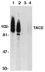
Left: Western blot analysis of TACE in HeLa and Jurkat whole cell lysate in the absence or presence of blocking peptide (Cat. No. 1131P) with TACE antibody at 1:500 dilution.
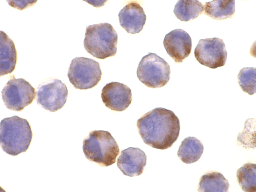
Below: Immunocytochemistry of TACE in HeLa cells with TACE antibody at 10 µg/ml.
Other Product Images
 Source : TACE antibody was raised against a peptide corresponding to amino acids near the carboxy terminus of human TACE This sequence differs from those of mouse and rat TACE by one amino acid.
Purification : Affinity chromatography purified via peptide column
Clonality and Clone : This is a polyclonal antibody.
Host : TACE antibody was raised in rabbit. Please use anti-rabbit secondary antibodies.
Immunogen : Human TACE (C-Terminus) Peptide (Cat. No. 1131P)
Application : TACE antibody can be used for detection of TACE by Western blot at 1:500 to 1:2000 dilution. 80 to 130 kDa bands can be detected, which may represent mature protein, precursor, and glycosylated TACE.
Tested Application(s) : E, WB, ICC
Buffer : Antibody is supplied in PBS containing 0.02% sodium azide.
Blocking Peptide : Cat. No. 1131P - TACE Peptide
Long-Term Storage : TACE antibody can be stored at 4ºC, stable for one year. As with all antibodies care should be taken to avoid repeated freeze thaw cycles. Antibodies should not be exposed to prolonged high temperatures.
Positive Control
1. Cat. No. 1201 - HeLa Cell Lysate
2. Cat. No. 1205 - Jurkat Whole Cell Lysate
Species Reactivity :H, M, R
GI Number : 73747889
Accession Number : NP_003174
Short Description : (CT) TNFalpha converting enzyme
References
1. Black RA, Rauch CT , Kozlosky CJ, et al. A metalloproteinase disintegrin that releases tumour-necrosis factor-alpha from cells. Nature 1997;385:729-733
2. Moss ML, Jin SL, Milla ME, et al. Cloning of a disintegrin metalloproteinase that processes precursor tumour-necrosis factor-alpha. Nature 1997;385:733-736
3. Mizui Y, Yamazaki K, Sagane K, Tanaka I. cDNA cloning of mouse tumor necrosis factor-alpha converting enzyme (TACE) and partial analysis of its promoter. Gene 1999;233:67-74
4. Peschon JJ, Slack JL, Reddy P, et al. An essential role for ectodomain shedding in mammalian development. Science 1998;282:1281-4
Survivin Antibody
Catalog# : 2235
Apoptosis, or programmed cell death, is related to many diseases, such as cancer. Apoptosis is triggered by a variety of stimuli including members in the TNF family and prevented by the inhibitor of apoptosis (IAP) proteins. IAP proteins form a conserved gene family that binds to and inhibits cell death proteases. A novel IAP protein was recently identified and designated survivin, apoptosis inhibitor 4 (API4), and TIAP (1-3). Survivin/TIAP interacted with the processed form of caspase-3 and inhibited its proteolytic activity. Survivin/TIAP is predominantly expressed in tissues of embryos, transformed cell lines, and many human cancers and lymphomas (1,3).
Additional Names : Survivin (CT), TIAP
 Description
Left: Western blot analysis of survivin in MOLT4 cell lysate in the absence (A) or presence (B) of blocking peptide with survivin antibody at 1 µg/ml.
Below: Immunocytochemistry staining of MOLT4 cells using survivin at 10 µg/ml.
Other Product Images
 Source : Survivin antibody was raised against a peptide corresponding to amino acids near the carboxy-terminus of human survivin.
Purification : Affinity chromatography purified via peptide column
Clonality and Clone : This is a polyclonal antibody.
Host : Survivin antibody was raised in rabbit. Please use anti-rabbit secondary antibodies.
Immunogen : Human Survivin (C-Terminus) Peptide (Cat. No. 2235P)
Application : Survivin antibody can be used for detection of survivin by Western blot at 0.5 to 1 µg/ml.An approximate 17 kDa band should be detected.
Tested Application(s) : E, WB, ICC
Buffer : Antibody is supplied in PBS containing 0.02% sodium azide.
Blocking Peptide : Cat. No. 2235P - Survivin Peptide
Long-Term Storage : Survivin antibody can be stored at 4ºC, stable for one year. As with all antibodies care should be taken to avoid repeated freeze thaw cycles. Antibodies should not be exposed to prolonged high temperatures.
Positive Control
1. Cat. No. 1205 - MOLT4 Cell Lysate
Species Reactivity :H
GI Number : 59859878
Accession Number : NP_001159
Short Description : (CT) A new inhibitor of apoptosis
References
1. Ambrosini G, Adida C, Altieri DC. A novel anti-apoptosis gene, survivin, expressed in cancer and lymphoma. Nat Med 1997;3:917-21
2. Li F, Ambrosini G, Chu EY, Plescia J, Tognin S, Marchisio PC, Altieri DC . Control of apoptosis and mitotic spindle checkpoint by survivin. Nature 1998;396:580-4
3. Kobayashi K, Hatano M, Otaki M, Ogasawara T, Tokuhisa T. Expression of a murine homologue of the inhibitor of apoptosis protein is related to cell proliferation. Proc Natl Acad Sci USA 1999;96:1457-62 (WD0600)
Survivin Antibody
Catalog# : 2233
Apoptosis, or programmed cell death, is related to many diseases, such as cancer. Apoptosis is triggered by a variety of stimuli including members in the TNF family and prevented by the inhibitor of apoptosis (IAP) proteins. IAP proteins form a conserved gene family that binds to and inhibits cell death proteases. A novel IAP protein was recently identified and designated survivin, apoptosis inhibitor 4 (API4), and TIAP (1-3). Survivin/TIAP interacted with the processed form of caspase-3 and inhibited its proteolytic activity. Survivin/TIAP is predominantly expressed in tissues of embryos, transformed cell lines, and many human cancers and lymphomas .
Additional Names : Survivin (NT), TIAP
 Description
Left: Western blot analysis of survivin in MOLT4 cell lysate in the absence (A) or presence (B) of blocking peptide with survivin antibody at 1 µg/ml.
Below: Immunocytochemistry of Survivin in Jurkat cells with Survivin antibody at 5 µg/ml.
Other Product Images
 Source : Survivin antibody was raised against a peptide corresponding to amino acids near the amino terminus of human survivin.
Purification : Affinity chromatography purified via peptide column
Clonality and Clone : This is a polyclonal antibody.
Host : Survivin antibody was raised in rabbit. Please use anti-rabbit secondary antibodies.
Immunogen : Human Survivin (N-Terminus) Peptide (Cat. No. 2233P)
Application : Survivin antibody can be used for detection of survivin by Western blot at 0.5 to 1 µg/ml.An approximate 17 kDa band should be detected.
Tested Application(s) : E, WB, ICC
Buffer : Antibody is supplied in PBS containing 0.02% sodium azide.
Blocking Peptide : Cat. No. 2233P - Survivin Peptide
Long-Term Storage : Survivin antibody can be stored at 4ºC, stable for one year. As with all antibodies care should be taken to avoid repeated freeze thaw cycles. Antibodies should not be exposed to prolonged high temperatures.
Positive Control
1. Cat. No. 1205 - MOLT4 Cell Lysate
2. Cat. No. 1205 - Jurkat Cell Lysate
Species Reactivity :H
GI Number : 59859878
Accession Number : NP_001159
Short Description : (NT) A new inhibitor of apoptosis
References
1. Ambrosini G, Adida C, Altieri DC. A novel anti-apoptosis gene, survivin, expressed in cancer and lymphoma. Nat Med 1997;3:917-21
2. Li F, Ambrosini G, Chu EY, Plescia J, Tognin S, Marchisio PC, Altieri DC . Control of apoptosis and mitotic spindle checkpoint by survivin. Nature 1998;396:580-4
3. Kobayashi K, Hatano M, Otaki M, Ogasawara T, Tokuhisa T. Expression of a murine homologue of the inhibitor of apoptosis protein is related to cell proliferation. Proc Natl Acad Sci USA 1999;96:1457-62 (WD0600)
sRANK Ligand Antibody
Catalog# : 3963
The receptor activator of NF-kappaB ligand (RANK-L) is a recently discovered member of the TNF-ligand family involved in the regulation of the T cell-dependent immune response, lymph node organogenesis and bone formation. RANK-L exists as both a normal, transmembrane form and a truncated, soluble form (sRANK-L), both of which can stimulate the receptor. Activation of T cells, such as by treatment with interleukin-7, induces RANK-L production and leads to an increase of osteoclast formation and bone loss. Finally, sRANK-L can activate the antiapoptotic kinase Akt through a signaling complex involving Src kinase and TRAF6, suggesting sRANK-L may also play a role in regulating apoptosis. This antibody will recognize both the soluble form and the uncleaved transmembrane form of RANK-L.
Additional Names : sRANK Ligand, sRANK-L, TNFSF11, TRANCE, OPGL
 Description
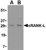
Left: Western blot analysis of sRANK-L in rat liver tissue lysate with sRANK-L antibody at (A) 0.25 and (B) 0.5 µg/ml.
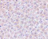
Below: Immunohistochemistry of sRANK-L in human liver tissue with sRANK-L antibody at 5 µg/ml.
Other Product Images
 Source : sRANK-L antibody was raised against a 14 amino acid peptide from near the center of human sRANK-L .
Purification : Affinity chromatography purified via peptide column
Clonality and Clone : This is a polyclonal antibody.
Host : sRANK Ligand antibody was raised in rabbit. Please use anti-rabbit secondary antibodies.
Application : sRANK-L antibody can be used for detection of sRANK-L by Western blot at 0.25 – 0.5 µg/ml.
Tested Application(s) : E, WB, IHC
Buffer : Antibody is supplied in PBS containing 0.02% sodium azide.
Blocking Peptide : Cat.No. 3963P - sRANK Ligand Peptide
Long-Term Storage : sRANK Ligand antibody can be stored at 4ºC, stable for one year. As with all antibodies care should be taken to avoid repeated freeze thaw cycles. Antibodies should not be exposed to prolonged high temperatures.
Positive Control
1. Cat. No. 1464 - Rat Liver Tissue Lysate
2. Cat. No. 1304 - Human Liver Tissue Lysate
Species Reactivity :H, M, R
GI Number : 4507595
Accession Number : NP_003692
Short Description : a member of the TNF-ligand family
References
1. Wong BR, Rho J, Arron J, et al. TRANCE is a novel ligand of the tumor necrosis factor receptor family that activates c-Jun N-terminal kinase in T cells. J. Biol. Chem. 1997; 272:25190-4.
2. Kong YY, Yoshida H, Sarosi I, et al. OPGL is a key regulator of osteoclastogenesis, lymphocyte development and lymph-node organogenesis. Nature 1999; 397:315-23.
3. Weitzmann MN, Cenci S, Rifas L, et al. Interleukin-7 stimulates osteoclast formation by up-regulating the T-cell production of soluble osteoclastogenic cytokines. Blood 2000; 96:1873-8.
4. Bharti AC, Takada Y, Shishodia S, et al. Evidence that receptor activator of the nuclear factor (NF)-kappaB ligand can suppress cell proliferation and induce apoptosis through activation of a NF-kappaB-independent and TRAF6-dependent mechanism. J. Biol. Chem. 2004; 279:6065-76.
SPG15 Antibody
Catalog# : 5023
Hereditary spastic paraplegias (HSPs) are genetically and phenotypically heterogeneous disorders. Spastic paraplegia with thinning of the corpus callosum (ARHSP-TCC) is a relatively frequent form of complicated hereditary spastic paraplegia in which mental retardation and muscle stiffness at onset are followed by slowly progressive paraparesis and cognitive deterioration. SPG15 is the second gene known to be responsible for ARHSP-TCC in the Italian population. Mutations in this gene are associated with autosomal recessive spastic paraplegia-15. SPG15 encodes a protein containing a FYVE zinc finger binding domain which is thought to target these proteins to membrane lipids through interaction with phospholipids in the membrane. SPG15 mRNA is widely distributed in human tissues, as well as in rat embryos, suggesting a possible role for this protein during embryonic development. SPG15 co-localizes partially with endoplasmic reticulum and endosome markers, suggesting a role in intracellular trafficking. Multiple isoforms of SPG15 are known to exist.
Additional Names : SPG15, Autosomal recessive spastic paraplegia 15, ZFYVE26

Description
Left: Western blot analysis of SPG15 in rat heart tissue lysate with SPG15 antibody at (A) 0.5 and (B) 1 µg/ml.
Source : SPG15 antibody was raised against a 16 amino acid peptide near the carboxy terminus of human SPG15.
Purification : Affinity chromatography purified via peptide column
Clonality and Clone : This is a polyclonal antibody.
Host : SPG15 antibody was raised in rabbit. Please use anti-rabbit secondary antibodies.
Application : SPG15 antibody can be used for detection of SPG15 by Western blot at 0.5 – 1 µg/ml.
Tested Application(s) : E, WB
Buffer : Antibody is supplied in PBS containing 0.02% sodium azide.
Blocking Peptide : Cat.No. 5023P - SPG15 Peptide
Long-Term Storage : SPG15 antibody can be stored at 4ºC, stable for one year. As with all antibodies care should be taken to avoid repeated freeze thaw cycles. Antibodies should not be exposed to prolonged high temperatures.
Positive Control
1. Cat. No. 1461 - Rat Heart Tissue Lysate
Species Reactivity :H, M, R
GI Number : 119601358
Accession Number : EAW80952
Short Description : Programmed cell death protein 5
References
1. Hughes CA, Byrne PC, Webb S, et al. SPG15, a new locus for autosomal recessive complicated HSP on chromosome 14q. Neurology 2001; 56:1230-3.
2. Denora PS, Muglia M, Casali C, et al. Spastic paraplegia with thinning of the corpus callosum and white matter abnormalities: further mutations and relative frequency in ZFYVE26/SPG15 in the Italian population. J. Neurol. Sci. 2009; 277:22-5.
3. Hanein S, Martin E, Boukhris A, et al. Identification of the SPG15 gene, encoding spastizin, as a frequent cause of complicated autosomal-recessive spastic paraplegia, including Kjellin syndrome. Am. J. Hum. Genet. 2008; 82:992-1002.
4. Boukhris A, Feki I, Denis E, et al. Spastic paraplegia 15: linkage and clinical description of three Tunisian families. Mov. Disord. 2008; 23:429-33.
A primary amino group can be used to attach a variety of modifiers (such as fluorescent dyes) to an oligonucleotide or used to attach an oligonucleotide to a solid surface, for example, a primary amine (NH2) at at the 5’-position is used to functionalize the corresponding terminus of the nucleotide for conjugation with e.g. an activated NHS ester or isothiocyanate fluorescent label. The 3'-NH2 modification allows the prevention of the 3' to 5' exonuclease degradation of oligonucleotides in biological fluids such as cell culture medium.
C6, C12 methylene (CH2) or longer and all of them are hydrophobic. Uni-LinkTM Amino Modifier provides a free primary amine attached to the 5'-end of an oligo via a six carbon aliphatic spacer arm. It is functionally interchangeable with
Amino Modifier C6 Amino-Modifier dA, Amino-Modifier dC, Amino-Modifier dG and both Amino-Modifier dT products can be added in place of a dA, dC, dG and dT residue, respectively, during oligonucleotide synthesis.
Purification
Desalt or cartridge (RP1) purification is acceptable for most common phosphoramidite modifiers. However, additional purification is strongly recommended for modifiers that require NHS-ester chemistry to conjugate a dye through an amine linker such as Molecular Probes dyes.
Minimum Guarantee Purified Yield
Due to the chemistry of many modifications, yield will be approximately 40 - 60 % less than a standard oligo. Please see our yield chart for details.
Quality Controls
Every oligo synthesized is strictly controlled for quality by using either MALDI-TOF mass spectrometry or polyacrylamide gel electrophoresis (PAGE) analysis. Final yields are determined using UV absorbance at OD260 In addition, we perform QC methods tailored to specific modifications, such as OD ratio measurement where appropriate.
For additional information, please send your request to biosyn@biosyn.com or call us at 1-800-227-0627.

 Description
 Source : TCCR antibody was raised against a synthetic peptide corresponding to amino acids near the carboxy terminus of human TCCR precursor This sequence is identical to that of mouse TCCR.