Showing posts with label Organic Laboratory Technique. Show all posts

Monday, May 9, 2011

SCRAPPER Antibody

SCRAPPER Antibody

Catalog# :4451

Members of the F-box protein family, such as Scrapper, are characterized by an approximately 40-amino acid F-box motif. SCF complexes, formed by SKP1, cullin, and F-box proteins, act as protein-ubiquitin ligases. Scrapper is selectively expressed in the brain, broadly expressed within the mouse CNS and is abundant at presynaptic membrane. Scrapper has orthologs in C. elegans, D. melanogaster, and mammals which suggests that it might function as an important membrane-localized E3 ligase in various species. Scrapper is a major presynaptic E3 ubiquitin ligase that acts through RIM1a via degradation and the ubiquitin-proteasome-system (UPS) pathway to critically regulate synaptic transmission. This identifies protein degradation as a mechanism for holding synaptic communication in check.

Additional Names : SCRAPPER (NT), F-box / Leucine rich repeat protein 20, FBXL20, FBl2, FBL20
Description
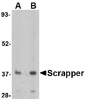
Left: Western blot analysis of SCRAPPER in A20 cell lysate with SCRAPPER antibody at (A) 0.5 and (B) 1 µg/ml.

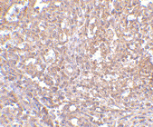
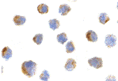
Below:Immunohistochemical staining of human brain tissue using Scrapper antibody at 2.5 µg/ml.


Source :SCRAPPER antibody was raised against a 18 amino acid peptide near the amino terminus of the human SCRAPPER.
Purification : Affinity chromatography purified via peptide column
Clonality and Clone : This is a polyclonal antibody.
Host : SCRAPPER antibody was raised in rabbit.
Please use anti-rabbit secondary antibodies.
Application : SCRAPPER antibody can be used for detection of SCRAPPER by Western blot at 0.5 – 1 µg/ml.
Tested Application(s) : E, WB, IHC
Buffer : Antibody is supplied in PBS containing 0.02% sodium azide.
Blocking Peptide :Cat.No. 4451P - SCRAPPER Peptide
Long-Term Storage : SCRAPPER antibody can be stored at 4ºC, stable for one year. As with all antibodies care should be taken to avoid repeated freeze thaw cycles. Antibodies should not be exposed to prolonged high temperatures.
Positive Control :
1. Cat. No. 1288 - A20 Cell Lysate
Species Reactivity : H, M, R
GI Number : 27734755
Accession Number : NP_116264
Short Description : (NT) F-box / Leucine rich repeat protein 20
References

1. Ho MS, Tsai PI, and Chien CT. F-box proteins: the key to protein degradation. J. Biomed. Sci. 2006; 13:181-91.
2. Dobie F and Craig AM. A fight for neurotransmission: SCRAPPER trashes RIM. Cell 2007; 130:775-7.
3. Yao I, Takagi H, Ageta H, et al. SCRAPPER-dependent ubiquitination of active zone protein RIM1 regulates synaptic vesicle release. Cell 2007; 130:943-57.
4. Ding M, Chao D, Wang G, et al. Spatial regulation of an E3 ubiquitin ligase directs selective synapse elimination. Science 2007; 317:947-51.

Sunday, May 8, 2011

SCRAPPER Antibody

SCRAPPER Antibody

Catalog# :4449

Members of the F-box protein family, such as Scrapper, are characterized by an approximately 40-amino acid F-box motif. SCF complexes, formed by SKP1, cullin, and F-box proteins, act as protein-ubiquitin ligases. Scrapper is selectively expressed in the brain, broadly expressed within the mouse CNS and is abundant at presynaptic membrane. Scrapper has orthologs in C. elegans, D. melanogaster, and mammals which suggests that it might function as an important membrane-localized E3 ligase in various species. Scrapper is a major presynaptic E3 ubiquitin ligase that acts through RIM1a via degradation and the ubiquitin-proteasome-system (UPS) pathway to critically regulate synaptic transmission. This identifies protein degradation as a mechanism for holding synaptic communication in check.

Additional Names : SCRAPPER (CT), F-box / Leucine rich repeat protein 20, FBXL20, FBl2, FBL20

Description
Left: Western blot analysis of SCRAPPER in A20 cell lysate with SCRAPPER antibody at (A) 0.5 and (B) 1 µg/ml.

Below: Immunohistochemical staining of human brain tissue using Scrapper antibody at 2.5 µg/ml.

Other Product Images

Source :SCRAPPER antibody was raised against a 18 amino acid peptide near the carboxy terminus of the human SCRAPPER.
Purification : Affinity chromatography purified via peptide column
Clonality and Clone : This is a polyclonal antibody.
Host : SCRAPPER antibody was raised in rabbit.
Please use anti-rabbit secondary antibodies.
Application : SCRAPPER antibody can be used for detection of SCRAPPER by Western blot at 0.5 – 1 µg/ml.
Tested Application(s) : E, WB, IHC
Buffer : Antibody is supplied in PBS containing 0.02% sodium azide.
Blocking Peptide :Cat.No. 4449P - SCRAPPER Peptide
Long-Term Storage : SCRAPPER antibody can be stored at 4ºC, stable for one year. As with all antibodies care should be taken to avoid repeated freeze thaw cycles. Antibodies should not be exposed to prolonged high temperatures.
Positive Control :
1. Cat. No. 1288 - A20 Cell Lysate
Species Reactivity : H, M, R
GI Number : 27734755
Accession Number : NP_116264
Short Description : (CT) F-box / Leucine rich repeat protein 20
References

1. Ho MS, Tsai PI, and Chien CT. F-box proteins: the key to protein degradation. J. Biomed. Sci. 2006; 13:181-91.
2. Dobie F and Craig AM. A fight for neurotransmission: SCRAPPER trashes RIM. Cell 2007; 130:775-7.
3. Yao I, Takagi H, Ageta H, et al. SCRAPPER-dependent ubiquitination of active zone protein RIM1 regulates synaptic vesicle release. Cell 2007; 130:943-57.
4. Ding M, Chao D, Wang G, et al. Spatial regulation of an E3 ubiquitin ligase directs selective synapse elimination. Science 2007; 317:947-51.
SCAMP4 Antibody

SCAMP4 Antibody

Catalog# :5289

Secretory carrier membrane proteins (SCAMPs) are integral membrane proteins seen in secretory and endocytic carriers that are thought to play roles in membrane trafficking. One member of this family, SCAMP4, lacks the putative calcium binding and leucine zipper structures as well as the protein-protein interacting NPF domains common to most SCAMP family members. In rats, SCAMP4 mRNA is repressed by progesterone in brain regions associated with female sexual behavior. At least three isoforms of SCAMP4 are known to exist. This SCAMP4 antibody is predicted to not cross-react with any other member of the secretory carrier membrane protein family.

Additional Names : SCAMP4, Secretory carrier membrane protein 4

Description
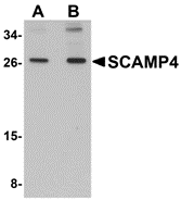
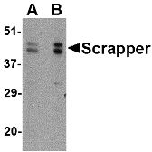
Left: Western blot analysis of SCAMP4 in 3T3 cell lysate with SCAMP4 antibody at (A) 1 and (B) 2 µg/ml.

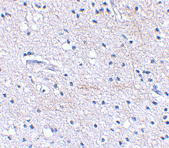
Below:Immunohistochemistry of SCAMP4 in human brain tissue with SCAMP4 antibody at 2.5 μg/ml.

Other Product Images

Source :SCAMP4 antibody was raised against a 15 amino acid peptide near the carboxy terminus of human SCAMP4.
Purification : Affinity chromatography purified via peptide column
Clonality and Clone : Polyclonal
Host : SCAMP4 antibody was raised in rabbit.
Please use anti-rabbit secondary antibodies.
Application : SCAMP4 antibody can be used for detection of SCAMP4 by Western blot at 1 - 2 µg/ml.
Tested Application(s) : E, WB, IHC
Buffer : Antibody is supplied in PBS containing 0.02% sodium azide.
Blocking Peptide :Cat.No. 5289P - SCAMP4 Peptide
Long-Term Storage : SCAMP4 antibody can be stored at 4ºC, stable for one year. As with all antibodies care should be taken to avoid repeated freeze thaw cycles. Antibodies should not be exposed to prolonged high temperatures.
Positive Control :
1. Cat. No. 1282 - 3T3 (NIH) Cell Lysate
Species Reactivity : H, M, R
GI Number : 17738287
Accession Number : NP_524558
Short Description : Secretory carrier membrane protein 4
References

1. Hubbard C, Singleton D, Rauch M, et al. The secretory carrier membrane protein family: structure and membrane topology. Mol. Biol. Cell 2000; 11:2933-47.
2. Krebs CJ and Pfaff DW. Expression of the SCAMP-4 gene, a new member of the secretory carrier membrane protein family, is repressed by progesterone in brain regions associated with female sexual behavior. Brain Res. Mol. Brain Res. 2001; 88:144-54.

Monday, April 25, 2011

LRFN3 Antibody

LRFN3 Antibody

Catalog# :5069

LRFN3 is one of a family of five transmembrane glycoproteins that are highly expressed in neuronal tissues. LRFN proteins share leucine-rich repeat (LRR)-immunoglobulin-like (Ig)-fibronectin type III (Fn)-transmembrane domain structure with other members of the LRR-Ig-Fn protein superfamily such as the Slitrk family of proteins. Expression of LRFN1, -3, and -4 mRNA was detected in embryonic neuronal cells, while Lrfn2 and Lrfn5 expression was primarily restricted to more mature cells. LRFN1, -2, and -4 bound to PDZ domains of postsynaptic PSD95, re-distributing PSD95 to the cell periphery. It has been suggested that the Lrfn proteins play a role in the developing and/or mature vertebrate nervous system. This antibody is predicted to not cross-react with other members of the LRFN family.

Additional Names : LRFN3, Leucine-rich repeat and fibronectin type III domain-containing protein 3, synaptic adhesion-like molecule 4, SALM4, FIGLER1
Description
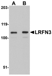
Left: Western blot analysis of LRFN3 in K562 lysate with LRFN3 antibody at (A) 1 and (B) 2 µg/ml.

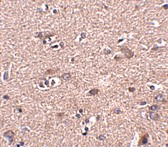
Below:Immunohistochemistry of LRFN3 in mouse brain tissue with LRFN3 antibody at 2.5 µg/ml.

Other Product Images

Source :LRFN3 antibody was raised against a 16 amino acid peptide near the center of human LRFN3.
Purification : Affinity chromatography purified via peptide column
Clonality and Clone : This is a polyclonal antibody.
Host : LRFN3 antibody was raised in rabbit.
Please use anti-rabbit secondary antibodies.
Application : LRFN3 antibody can be used for detection of LRFN3 by Western blot at 1 – 2 µg/ml.
Tested Application(s) : E, WB, IHC
Buffer : Antibody is supplied in PBS containing 0.02% sodium azide.
Blocking Peptide :Cat.No. 5069P - LRFN3 Peptide
Long-Term Storage : LRFN3 antibody can be stored at 4ºC, stable for one year. As with all antibodies care should be taken to avoid repeated freeze thaw cycles. Antibodies should not be exposed to prolonged high temperatures.
Positive Control :
1. Cat. No. 1204 - K562 Cell Lysate
Species Reactivity : H, M, R
GI Number : 13375646
Accession Number : NP_078785
Short Description : Leucine-rich repeat and fibronectin type III domain-containing protein 5
References
1. Morimura N, Inoue T, Katayama K, et al. Comparative analysis of structure, expression and PSD95-binding capacity of Lrfn, a novel family of neuronal transmembrane proteins. Gene 2006; 380:72-83.
2. Kobe B and Kajava AV. The leucine-rich repeat as a protein recognition motif. Curr. Opin. Struct. Biol. 2001; 11:725-32.
3. Aruga J and Mikoshiba K. Identification and characterization of Slitrk, a novel transmembrane protein family controlling neurite outgrowth. Mol. Cell Neurosci. 2003; 24:117-29.

LRFN2 Antibody

LRFN2 Antibody

Catalog# :5067

LRFN2 is one of a family of five transmembrane glycoproteins that are highly expressed in neuronal tissues. LRFN proteins share leucine-rich repeat (LRR)-immunoglobulin-like (Ig)-fibronectin type III (Fn)-transmembrane domain structure with other members of the LRR-Ig-Fn protein superfamily such as the Slitrk family of proteins. Expression of LRFN1, -3, and -4 mRNA was detected in embryonic neuronal cells, while Lrfn2 and Lrfn5 expression was primarily restricted to more mature cells. LRFN1, -2, and -4 bound to PDZ domains of postsynaptic PSD95, re-distributing PSD95 to the cell periphery. It has been suggested that the Lrfn proteins play a role in the developing and/or mature vertebrate nervous system. At least two isoforms of LRFN2 are known to exist. This antibody is predicted to not cross-react with other members of the LRFN family.

Additional Names : LRFN2, Leucine-rich repeat and fibronectin type III domain-containing protein 2, synaptic adhesion-like molecule 1
Description
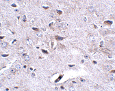
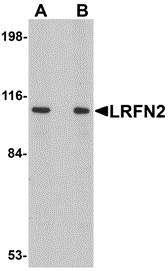
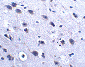
Left:Western blot analysis of LRFN2 in rat brain lysate with LRFN2 antibody at (A) 1 and (B) 2 μg/ml

Below:Immunohistochemistry of LRFN2 in mouse brain tissue with LRFN2 antibody at 2.5 μg/ml.

Other Product Images

Source :LRFN2 antibody was raised against a 14 amino acid peptide near the carboxy terminus of human LRFN2.
Purification : Affinity chromatography purified via peptide column
Clonality and Clone : This is a polyclonal antibody.
Host : LRFN2 antibody was raised in rabbit.
Please use anti-rabbit secondary antibodies.
Application : LRFN2 antibody can be used for detection of LRFN2 by Western blot at 1 – 2 µg/ml.
Tested Application(s) : E, WB, IHC
Buffer : Antibody is supplied in PBS containing 0.02% sodium azide.
Blocking Peptide :Cat.No. 5067P - LRFN2 Peptide
Long-Term Storage : LRFN2 antibody can be stored at 4ºC, stable for one year. As with all antibodies care should be taken to avoid repeated freeze thaw cycles. Antibodies should not be exposed to prolonged high temperatures.
Positive Control :
1. Cat. No. 1463 - Rat Brain Tissue Lysate
Species Reactivity : H, M, R
GI Number : 62286951
Accession Number : Q9ULH4
Short Description : Leucine-rich repeat and fibronectin type III domain-containing protein 4
References
1. Morimura N, Inoue T, Katayama K, et al. Comparative analysis of structure, expression and PSD95-binding capacity of Lrfn, a novel family of neuronal transmembrane proteins. Gene 2006; 380:72-83.
2. Kobe B and Kajava AV. The leucine-rich repeat as a protein recognition motif. Curr. Opin. Struct. Biol. 2001; 11:725-32.
3. Aruga J and Mikoshiba K. Identification and characterization of Slitrk, a novel transmembrane protein family controlling neurite outgrowth. Mol. Cell Neurosci. 2003; 24:117-29.

Monday, March 21, 2011

RNA Polymerase


For over 20 years, Bio Synthesis has provided custom oligonucleotide production services worldwide, including researcher at university, biotechnology and pharmaceutical institutions. We offer wide variety of oligo modifications such as RNA syntehsis from small discovery research scale to multi-gram ASR/cGMP for clinical diagnostic production.

RNA Polymerase

RNA polymerase (RNAP or RNApol) is an enzyme that makes an RNA copy of a DNA or RNA template. In cells, RNAP is needed for constructing RNA chains from DNA genes, a process called transcription. RNA polymerase enzymes are essential to life and are found in all organisms and many viruses. In chemical terms, RNAP is a nucleotidyl transferase that polymerizes ribonucleotides at the 3' end of an RNA transcript.

RNA Polymerase In Archaea

Archaea have a single RNAP that is closely related to the three main eukaryotic polymerases. Thus, it has been speculated that the archaeal polymerase resembles the ancestor of the specialized eukaryotic polymerases.

RNA Protection Assays

A nuclease protection assay is a laboratory test used in biochemistry and genetics to identify individual RNA molecules in a heterogeneous RNA sample extracted from cells. The technique can identify one or more RNA molecules of known sequence even at low total concentration.

Thursday, March 3, 2011

ORAI1 Antibody

ORAI1 Antibody

Catalog# :4281

Antigen stimulation of immune cells triggers Ca++ entry through Ca++ release-activated Ca++ (CRAC) channels. ORAI1 is a recently identified four-transmembrane spanning protein that is an essential component of CRAC. A missense mutation in this protein in humans is the cause of one form of hereditary severe combined immune deficiency (SCID) which results in ablated T-cell Ca++ entry. It has been suggested that ORAI1 functions as a highly selective Ca++ plasma membrane channel that is gated through interactions with STIM1, the store-activated endoplasmic reticulum Ca++ sensor. ORAI1 often migrates at a higher than expected molecular weight in SDS-PAGE. This antibody is predicted to have no cross-reactivity to ORAI2 or ORAI3.

Additional Names : ORAI1 (CT), Transmembrane protein 142A, TMEM142A, Calcium release-activated calcium channel protein 1
Description
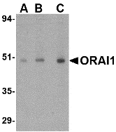
Left: Western blot analysis of ORAI1 in human ovary tissue lysate with ORAI1 antibody at (A) 0.5, (B) 1 and (C) 2 µg/ml.

Below:Immunohistochemistry of ORAI1 in human spleen tissue with ORAI1 antibody at 2.5 µg/ml.

Other Product Images
Source :ORAI1 antibody was raised against a 16 amino acid peptide from near the carboxy terminus of human ORAI1.
Purification : Affinity chromatography purified via peptide column
Clonality and Clone : This is a polyclonal antibody.
Host : ORAI1 antibody was raised in rabbit.
Please use anti-rabbit secondary antibodies.
Application : ORAI1 antibody can be used for detection of ORAI1 by Western blot at 0.5 – 1 µg/ml.
Tested Application(s) : E, WB, IHC
Buffer : Antibody is supplied in PBS containing 0.02% sodium azide.
Blocking Peptide :Cat.No. 4281P - ORAI1 Peptide
Long-Term Storage : ORAI1 antibody can be stored at 4ºC, stable for one year. As with all antibodies care should be taken to avoid repeated freeze thaw cycles. Antibodies should not be exposed to prolonged high temperatures.
Positive Control :
1. Cat. No. 1316 - Human Ovary Tissue Lysate
Species Reactivity : H, M, R
GI Number : 97180269
Accession Number : Q96D31
Short Description : (CT) a T cell calcium channel
References
1. Lewis RS. Calcium signaling mechanisms in T lymphocytes. Annu. Rev. Immunol. 2001; 19:497-521.
2. Feske S, Gwack Y, Prakriya M, et al. A mutation in Orai1 causes immune deficiency by abrogating CRAC channel function. Nature 2006; 441:179-85.
3. Soboloff J, Spassova MA, Dziadek MA, et al. Calcium signals mediated by STIM and Orai proteins – a new paradigm in inter-organelle communication. Biochim. Biophys. Acta. 2006; 1763:1161-8.

Friday, December 10, 2010

NUP107 Antibody

NUP107 Antibody

Catalog# :4701

The nuclear pore complex (NPC) is a protein assembly localized at the nuclear rim and mediates macromolecular transport between the nucleus and the cytoplasm. The mammalian nucleoporin (NUP)-107 is part of the hetero-oligomeric complex that also contains NUP160, NUP133, NUP96, and mammalian homolog of yeast sec13p. While the majority of the NUP107-160 nuclear pore sub-complex localizes to the nuclear pore, a small fraction is observed at kinetochores and pro-metaphase spindle poles in mitotic cells in association with proteins such as Mad1, Mad2, Bub3 and Cdc20. Immunodepletion of the NUP107-160 complex resulted in defective spindle assembly indicating that it has multiple functions. NUP107 has recently been identified as an HIV dependency factor (HDF), suggesting that NUP107 may be an important drug target in HIV treatment. Multiple isoforms of NUP107 are known to exist.


Additional Names : NUP107 (CT), Nucleoporin 107, nuclear pore complex protein 107

Description
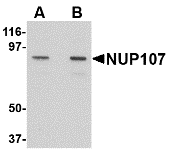
Left: Western blot analysis of NUP107 in A549 cell lysate with NUP107 antibody at (A) 1 and (B) 2 µg/ml.

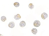
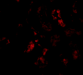
Below: Immunocytochemistry of NUP107 in A549 cells with NUP107 antibody at 2.5 μg/ml.

Other Product Images
Source :
NUP107 antibody was raised against a 15 amino acid peptide from near the carboxy terminus of human NUP107.
Purification : Affinity chromatography purified via peptide column
Clonality and Clone : This is a polyclonal antibody.
Host : NUP107 antibody was raised in rabbit. Please use anti-rabbit secondary antibodies.
Application : NUP107 antibody can be used for detection of NUP107 by Western blot at 1 – 2 µg/ml.
Tested Application(s) : E, WB
Buffer : Antibody is supplied in PBS containing 0.02% sodium azide.
Blocking Peptide :Cat.No. 4701P - NUP107 Peptide
Long-Term Storage : NUP107 antibody can be stored at 4ºC, stable for one year. As with all antibodies care should be taken to avoid repeated freeze thaw cycles. Antibodies should not be exposed to prolonged high temperatures.
Positive Control :
1. Cat. No. 1203 - A549 Cell Lysate
Species Reactivity : H, R
GI Number : 12230339
Accession Number : P57740
Short Description : (CT) Nucleoporin 107
References

1. Tran EJ and Wente SR. Dynamic nuclear pore complex: life on the edge. Cell 2006; 125:1041-53.
2. Boehmer T, Enninga J, Dales S, et al. Depletion of a single nucleoporin, Nup107, prevents the assembly of a subset of nucleoporins into the nuclear pore complex. Proc. Natl. Acad. Sci. USA 2003; 100:981-5.
3. Orjalo AV, Arnaoutov A, Shen Z, et al. The Nup107-160 nucleoporin complex is required for correct bipolar spindle assembly. Mol. Bio. Cell 2006; 17:3806-18.
4. Brass AL, Dykxhoorn DM, Benita Y, et al. Identification of host proteins required for HIV infection through a functional genomic screen. Science 2008; 319:921-6.

Monday, November 8, 2010

JMJD6 Antibody

JMJD6 Antibody

Catalog# : 5383

The jumonji domain-containing protein (JMJD) family is defined by the presence of the JmjC domain that is observed in several diverse species. JMJD6 was initially identified as a membrane protein that participates in phagocytosis but recent studies have shown that it has other functions when it is expressed in the cytosol and nucleus. JMJD6 is thought to play important roles in regulation of development and differentiation as knockdown experiments in mice resulted in neonatal lethality with severe defects in the morphology of numerous organs. JMJD6 also can catalyze the lysyl-hydroxylation of U2AF65, a protein involved with RNA splicing, suggesting that some of the functions attributed to JMJD6 may be due to its regulatory activity of RNA splicing.
Additional Names : JMJD6, Jumonji domain-containing protein 6, phosphatidylserine receptor, PTDSR, PSR
Description
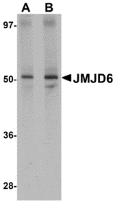
Left: Western blot analysis of JMJD6 in human brain tissue lysate with JMJD6 antibody at (A) 1 and (B) 2 µg/ml.


Source : JMJD6 antibody was raised against a 15 amino acid peptide from near the amino terminus of human JMJD6.
Purification : Affinity chromatography purified via peptide column
Clonality and Clone : Polyclonal
Host : JMJD6 antibody was raised in rabbit. Please use anti-rabbit secondary antibodies.
Application : JMJD6 antibody can be used for detection of JMJD6 by Western blot at 1 - 2 µg/ml.
Tested Application(s) : E, WB
Buffer : Antibody is supplied in PBS containing 0.02% sodium azide.
Blocking Peptide : Cat.No. 5383P - JMJD6 Peptide
Long-Term Storage : JMJD6 antibody can be stored at 4ºC, stable for one year. As with all antibodies care should be taken to avoid repeated freeze thaw cycles. Antibodies should not be exposed to prolonged high temperatures.
Positive Control
1. Cat. No. 1303 - Human Brain Tissue Lysate
Species Reactivity :H, M, R
GI Number : 67461014
Accession Number : Q6NYC1
Short Description : Jumonji domain-containing protein 6
References

1. Takeuchi T, Watanabe Y, Takano-Shimizu T, et al. Roles of jumonji and jumonji family genes in chromatin regulation and development. Dev. Dyn. 2006; 235:2449-59.
2. Fadok VA, Bratton DL, Rose DM, et al. A receptor for phosphatidylserine-specific clearance of apoptotic cells. Nature 2000; 405:85-90.
3. Zakharova L, Dadsetan S, and Fomina AF. Endogenous JMJD6 gene product is expressed at the cell surface and regulates phagocytosis in immature monocyte-like activated THP-1 cells. J. Cell. Phys. 2009; 221:84-91.
4. Bose J, Gruber AD, Helming L, et al. The phosphatidylserine receptor has essential functions during embryogenesis but not in apoptotic cell removal. J. Biol. 2004; 3:15.

Tuesday, November 2, 2010

GNPDA2 Antibody


GNPDA2 Antibody

Catalog# : 5061

Glucosamine-6-phosphate deaminase (GNPDA) is an allosteric enzyme that catalyzes the reversible conversion of D-glucosamine-6-phosphate into D-fructose-6-phosphate and ammonium. GNPDA2 is the second mammalian glucosamine-6-phosphate deaminase discovered, and is closer in structure and activity to the E. coli enzyme than GNPDA1. GNPDA1 possesses greater affinity for ammonium than either GNPDA2 or the E. coli enzyme suggesting that the forward reaction of D-glucosamine-6-phosphate into D-fructose-6-phosphate and ammonium is catalyzed at a slower rate than GNPDA2. GNPDA2 antibody is predicted to not cross-react with GNPDA1.
Additional Names : GNPDA2, Glucosamine-6-phosphate deaminase 2, GNPI

Description
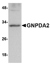
Left: Western blot analysis of GNPDA2 in human kidney lysate with GNPDA2 antibody at 1 µg/ml.

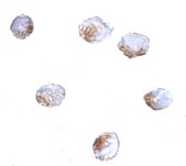
Below: Immunocytochemistry of GNPDA2 in 293 cells with GNPDA2 antibody at 4 µg/ml.

Other Product Images
Source : GNPDA2 antibody was raised against an 8 amino acid peptide near the carboxy terminus of human GNPDA2.
Purification : Affinity chromatography purified via peptide column
Clonality and Clone : This is a polyclonal antibody.
Host : GNPDA2 antibody was raised in rabbit. Please use anti-rabbit secondary antibodies.
Application : GNPDA2 antibody can be used for detection of GNPDA2 by Western blot at 1 – 2 µg/ml.
Tested Application(s) : E, WB, ICC
Buffer : Antibody is supplied in PBS containing 0.02% sodium azide.
Blocking Peptide : Cat.No. 5061P - GNPDA2 Peptide
Long-Term Storage : GNPDA2 antibody can be stored at 4ºC, stable for one year. As with all antibodies care should be taken to avoid repeated freeze thaw cycles. Antibodies should not be exposed to prolonged high temperatures.
Positive Control
1. Cat. No. 1305 - Human Kidney Tissue Lysate
Species Reactivity :H, M
GI Number : 19923881
Accession Number : NP_612208
Short Description : Glucosamine-6-phosphate deaminase 2
References

1. Wolosker H, Kline D, Bian Y, et al. Molecularly cloned mammalian glucosamine-6-phosphate deaminase localizes to transporting epithelium and lacks oscillin activity. FASEB J. 1998; 12:91-9.
2. Zhang J, Zhang W, Zou D, et al. Cloning and functional characterization of GNPI, a novel human homolog of glucosamine-6-phosphate isomerase/oscillin. J. Cell Biochem. 2003; 88:932-40.
3. Arreola R, Valderrama B, Morante ML, et al. Two mammalian glucosamine-6-phosphate deaminases: a structural and genetic study. FEBS Lett. 2003; 551:63-70.

Wednesday, October 27, 2010

CSN8 Antibody

CSN8 Antibody

Catalog# : 4601

The COP9 signalosome (CSN) is an evolutionarily conserved protein complex of the eight subunits that interacts with deubiquitinating enzymes and protein kinases and is highly homologous to the lid sub-complex of 26S proteasome. The CSN complex is an essential regulator of the ubiquitin conjugation pathway by mediating the deneddylation of the SCF-type E3 ligase complexes, which leads to a decrease in ubiquitin ligase activity of SCF-comlpexes such as SCF, CSA or DDB2. It is also involved in phosphorylation of p53, c-jun/JUN, ITPK1 and IRF8/ICSBP, possibly via its association with CK2 and PKD kinases. CSN8 encodes the smallest and the least conserved but first identified subunit of CSN. Recent studies show CSN8 is essential for Drosophila development and is essential for peripheral T cell homeostasis and antigen receptor-induced entry into the cell cycle from quiescence.
Additional Names : CSN8 (CT), COP9 signalosome complex subunit 8, SGN8, JAB1-containing signalosome subunit 8
Description
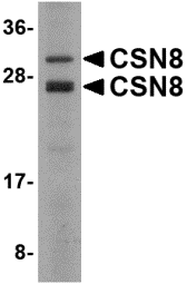
Left: Western blot analysis of CSN8 in Human liver lysate with CSN8 antibody at 2 µg/ml.


Source : CSN8 antibody was raised against a 15 amino acid peptide near the carboxy terminus of the human CSN8.
Purification : Affinity chromatography purified via peptide column
Clonality and Clone : This is a polyclonal antibody.
Host : CSN8 antibody was raised in rabbit. Please use anti-rabbit secondary antibodies.
Application : CSN8 antibody can be used for detection of CSN8 by Western blot at 1 - 2 µg/ml.
Tested Application(s) : E, WB
Buffer : Antibody is supplied in PBS containing 0.02% sodium azide.
Blocking Peptide : Cat.No. 4601P - CSN8 Peptide
Long-Term Storage : CSN8 antibody can be stored at 4ºC, stable for one year. As with all antibodies care should be taken to avoid repeated freeze thaw cycles. Antibodies should not be exposed to prolonged high temperatures.
Positive Control
1. Cat. No. 1304 - Human Liver Tissue Lysate
Species Reactivity :H, M, R
GI Number : 5729779
Accession Number : NP_006701
Short Description : (CT) COP9 signalosome complex subunit 8
References

1. Wei N and Deng XW. The COP9 signalosome. Annu. Rev. Cell Dev. Biol. 2003; 19:261-86.
2. Bech-Otschir D, Seeger M, and Dubiel W. The COP9 signalosome: at the interface between signal transduction and ubiquitin-dependent proteolysis. J. Cell Sci. 2002; 115:467-73.
3. Groisman R, Polanowska J, Kuraoka I, et al. The ubiquitin ligase activity in the DDB2 and regulated by the COP9 signalosome in response to DNA damage. Cell 2003; 113:357-67.
4. Uhle S, Medalia O, Waldron R, et al. Protein kinase CK2 and protein kinase D are associated with the COP9 signalosome. EMBO J. 2003; 22:1302-12.

Tuesday, October 19, 2010

ASAH1 Antibody

ASAH1 Antibody

Catalog# : 4741
Sphingolipids are hydrolyzed by ceramidases to yield sphingosine and fatty acids. These ceramidases are classified according to the pH range that supports their optimal activity. ASAH1 is an acid ceramidase and key regulator of ceramide metabolism. Mutations in this gene results in Farber Lipogranulomatosis, a fatal human genetic disorder that results in the painful swelling of the joints and tendons and pulminary insufficiency, while a complete knockout of its expression is lethal in mice. Recent studies have shown elevated levels of ASAH1 in Alzheimer’s disease (AD) patients correlating with a reduction in sphingomyelin and elevation of ceramide. Pretreatment of cultured neurons with recombinant AHAH1 prevented the cells from undergoing A-beta (Ab)-induced apoptosis. Multiple isoforms of this protein are known to exist.
Additional Names : ASAH1, N-acylsphingosine amidohydrolase (non-lysosomal ceramidase) 1, acid ceramidase

Description
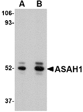
Left: Western blot analysis of ASAH1 in mouse heart tissue lysate with ASAH1 antibody at (A) 1 and (B) 2 µg/ml.

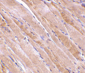
Below: Immunohistochemistry of ASAH1 in human heart tissue with ASAH1 antibody at 2.5 μg/ml.

Other Product Images

Source : ASAH1 antibody was raised against a 16 amino acid peptide near the carboxy terminus of the human ASAH1.
Purification : Affinity chromatography purified via peptide column
Clonality and Clone : This is a polyclonal antibody.
Host : ASAH1 antibody was raised in rabbit. Please use anti-rabbit secondary antibodies.
Application : ASAH1 antibody can be used for detection of ASAH1 by Western blot at 1 µg/ml.
Tested Application(s) : E, WB
Buffer : Antibody is supplied in PBS containing 0.02% sodium azide.
Blocking Peptide : Cat.No. 4741P - ASAH1 Peptide
Long-Term Storage : ASAH1 antibody can be stored at 4ºC, stable for one year. As with all antibodies care should be taken to avoid repeated freeze thaw cycles. Antibodies should not be exposed to prolonged high temperatures.
Positive Control
1. Cat. No. 1401 - Mouse Heart Tissue Lysate
Species Reactivity :H, M, R
GI Number : 119584199
Accession Number : EAW63795
Short Description : an acid ceramidase
References
1. Nilsson A and Duan RD. Alkaline sphingomyelinases and ceramidases of the gastrointestinal tract. Chem. Phys. Lipids 1999; 102:97-105.
2. Koch J, Gartner S, Li CM, et al. Molecular cloning and characterization of a full-length complementary DNA encoding human acid ceramidase. Identification of the first molecular lesion causing Farber’s disease. J. Biol. Chem. 1996; 271:33110-5.
3. Li CM, Park JH, Simonaro CM, et al. Insertional mutagenesis of the mouse acid ceramidase gene leads to early embryonic lethality in homozygotes and progressive lipid storage disease in heterozygotes. Genomics 2002; 79:218-24.
4. He X, Huang Y, Li B. Deregulation of sphingolipid metabolism in Alzheimer’s disease. Neurobiol. Aging 2008; epub.

Friday, October 15, 2010

Custom Peptide

Bio-Synthesis, founded in 1984, is a U.S. based company whose primary emphasis is providing high quality custom products such as custom oligonucleotide, custom peptides, custom antibody, and services for life science research, diagnostics, and therapeutic applications. Our integrated manufacturing programs enable a seamless transition from the inception of an idea through product launch by providing our client with a comprehensive portfolio of services that span across genomic, proteomic, and cell biology.

As a leading supplier of genomic and proteomic research products, BSI is able to offer its customers integrated solutions for custom DNA synthesis, custom RNA synthesis, custom polyclonal antibody production, custom peptide synthesis, molecular diagnostic detection kits, DNA Real-Time PCR products, DNA microarray services and a wide range of custom DNA-based diagnostic kits and consumables.

The proteomic division is experienced in the design of peptides, both for antibody production and bioactive peptide synthesis, e.g cosmetic peptides. While the custom peptide synthesis division has the ability to produce thousands of modified or non-modified peptides per day for research applications, BSI also provides diagnostic/ therapeutic GMP custom peptide production. Synthesis of custom peptides results in quantities ranging from milligrams to multi-kilograms. The staff in the custom antibody division provide expert support in the selection of peptide antigens for target antibody production and offer convenient custom-bundled packages with a wide range of host animals. In addition, peptide, antibody and protein arrays are also amongst its expertise.

BSI is a U.S. based company whose primary emphasis is on the synthesis of high quality custom products for a variety of applications and a full-service contract manufacturing organization (CMO) delivering research, development and production services for cGMP clinical batch manufacturing.

Wednesday, October 13, 2010

Emerin Antibody

Emerin Antibody

Catalog# : 4031

Emerin is a serine-rich nuclear membrane protein and a member of the nuclear lamina-associated protein family that includes proteins such as LAP2 and MAN1. Each family member, including Emerin, has an ~40 amino acid LEM-domains that binds barrier-to-autointegration (BANF1), a conserved chromatin protein that also serves as a host cell component of retroviral integration complexes, including that of HIV. Emerin is anchored at the inner membrane of the nuclear envelope where it binds to nuclear intermediate filaments that are formed by lamin proteins. Dreifuss-Emery muscular dystrophy is an X-linked inherited degenerative myopathy resulting from mutation in the emerin gene.
Additional Names : Emerin, Emery-Dreifuss muscular dystrophy, EDMD, EMD
Description
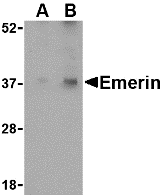
Left: Western blot analysis of Emerin in human skeletal muscle tissue lysate with Emerin antibody at (A) 0.5 and (B) 1 µg/ml.

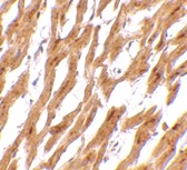
Below: Immunohistochemistry of emerin in human skeletal muscle tissue with emerin antibody at 2.5 µg/ml.

Other Product Images
Source : Emerin antibody was raised against a 19 amino acid peptide from near the amino terminus of human Emerin.
Purification : Affinity chromatography purified via peptide column
Clonality and Clone : This is a polyclonal antibody.
Host : Emerin antibody was raised in rabbit. Please use anti-rabbit secondary antibodies.
Application : Emerin antibody can be used for detection of Emerin by Western blot at 0.5 – 1 µg/ml.
Tested Application(s) : E, WB, IHC
Buffer : Antibody is supplied in PBS containing 0.02% sodium azide.
Blocking Peptide : Cat.No. 4031P - Emerin Peptide
Long-Term Storage : Emerin antibody can be stored at 4ºC, stable for one year. As with all antibodies care should be taken to avoid repeated freeze thaw cycles. Antibodies should not be exposed to prolonged high temperatures.
Positive Control
1. Cat. No. 1375 - Human Skeletal Muscle Tissue Lysate
Species Reactivity :H, M, R
GI Number : 4557553
Accession Number : NP_000108
Short Description : a serine-rich nuclear membrane member of the nuclear lamina-associated protein family
References
1. Schirmer EC, Florens L, Guan T, et al. Nuclear membrane proteins with potential disease links found by subtractive proteomics. Science 2003; 301:1380-2.
2. Cai M, Huang Y, Ghirlando R, et al. Solution structure of the constant region of nuclear envelope protein LAP2 reveals two LEM-domain structures: one binds BAF and the other binds DNA. EMBO J. 2001; 20:4399-407.
3. Chen H and Engelman A. The barrier-to-autointegration protein is a host factor for HIV type 1 integration. Proc. Natl. Acad. Sci. USA 1998; 95:15270-4.
4. Hutchison CJ. Lamins: building blocks or regulators of gene expression? Nat. Rev. Mol. Cell Biol. 2002; 3:848-58.

DDX3 Antibody

DDX3 Antibody

Catalog# : 3759

DDX3 contains all of the motifs of the DEAD-box family of RNA helicases, including the Asp-Glu-Ala-Asp sequence that gives the protein family its name and distinguishes it from other RNA helicases (1,2). DDX3 is localized to the X chromosome and has a highly conserved functional homolog (DBY) on the Y chromosome (3). DDX3 is thought to be involved in RNA splicing, RNA transport, and translation initiation (4). It has also been shown to be involved in cell growth control and is deregulated in hepatitis virus-associated hepatocellular carcinoma (5). Recent experiments suppressing DDX3 expression blocked HIV-1 RNA export from the nucleus, suggesting that DDX3 functions as a shuttling protein that transports the HIV-1 protein Rev and its cofactor CRM1 from the nucleus to the cytoplasm (6).
Additional Names : DDX3 (IN), Dead box polypeptide 3 X-linked, DBX, Helicase-like protein 2
Description
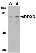
Left: Western blot analysis of DDX3 in HepG2 cell lysate with DDX3 antibody at (A) 1 and (B) 2 µg/ml.

Below: Immunocytochemistry of DDX3 in HepG2 cells with DDX3 antibody at 10 µg/ml.

Other Product Images

Source : DDX3 antibody was raised against a 16 amino acid peptide from near the center of human DDX3.
Purification : Affinity chromatography purified via peptide column
Clonality and Clone : This is a polyclonal antibody.
Host : DDX3 antibody was raised in rabbit. Please use anti-rabbit secondary antibodies.
Immunogen : Human DDX3 (Intermediate Domain) Peptide (Cat. No. 3759P)
Application : DDX3 antibody can be used for the detection of DDX3 by Western blot at 1 – 2 µg/ml.DDX3 antibody will detect both DDX3 and DBY.
Tested Application(s) : E, WB, ICC
Buffer : Antibody is supplied in PBS containing 0.02% sodium azide.
Blocking Peptide : Cat. No. 3759P - DDX3 Peptide
Long-Term Storage : DDX3 antibody can be stored at 4ºC, stable for one year. As with all antibodies care should be taken to avoid repeated freeze thaw cycles. Antibodies should not be exposed to prolonged high temperatures.
Positive Control
1. Cat. No. 1211 - HepG2 Cell Lysate
Species Reactivity :H, M, R
GI Number : 3523150
Accession Number : AAC34298
Short Description : (IN) An RNA helicase that may be involved in HIV-1 nuclear export
References
1. Linder P, Lasko PF, Ashburner M, et al. Birth of the D-E-A-D box. Nature 1989; 337:121-2.
2. Park SH, Lee SG, Kim Y, et al. Assignment of a human putative RNA helicase gene, DDX3, to human X chromosome bands p11.3-->p11.23. Cytogenet. Cell Genet. 1998; 81:178-9.
3. Lahn BT and Page DC. Functional coherence of the human Y chromosome. Science 1997; 278:675-80.
4. Abdelhaleem M. RNA helicases: regulators of differentiation. Clin. Biochem. 2005; 38:499-503

Thursday, October 7, 2010

BANF1 Antibody

BANF1 Antibody

Catalog# : 4019

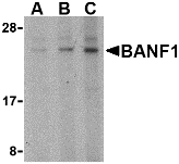
Barrier-to-autointegration factor 1 (BANF1) is a conserved chromatin protein that non-specifically binds double-stranded DNA. BAF also interacts with a family of nuclear proteins that include LAP2, emerin, and MAN1. It is also a host cell component of retroviral pre-integration complexes (PICs), including that of HIV. BAF will bind to p55 Gag (the structural precursor of HIV-1 virions) as well as its cleaved product matrix. In addition to being a host cell component of the PIC, it is thought that BAF is also present at low levels in incoming virions, and thus might contribute to the assembly or activity of HIV-1 PICs through direct binding to matrix as well as DNA. Despite its predicted molecular weight, BANF1 often migrates at a higher size in SDS-PAGE.
Additional Names : BANF1 (NT), Barrier to autointegration factor 1, BAF, breakpoint cluster region protein 1
Description
Left: Western blot analysis of BANF1 in mouse kidney tissue lysate with BANF1 antibody at (A) 0.5, (B) 1 and (C) 2 µg/ml.

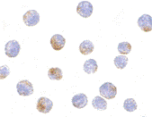
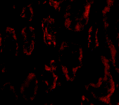
Below: Immunofluorescence of BANF1 in human kidney tissue with BANF1 antibody at 20 µg/ml.

Other Product Images
Source : BANF1 antibody was raised against a 19 amino acid peptide from near the amino terminus of human BANF1.
Purification : Affinity chromatography purified via peptide column
Clonality and Clone : This is a polyclonal antibody.
Host : BANF1 antibody was raised in rabbit. Please use anti-rabbit secondary antibodies.
Application : BANF1 antibody can be used for detection of BANF1 by Western blot at 0.5 – 1 µg/ml.
Tested Application(s) : E, WB, IF
Buffer : Antibody is supplied in PBS containing 0.02% sodium azide.
Blocking Peptide : Cat.No. 4019P - BANF1 Peptide
Long-Term Storage : BANF1 antibody can be stored at 4ºC, stable for one year. As with all antibodies care should be taken to avoid repeated freeze thaw cycles. Antibodies should not be exposed to prolonged high temperatures.
Positive Control
1. Cat. No. 1405 - Mouse Kidney Tissue Lysate
Species Reactivity :H, M, R
GI Number : 13543577
Accession Number : AAH05942
Short Description : (NT) Barrier to autointegration factor
References

1. Furukawa K. LAP2 binding protein 1 (L2BP1/BAF) is a candidate mediator of LAP2-chromatin interaction. J. Cell Sci. 1999; 112:2485-92.
2. Cai M, Huang Y, Ghirlando R, et al. Solution structure of the constant region of nuclear envelope protein LAP2 reveals two LEM-domain structures: one binds BAF and the other binds DNA. EMBO J. 2001; 20:4399-407.
3. Chen H and Engelman A. The barrier-to-autointegration protein is a host factor for HIV type 1 integration. Proc. Natl. Acad. Sci. USA 1998; 95:15270-4.

BANF1 Antibody

BANF1 Antibody

Catalog# : 4017
Barrier-to-autointegration factor 1 (BANF1) is a conserved chromatin protein that non-specifically binds double-stranded DNA. BANF1 also interacts with a family of nuclear proteins that include LAP2, emerin, and MAN1. It is also a host cell component of retroviral pre-integration complexes (PICs), including that of HIV. BANF1 will bind to p55 Gag (the structural precursor of HIV-1 virions) as well as its cleaved product matrix. In addition to being a host cell component of the PIC, it is thought that BANF1 is also present at low levels in incoming virions, and thus might contribute to the assembly or activity of HIV-1 PICs through direct binding to matrix as well as DNA. Despite its predicted molecular weight, BANF1 often migrates at a higher size in SDS-PAGE.
Additional Names : BANF1 (CT), Barrier to autointegration factor 1, BAF, breakpoint cluster region protein 1
Description
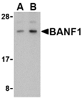
Left: Western blot analysis of BANF1 in human kidney tissue lysate with BANF1 antibody at (A) 0.5 and (B) 1 µg/ml.

Below: Immunofluorescence of BANF1 in human kidney tissue with BANF1 antibody at 20 µg/ml.

Other Product Images
Source : BANF1 antibody was raised against a 15 amino acid peptide from near the carboxy terminus of human BANF1.
Purification : Affinity chromatography purified via peptide column
Clonality and Clone : This is a polyclonal antibody.
Host : BANF1 antibody was raised in rabbit. Please use anti-rabbit secondary antibodies.
Application : BANF1 antibody can be used for detection of BANF1 by Western blot at 0.5 – 1 µg/ml.
Tested Application(s) : E, WB, IF
Buffer : Antibody is supplied in PBS containing 0.02% sodium azide.
Blocking Peptide : Cat.No. 4017P - BANF1 Peptide
Long-Term Storage : BANF1 antibody can be stored at 4ºC, stable for one year. As with all antibodies care should be taken to avoid repeated freeze thaw cycles. Antibodies should not be exposed to prolonged high temperatures.
Positive Control
1. Cat. No. 1305 - Human Kidney Tissue Lysate
Species Reactivity :H, M
GI Number : 13543577
Accession Number : AAH05942
Short Description : (CT) Barrier to autointegration factor
References
1. Furukawa K. LAP2 binding protein 1 (L2BP1/BAF) is a candidate mediator of LAP2-chromatin interaction. J. Cell Sci. 1999; 112:2485-92.
2. Cai M, Huang Y, Ghirlando R, et al. Solution structure of the constant region of nuclear envelope protein LAP2 reveals two LEM-domain structures: one binds BAF and the other binds DNA. EMBO J. 2001; 20:4399-407.
3. Chen H and Engelman A. The barrier-to-autointegration protein is a host factor for HIV type 1 integration. Proc. Natl. Acad. Sci. USA 1998; 95:15270-4.

TNFRSF14 Antibody

TNFRSF14 Antibody

Catalog# : 3773

Tumor necrosis factor receptor (TNFR) superfamily members are defined by cysteine-rich domains in their extracellular regions that bind TNF-related ligands that share a common structural homology in their extracellular domain. TNFRSF14 was initially identified as the Herpesvirus entry mediator and upon binding to the herpes simplex virus (HSV) envelope glycoprotein D or either of its natural ligands LIGHT and lymphotoxin alpha (LT), activates the transcription factors NF-kappaB and AP-1. Activation of this signal transduction pathway in T cells stimulates T cell proliferation and cytokine production, leading to inflammation and enhanced CTL-mediated tumor immunity, suggesting that these proteins may be useful as potential targets for controlling cellular immune responses.
Additional Names : TNFRSF14 (CT), Tumor necrosis factor receptor superfamily 14, Herpesvirusentry mediator, HVEM, TR2
Description
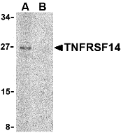
Left: Western blot analysis of TNFRSF14 in Raji cell lysate with TNFRSF14 antibody at 2 µg/ml in (A) the absence and (B) the presence of blocking peptide.

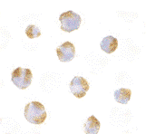
Below: Immunocytochemistry of TNFRSF14 in Raji with TNFRSF14 antibody at 10 µg/ml.

Other Product Images
Source : TNFRSF14 antibody was raised against a 16 amino acid peptide from near the carboxy terminus of human TNFRSF14.
Purification : Affinity chromatography purified via peptide column
Clonality and Clone : This is a polyclonal antibody.
Host : TNFRSF14 antibody was raised in rabbit. Please use anti-rabbit secondary antibodies.
Application : TNFRSF14 antibody can be used for the detection of TNFRSF14 by Western blot at 1 - 2 µg/ml.
Tested Application(s) : E, WB, ICC
Buffer : Antibody is supplied in PBS containing 0.02% sodium azide.
Blocking Peptide : Cat.No. 3773P - TNFRSF14 Peptide
Long-Term Storage : TNFRSF14 antibody can be stored at 4ºC, stable for one year. As with all antibodies care should be taken to avoid repeated freeze thaw cycles. Antibodies should not be exposed to prolonged high temperatures.
Positive Control
1. Cat. No. 1207 - Raji Cell Lysate
Species Reactivity :H
GI Number : 13878821
Accession Number : Q92956
Short Description : (CT) Herpesvirus entry mediator
References
1. Watts TH. TNF/TNFR family members in costimulation of T cell responses. Annu. Rev. Immunol. 2005; 23:23-68.
2. Montgomery RI, Warner MS, Lum BJ, et al. Herpes simplex virus-1 entry into cells mediated by a novel member of the TNF/NGF receptor family. Cell 1996; 87:427-36.
3. Marsters SA, Ayres TM, Skubatch M, et al. Herpesvirus entry mediator, a member of the tumor necrosis factor receptor (TNFR) family, interacts with members of the TNFR-associated factor family and activates the transcription factors NF-kappaB and AP-1. J. Biol. Chem. 1997; 272:14029-32.
4. Mauri DN, Ebner R, Montgomery RI, et al. LIGHT, a new member of the TNF superfamily, and lymphotoxin alpha are ligands for herpesvirus entry mediator. Immunity 1998; 8:21-30.

NGFR Antibody

NGFR Antibody

Catalog# : 3593

The tumor necrosis factor (TNF) and TNF receptor (TNFR) gene superfamilies regulate numerous biological functions including cell proliferation, differentiation, and survival through regulating the activation of the transcription factor NF-kappaB and various mitogen-activated protein kinases (reviewed in 1). Nerve growth factor receptor (NGFR) was one of the earliest characterized members of this family (2). Also known as the low-affinity receptor p75NTR, this receptor is involved in several diverse functions such as apoptosis, neurite outgrowth during development, and myelination (reviewed in 3). Its ligands include NGF, brain-derived neurotrophic factor (BDNF), NT3, and NT4 (4). NGFR can also associate with other NGF receptors such as Trk through the cytosolic and transmembrane domains and thus can function as a co-receptor that refines Trk affinity and specificity for neurotrophins (5). Finally, upon binding of various neurotrophins, NGFR associates with tumor necrosis factor receptor-6 (TRAF6), suggesting that it can potentially function as a signal transducer for NGF signals through NGFR (6).
Additional Names : NGFR, p75NTR, TNFRSF16
Description
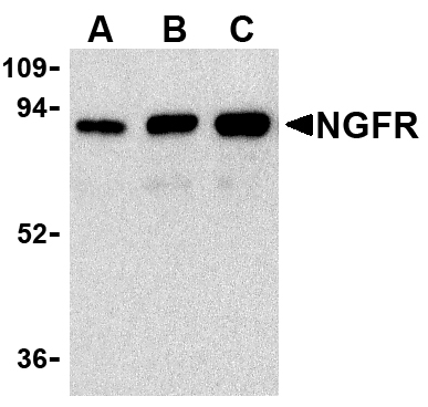
Left: Western blot analysis of (A) 25 ng, (B) 50 ng, and (C) 100 ng of purified recombinant NGFR with NGFR antibody at 1 µg/ml.

Below: Immunocytochemistry of NGFR in A20 cells with NGFR antibody at 10 µg/ml.

Other Product Images
Source : NGFR antibody was raised against purified recombinant human NGFR.
Purification : Affinity chromatography purified via peptide column
Clonality and Clone : This is a polyclonal antibody.
Host : NGFR antibody was raised in rabbit. Please use anti-rabbit secondary antibodies.
Immunogen : Recombinant human NGFR
Application : NGFR antibody can be used for the detection of NGFR by Western blot at 1 µg/ml.
Tested Application(s) : E, WB, ICC
Buffer : Antibody is supplied in PBS containing 0.02% sodium azide.
Blocking Peptide : Cat.No. 3593P - NGFR Peptide
Long-Term Storage : NGFR antibody can be stored at 4ºC, stable for one year. As with all antibodies care should be taken to avoid repeated freeze thaw cycles. Antibodies should not be exposed to prolonged high temperatures.
Positive Control
1. Cat. No. 1288 - A-20 Cell Lysate
Species Reactivity :H, M
GI Number : 4505393
Accession Number : NP_002498
Short Description : low affinity nerve growth factor receptor
References
1. Gaur U, Aggarwal BB. Regulation of proliferation, survival and apoptosis by members of the TNF superfamily. Biochem. Pharmacol. 2003; 66:1403-8.
2. Johnson D, Lanahon A, Buck CR, et al. Expression and structure of the human NGF receptor. Cell 1986; 47:545-54.
3. Gentry JJ, Barker, PA, and Carter BD. The p75 neuro-trophin receptor: multiple interactors and numerous functions. Prog. Brain Res. 2004;146:25-39.
4. Nykjaer A, Willnow TE, and Petersen CM. p75NTR – live or let die. Curr. Opin. Neurobio. 2005; 15:49-57.

Wednesday, October 6, 2010

IRF8 Antibody

IRF8 Antibody

Catalog# : 3401

Interferons (IFN)s are involved in a multitude of immune interactions during viral infections and play a major role in both the induction and regulation of innate and adaptive antiviral mechanisms (1 for review). During infection, host-virus interactions signal downstream molecules such as transcription factors such as IFN regulatory factor-3 (IRF3) which can act to stimulate transcription of IFN-alpha/beta genes. Unlike IRF3, IRF8 appears to act as a negative regulator of IFN-induced genes in most cases (2,3), but IRF8 mediates activation of NF-kappaB by the toll-like receptor 9 (TLR9) after stimulation by unmethylated CpG DNA in dendritic cells (4,5). Finally, it has been shown that IRF8 decreases bcl-2 expression and thus may play a role in chronic myelogenous leukemia (6).
Additional Names : IRF8

Description
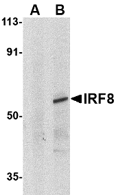
Left: Western blot analysis of IRF8 in human thymus tissue lysate with IRF8 antibody at 1µg/ml in (A) the presence and (B) absence of blocking peptide.


Source : IRF8 antibody was raised against a peptide corresponding to 16 amino acids near the carboxy-terminus of human IRF8.
Purification : Affinity chromatography purified via peptide column
Clonality and Clone : This is a polyclonal antibody.
Host : IRF8 antibody was raised in rabbit. Please use anti-rabbit secondary antibodies.
Immunogen : Human IRF8 Peptide (Cat. No. 3401P)
Application : IRF8 antibody can be used for detection of IRF8 by Western blot at 1 - 2 µg/ml.
Tested Application(s) : E, WB
Buffer : Antibody is supplied in PBS containing 0.02% sodium azide.
Blocking Peptide : Cat. No. 3401P - IRF8 Peptide
Long-Term Storage : IRF8 antibody can be stored at 4ºC, stable for one year. As with all antibodies care should be taken to avoid repeated freeze thaw cycles. Antibodies should not be exposed to prolonged high temperatures.
Positive Control
1. Cat. No. 1314 - Human Thymus Tissue Lysate
Species Reactivity :H, M, R
GI Number : 4504567
Accession Number : NP_002154
Short Description : interferon regulatory factor 8
References

1. Malmgaard L. Induction and regulation of IFNs during viral infections. J. Interferon & Cyto. Res. 2004; 24:439-54
2. Weisz A, Marx P, Sharf R, et al. The human interferon consensus sequence binding protein (H-ICSBP) is a negative regulator of enhancer elements common to interferon inducible genes. J. Biol. Chem. 1992; 267:25589-96.
3. Nelson N, Marks MS, Driggers PH, et al. Interferon consensus sequence-binding protein, a member of the interferon regulatory factor family, suppresses interferon-induced gene transcription. Mol. Cell. Biol. 1993; 13:588-99.
4. Tsujimura H, Tamura T, Kong HJ, et al. Toll-like receptor 9 signaling activates NF-kappaB through IFN regulatory factor-8/IFN consensus sequence binding protein in dendritic cells. J. Immunol. 2004; 172:6820-7.