TL-1RAcP Antibody
Catalog# : 2131
The pro-inflammatory cytokine IL-1 induced cellular response requires two subunits of its receptor, IL-1 receptor I(IL-1RI) and IL-1 receptor accessory protein (IL-1RAcP) (1).IL-1 RAcP forms a complex with IL-1RI in response to IL-1 treatment. The IL-1 receptor-associated kinase (IRAK), which mediates activation of NF-kappaB inducing kinae (NIK) and of NF-kappaB, recruites to the IL-1R complex through IL-1 RAcP(2). IL-1 activation of stress-activated protein kinase and of acid sphingomyelinase also requires IL-1RAcP(3,4). Like IL-1RI, IL-1RAcP subunit is essential for IL-1 mediated cellular response. IL-1RAcP is expressed in many tissues.
Additional Names : IL-1RAcP (C1), IL-1RAcP
 Description
Left: Western blot analysis of IL-1RAcP in HeLa whole cell lysate with IL-1RAcP antibody at 1:500 dilution.
Below: Immunocytochemistry of IL-1RAcP in HeLa cells with IL-1RAcP antibody at 5 µg/ml.
Other Product Images
 Source : IL-1RAcP antibody was raised against a peptide corresponding to amino acids near the carboxy terminus of IL-1RAcP, which differs from the sequence of mouse origin by three amino acids.
Purification : Affinity chromatography purified via peptide column
Clonality and Clone : This is a polyclonal antibody.
Host : IL-1RAcP antibody was raised in rabbit. Please use anti-rabbit secondary antibodies.
Immunogen : Human IL-1RAcP (C-Terminus) Peptide (Cat. No. 2131P)
Application : IL-1RAcP antibody can be used for detection of IL-1RAcP by Western blot at 1:500 to 1:1000 dilution. An approximately 66 kDa band can be detected. It has no cross activity to other member in the IL-1 receptor family.
Tested Application(s) : E, WB, ICC
Buffer : Antibody is supplied in PBS containing 0.02% sodium azide.
Blocking Peptide : Cat. No. 2131P - IL-1RAcP Peptide
Long-Term Storage : IL-1RAcP antibody can be stored at 4ºC, stable for one year. As with all antibodies care should be taken to avoid repeated freeze thaw cycles. Antibodies should not be exposed to prolonged high temperatures.
Positive Control
1. Cat. No. 1201 - HeLa Cell Lysate
Species Reactivity :H
GI Number : 2599126
Accession Number : AF029213
Short Description : (C1) IL-1R accessory protein
References
1. Greenfeder SA, Nunes P, Kwee L, Labow M, Chizzonite RA, Ju G. Molecular cloning and characterization of a second subunit of the interleukin 1 receptor complex. J Biol Chem 1995;270:13757-65
2. Huang J, Gao X, Li S, Cao Z. Recruitment of IRAK to the interleukin 1 receptor complex requires interleukin 1 receptor accessory protein. Proc Natl Acad Sci U S A 1997;94:12829-32
3. Wesche H, Korherr C, Kracht M, Falk W, Resch K, Martin MU. The interleukin-1 receptor accessory protein (IL-1RAcP) is essential for IL-1-induced activation of interleukin-1 receptor-associated kinase (IRAK) and stress-activated protein kinases (SAP kinases). J Biol Chem 1997;272:7727-31
4. Hofmeister R, Wiegmann K, Korherr C, Bernardo K, Kronke M, Falk W. Activation of acid sphingomyelinase by interleukin-1 (IL-1) requires the IL-1 receptor accessory protein. J Biol Chem 1997;272:27730-6 (RD1299)
IL-1RAcP Antibody
Catalog# : 2129
The pro-inflammatory cytokine IL-1 induced cellular response requires two subunits of its receptor, IL-1 receptor I (IL-1RI) and IL-1 receptor accessory protein (IL-1RAcP) (1). IL-1RAcP forms a complex with IL-1RI in response to IL-1 treatment. The IL-1 receptor-associated kinase (IRAK), which mediates activation of NFkappa-B inducing kinae (NIK) and of NF-kappaB, recruits to the IL-1R complex through IL-1RAcP (2). IL-1 activation of stress-activated protein kinase and of acid sphingomyelinase also requires IL-1RAcP (3,4). Like IL-1RI, IL-1RAcP subunit is essential for IL-1 mediated cellular response. IL-1RAcP is expressed in many tissues.
Additional Names : IL-1RAcP (C2), IL-1RAcP
 Description
Left: Western blot analysis of IL-1RAcP in HeLa whole cell lysate with IL-1RAcP antibody (C2) at 1:1000 dilution.
Source : IL-1RAcP antibody was raised against a peptide corresponding to amino acids 525 to 540 of human IL-1RAcP, which is identical to those of mouse and rat origins.
Purification : Affinity chromatography purified via peptide column
Clonality and Clone : This is a polyclonal antibody.
Host : IL-1RAcP antibody was raised in rabbit. Please use anti-rabbit secondary antibodies.
Immunogen : Human IL-1RAcP (C-Terminus 2) Peptide (Cat. No. 2129P)
Application : IL-1RAcP antibody can be used for detection of IL-1RAcP by Western blot at 1:1000 to 1:2000 dilution. An approximately 66 kDa band can be detected. This polyclonal antibody has no cross activity to other members in the IL-1 receptor family.
Tested Application(s) : E, WB
Buffer : Antibody is supplied in PBS containing 0.02% sodium azide.
Blocking Peptide : Cat. No. 2129P - IL-1RAcP Peptide
Long-Term Storage : IL-1RAcP antibody can be stored at 4ºC, stable for one year. As with all antibodies care should be taken to avoid repeated freeze thaw cycles. Antibodies should not be exposed to prolonged high temperatures.
Positive Control
1. Cat. No. 1201 - HeLa Cell Lysate
Species Reactivity :H
GI Number : 2599126
Accession Number : AF029213
Short Description : (C2) IL-1R accessory protein
References
1. Greenfeder SA, Nunes P, Kwee L, Labow M, Chizzonite RA, Ju G. Molecular cloning and characterization of a second subunit of the interleukin 1 receptor complex. J Biol Chem 1995;270:13757-65
2. Huang J, Gao X, Li S, Cao Z. Recruitment of IRAK to the interleukin 1 receptor complex requires interleukin 1 receptor accessory protein. Proc Natl Acad Sci U S A 1997;94:12829-32
3. Wesche H, Korherr C, Kracht M, Falk W, Resch K, Martin MU. The interleukin-1 receptor accessory protein (IL-1RAcP) is essential for IL-1-induced activation of interleukin-1 receptor-associated kinase (IRAK) and stress-activated protein kinases (SAP kinases). J Biol Chem 1997;272:7727-31
4. Hofmeister R, Wiegmann K, Korherr C, Bernardo K, Kronke M, Falk W. Activation of acid sphingomyelinase by interleukin-1 (IL-1) requires the IL-1 receptor accessory protein. J Biol Chem 1997;272:27730-6 (RD1299)
GITRL Antibody
Catalog# : 3591
The tumor necrosis factor (TNF) and TNF receptor (TNFR) gene superfamilies regulate numerous biological functions including cell proliferation, differentiation, and survival through regulating the activation of the transcription factor NF-kappaB and various mitogen-activated protein kinases (reviewed in 1). The glucocorticoid-induced tumor necrosis factor receptor (GITR) is an emerging member of this family that is expressed on CD4+ CD25+ regulatory T cells and appears to have crucial immune regulation functions (2,3). Its ligand GITRL is expressed in endothelial and antigen-presenting cells (4) and can activate NF-kappaB, induce both pro- and anti-apoptotic effects, inhibit the suppressive activity of regulatory T cells, and co-stimulate responder T cells through GITR (5). Dominant negative forms of NIK and TRAF2 expressed in transfected 293 cells substantially inhibited NF-kappaB activation, suggesting that the GITRL-GITR pathway involves both NIK and TRAF2 (4).
Additional Names : GITRL, AITRL
 Description
Left: Western blot analysis of (A) 5 ng, (B) 25 ng, and (C) 50 ng of purified recombinant GITRL with ITRL antibody at 1 µg/ml.
Below: Immunocytochemistry of GITRL in THP-1 cells with GITRL antibody at 10 µg/ml.
Other Product Images
 Source : GITRL antibody was raised against purified recombinant human GITR ligand.
Purification : Affinity chromatography purified via peptide column
Clonality and Clone : This is a polyclonal antibody.
Host : GITRL antibody was raised in rabbit. Please use anti-rabbit secondary antibodies.
Immunogen : Recombinant human GITR ligand
Application : GITRL antibody can be used for the detection of GITRL by Western blot at 1 µg/ml.
Tested Application(s) : E, WB, ICC
Buffer : Antibody is supplied in PBS containing 0.02% sodium azide.
Blocking Peptide : Cat.No. 3591P - GITRL Peptide
Long-Term Storage : GITRL antibody can be stored at 4ºC, stable for one year. As with all antibodies care should be taken to avoid repeated freeze thaw cycles. Antibodies should not be exposed to prolonged high temperatures.
Positive Control
1.Cat. No. 1208 - THP-1 Cell Lysate
Species Reactivity :H, M
GI Number : 13124621
Accession Number : Q9UNG2
Short Description : glucocorticoid-induced TNF receptor ligand
References
1.Gaur U, Aggarwal BB. Regulation of proliferation, survival and apoptosis by members of the TNF superfamily. Biochem. Pharmacol. 2003; 66:1403-8.
2.Ronchetti S, Nocentini G, Riccardi C, et al. Role of GITR in activation response of T lymphocytes.Blood 2002; 100:350-2.
CTRP7 Antibody
Catalog# : 3579
Adipose tissue of an organism plays a major role in regulating physiologic and pathologic processes such as metabolism and immunity by producing and secreting a variety of bioactive molecules termed adipokines (reviewed in 1). One highly conserved family of adipokines is adiponectin/ACRP30 and its structural and functional paralogs, the C1q/tumor necrosis factor-?-related proteins (CTRPs) 1-7 (2). Unlike adiponectin, which is expressed exclusively by differentiated adipocytes, the CTRPs are expressed in a wide variety of tissues (3). These proteins are thought to act mainly on liver and muscle tissue to control glucose and lipid metabolism. An analysis of the crystal structure of adiponectin revealed a structural and evolutionary link between TNF and C1q-containing proteins, suggesting that these proteins arose from a common ancestral innate immunity gene (4). Like the other members of the adiponectin and CTRP protein family, the mature CTRP7 is secreted and can be found in the organism’s circulatory system (1).
Additional Names : CTRP7 (CT), CTRP7
 Description
Left: Western blot analysis of CTRP7 in 293 cell lysate with CTRP7 antibody at (A) 0.5, (B) 1, and (C) 2 µg/ml.
Below: Immunocytochemistry of CTRP7 in HL60 cells with CTRP7 antibody at 0.5 µg/ml.
Other Product Images
 Source : CTRP7 antibody was raised against a 15 amino acid peptide from near the carboxy-terminus of human CTRP7.
Purification : Affinity chromatography purified via peptide column
Clonality and Clone : This is a polyclonal antibody.
Host : CTRP7 antibody was raised in rabbit. Please use anti-rabbit secondary antibodies.
Immunogen : Human CTRP7 (C-Terminus) Peptide (Cat. No. 3579P)
Application : antibody can be used for the detection of CTRP7 by Western blot at 0.5 µg/ml.293 cell lysate can be used as positive control. Anti-CTRP7 is human, mouse, and rat reactive. These proteins are often highly modified post-translationally and migrate in SDS-PAGE at positions other than their predicted size.
Tested Application(s) : E, WB, ICC
Buffer : Antibody is supplied in PBS containing 0.02% sodium azide.
Blocking Peptide : Cat. No. 3579P - CTRP7 Peptide
Long-Term Storage : CTRP7 antibody can be stored at 4ºC, stable for one year. As with all antibodies care should be taken to avoid repeated freeze thaw cycles. Antibodies should not be exposed to prolonged high temperatures.
Positive Control
1. Cat. No. 1210 - 293 Cell Lysate
2. Cat. No. 1209 - HL60 Cell Lysate
Species Reactivity :H, M, R
GI Number : 18645144
Accession Number : AAH24015
Short Description : (CT) a novel adipokine
References
1. Fantuzzi G. Adipose tissue, adipokines, and inflammation. J. Allergy Clin. Immunol. 2005; 115:911-9.
2. Tsao T-S, Lodish HF, and Fruebis J. ACRP30, a new hormone controlling fat and glucose metabolism. Euro. J. Pharmacol. 2002; 440:213-21.
3. Wong GW, Wang J, Hug C, et al. A family of Acrp30/ adiponectin structural and functional paralogs. Proc. Natl. Acad. Sci. USA 2004; 101:10302-7.
4. Shapiro L and Scherer PE. The crystal structure of a complement-1q family protein suggests an evolutionary link to tumor necrosis factor. Curr. Biol. 1998; 8:335-8.
Bio-Synthesis offer custom construction of polymer bioconjugation by covalently attached synthetic PEG (polyethylene glycol) polymer to nucleic acids, peptides, enzymes, carbohydrates. These polymer bioconjugates, sometimes called "bio-hybrid" or "macromolecular chimeras" have many benefits. Benefits range beyond biomedical field and includes issues as diverse as bio-sensors, artificial enzymes, biometrics, and much more.
PEG (Polyethylene glycol) Polymer
PEG (polyethylene glyco), the most often used polymer. This chemical compound composed of repeating ethylene glycol units. It also known as poly ethylene oxide (PEO) and polyoxyethylene (POE) with molecular weights from 300g/mol to 10,000,000 g/mol.
PEG Advantage
The attachment of a PEG polymer to a therapeutic molecule called PEGylation. This technology is now widely used for the modification of proteins, peptides, antibody fragments, oligonucleotides and the like for use as drugs. There are advantages like: Increase bioavailability, Reduce toxicity an immunogenicity, Hydrophilic (aqueous-soluble), Highly flexible etc.
Type of PEG for Conjugation
There are various type of PEG like: Linear Activated PEGs, Multi-arm PEG conjugation, Branched PEG conjugation, Heterofuntional PEGs conjugation, Comb-Shaped Copolymers, PEG containing Biotin.
CTRP7 Antibody
Catalog# : 3673
Adipose tissue of an organism plays a major role in regulating physiologic and pathologic processes such as metabolism and immunity by producing and secreting a variety of bioactive molecules termed adipokines (reviewed in 1). One highly conserved family of adipokines is adiponectin/ACRP30 and its structural and functional paralogs, the C1q/tumor necrosis factor-?-related proteins (CTRPs) 1-7 (2). Unlike adiponectin, which is expressed exclusively by differentiated adipocytes, the CTRPs are expressed in a wide variety of tissues (3). These proteins are thought to act mainly on liver and muscle tissue to control glucose and lipid metabolism. An analysis of the crystal structure of adiponectin revealed a structural and evolutionary link between TNF and C1q-containing proteins, suggesting that these proteins arose from a common ancestral innate immunity gene (4). Like the other members of the adiponectin and CTRP protein family, the mature CTRP7 is secreted and can be found in the organism’s circulatory system (1).
Additional Names : CTRP7
 Description
Left: Western blot of recombinant CTRP7: (A) 5 ng, (B) 25 ng, and (C) 50 ng with CTRP2 antibody at 1 ug/ml.
Below: Immunocytochemistry of CTRP7 in 293 cells with CTRP7 antibody at 10 µg/ml.
Other Product Images
 Source : Rabbit anti-CTRP7 polyclonal antibody was raised against recombinant human CTRP7.
Purification : Affinity chromatography purified via peptide column
Clonality and Clone : This is a polyclonal antibody.
Host : CTRP7 antibody was raised in rabbit. Please use anti-rabbit secondary antibodies.
Immunogen : Recombinant mouse CTRP7 (Genbank accession No. AAY21932).
Application : CTRP7 antibody can be used for the detection of CTRP7 by Western blot at 0.5 µg/ml.These proteins are often highly modified post-translationally and migrate in SDS-PAGE at positions other than their predicted size.
Tested Application(s) : E, WB, ICC
Buffer : Antibody is supplied in PBS containing 0.02% sodium azide.
Blocking Peptide : Cat.No. 3673P - CTRP7 Peptide
Long-Term Storage : CTRP7 antibody can be stored at 4ºC, stable for one year. As with all antibodies care should be taken to avoid repeated freeze thaw cycles. Antibodies should not be exposed to prolonged high temperatures.
Positive Control
1. Cat. No. 1210 - 293 Cell Lysate
Species Reactivity :H, M, R
GI Number : 62913965
Accession Number : AAY21932
Short Description : a novel adipokine
References
1. Fantuzzi G. Adipose tissue, adipokines, and inflammation. J. Allergy Clin. Immunol. 2005; 115:911-9.
2. Tsao T-S, Lodish HF, and Fruebis J. ACRP30, a new hormone controlling fat and glucose metabolism. Euro. J. Pharmacol. 2002; 440:213-21.
3. Wong GW, Wang J, Hug C, et al. A family of Acrp30/ adiponectin structural and functional paralogs. Proc. Natl. Acad. Sci. USA 2004; 101:10302-7.
4. Shapiro L and Scherer PE. The crystal structure of a complement-1q family protein suggests an evolutionary link to tumor necrosis factor. Curr. Biol. 1998; 8:335-8.
CTRP2 Antibody
Catalog# : 3561
Adipose tissue of an organism plays a major role in regulating physiologic and pathologic processes such as metabolism and immunity by producing and secreting a variety of bioactive molecules termed adipokines (reviewed in 1). One highly conserved family of adipokines is adiponectin/ACRP30 and its structural and functional paralogs, the C1q/tumor necrosis factor-?-related proteins (CTRPs) 1-7 (2). Unlike adiponectin, which is expressed exclusively by differentiated adipocytes, the CTRPs are expressed in a wide variety of tissues (3). These proteins are thought to act mainly on liver and muscle tissue to control glucose and lipid metabolism. An analysis of the crystal structure of adiponectin revealed a structural and evolutionary link between TNF and C1q-containing proteins, suggesting that these proteins arose from a common ancestral innate immunity gene (4). Of the CTRPs, CTRP2 is most similar structurally and functionally to adiponectin. Recombinant CTRP2 rapidly activated AMPK and MAPK in cultured C2C12 cells, leading to increased glycogen accumulation and fatty acid oxidation (3).
Additional Names : CTRP2 (NT), CTRP2
 Description
Left: Western blot analysis of CTRP2 in Caco-2 cell lysate with CTRP2 antibody at 1 µg/ml in either the (A) absence or (B) presence of blocking peptide.
Source : CTRP2 antibody was raised against a 16 amino acid peptide from near the amino-terminus of human CTRP2.
Purification : Affinity chromatography purified via peptide column
Clonality and Clone : This is a polyclonal antibody.
Host : CTRP2 antibody was raised in rabbit. Please use anti-rabbit secondary antibodies.
Immunogen : Murine CTRP2 (N-Terminus) Peptide (Cat. No. 3561P)
Application : CTRP2 antibody can be used for the detection of CTRP2 by Western blot at 1 µg/ml.Caco-2 cell lysate can be used as positive control. Anti-CTRP2 is human and mouse reactive. These proteins are often highly modified post-translationally and migrate in SDS-PAGE at positions other than their predicted size.
Tested Application(s) : E, WB
Buffer : Antibody is supplied in PBS containing 0.02% sodium azide.
Blocking Peptide : Cat. No. 3561P - CTRP2 Peptide
Long-Term Storage : CTRP2 antibody can be stored at 4ºC, stable for one year. As with all antibodies care should be taken to avoid repeated freeze thaw cycles. Antibodies should not be exposed to prolonged high temperatures.
Positive Control
1. Cat. No. 1223 - Caco-2 Cell Lysate
Species Reactivity :H, M
GI Number : 94818738
Accession Number : NP_114114
Short Description : (NT) a novel adipokine
References
1. Fantuzzi G. Adipose tissue, adipokines, and inflammation. J. Allergy Clin. Immunol. 2005; 115:911-9.
2. Tsao T-S, Lodish HF, and Fruebis J. ACRP30, a new hormone controlling fat and glucose metabolism. Euro. J. Pharmacol. 2002; 440:213-21.
3. Wong GW, Wang J, Hug C, et al. A family of Acrp30/ adiponectin structural and functional paralogs. Proc. Natl. Acad. Sci. USA 2004; 101:10302-7.
4. Shapiro L and Scherer PE. The crystal structure of a complement-1q family protein suggests an evolutionary link to tumor necrosis factor. Curr. Biol. 1998; 8:335-8.
CTRP2 Antibody
Catalog# : 3671
Adipose tissue of an organism plays a major role in regulating physiologic and pathologic processes such as metabolism and immunity by producing and secreting a variety of bioactive molecules termed adipokines (reviewed in 1). One highly conserved family of adipokines is adiponectin/ACRP30 and its structural and functional paralogs, the C1q/tumor necrosis factor-?-related proteins (CTRPs) 1-7 (2). Unlike adiponectin, which is expressed exclusively by differentiated adipocytes, the CTRPs are are expressed in a wide variety of tissues (3). These proteins are thought to act mainly on liver and muscle tissue to control glucose and lipid metabolism. An analysis of the crystal structure of adiponectin revealed a structural and evolutionary link between TNF and C1q-containing proteins, suggesting that these proteins arose from a common ancestral innate immunity gene (4). Of the CTRPs, CTRP2 is most similar structurally and functionally to adiponectin. Recombinant CTRP2 rapidly activated AMPK and MAPK in cultured C2C12 cells, leading to increased glycogen accumulation and fatty acid oxidation (3).
Additional Names : CTRP2
 Description
Left: Western blot of recombinant CTRP2: (A) 5 ng, (B) 25 ng, and (C) 50 ng with CTRP2 at 1 ug/ml.
Below: Immunocytochemistry of CTRP2 in Jurkat cells with CTRP2 antibody at 10 µg/ml.
Other Product Images
 Source : CTRP2 antibody was raised against recombinant human CTRP2.
Purification : Affinity chromatography purified via peptide column
Clonality and Clone : This is a polyclonal antibody.
Host : CTRP2 antibody was raised in rabbit. Please use anti-rabbit secondary antibodies.
Immunogen : Recombinant mouse CTRP2 (Genbank accession No. AAH30324).
Application : CTRP2 antibody can be used for the detection of CTRP2 by Western blot at 1 µg/ml.Caco-2 cell lysate can be used as positive control. Anti-CTRP2 is human, mouse, and rat reactive. These proteins are often highly modified post-translationally and migrate in SDS-PAGE at positions other than their predicted size.
Tested Application(s) : E, WB, ICC
Buffer : Antibody is supplied in PBS containing 0.02% sodium azide.
Blocking Peptide : Cat.No. 3671P - CTRP2 Peptide
Long-Term Storage : CTRP2 antibody can be stored at 4ºC, stable for one year. As with all antibodies care should be taken to avoid repeated freeze thaw cycles. Antibodies should not be exposed to prolonged high temperatures.
Positive Control
1. Cat. No. 1205 - Jurkat Cell Lysate
Species Reactivity :H, M, R
GI Number : 94818738
Accession Number : NP_114114
Short Description : a novel adipokine
References
1. Fantuzzi G. Adipose tissue, adipokines, and inflammation. J. Allergy Clin. Immunol. 2005; 115:911-9.
2. Tsao T-S, Lodish HF, and Fruebis J. ACRP30, a new hormone controlling fat and glucose metabolism. Euro. J. Pharmacol. 2002; 440:213-21.
3. Wong GW, Wang J, Hug C, et al. A family of Acrp30/ adiponectin structural and functional paralogs. Proc. Natl. Acad. Sci. USA 2004; 101:10302-7.
4. Shapiro L and Scherer PE. The crystal structure of a complement-1q family protein suggests an evolutionary link to tumor necrosis factor. Curr. Biol. 1998; 8:335-8.
BCMA Antibody
Catalog# : 2397
Members in the TNF superfamily regulate immune responses and induce apoptosis. Two novel members in the TNF family were recently identified and designated BAFF/BLyS/TALL-1/THANK/zTNF4 (1-4) and April/TALL-2, respectively, (3,5). BAFF was characterized as a B cell activator since it induced B cell proliferation and immunoglobulin secretion (1,2). April regulates immunological and non-immunological cell growth. Three receptors, BCMA (for B cell maturation protein), TACI, and BAFF-R, for BAFF were recently identified (6,7). BCMA, like TACI, binds BAFF and April. BAFF and its receptors induce B cell development and survival, activate NF-kB and c-jun N-terminal kinase, and are involved in B cell associated autoimmune diseases.
Additional Names : BCMA
 Description
Left: Western blot analysis of BCMA in human spleen (A) tissue lysate, K562 (B), and U937 (C) cell lysates with BCMA antibody at 5 µg/ml.
Below: Immunohistochemistry of BCMA in human spleen cells with BCMA antibody at 10 µg/ml.
Other Product Images
 Source : BCMA antibody was raised against a synthetic peptide mapping at the carboxy terminus of human BCMA .
Purification : Affinity chromatography purified via peptide column
Clonality and Clone : This is a polyclonal antibody.
Host : BCMA antibody was raised in rabbit. Please use anti-rabbit secondary antibodies.
Immunogen : Human BCMA Peptide (Cat. No. 2397P)
Application : BCMA antibody can be used for detection of BCMA by Western blot at 2 to 5 µg/ml.
Tested Application(s) : E, WB, IHC
Buffer : Antibody is supplied in PBS containing 0.02% sodium azide.
Blocking Peptide : Cat. No. 2397P - BCMA Peptide
Long-Term Storage : BCMA antibody can be stored at 4ºC, stable for one year. As with all antibodies care should be taken to avoid repeated freeze thaw cycles. Antibodies should not be exposed to prolonged high temperatures.
Positive Control
1. Cat. No. 1204 - K562 Cell Lysate
2. Cat. No. 1215 - U937 Whole Cell Lysate
3. Cat. No. 1306 - Human Spleen Tissue Lysate
Species Reactivity :H
GI Number : 23238192
Accession Number : NP_001183
Short Description : Receptor for BAFF and APRIL
References
1. Moore PA, Belvedere O, Orr A, et al. BLyS: member of the tumor necrosis factor family and B lymphocyte stimulator. Science 1999;285:260-3
2. Schneider P, MacKay F, Steiner V, et al. BAFF, a novel ligand of the tumor necrosis factor family, stimulates B cell growth. J Exp Med 1999;189:1747-56
3. Shu HB, Hu WH, Johnson H. TALL-1 is a novel member of the TNF family that is down-regulated by mitogens. J Leukoc Biol 1999;65:680-3
4. Mukhopadhyay A, Ni J, Zhai Y, Yu GL, Aggarwal BB. Identification and characterization of a novel cytokine, THANK, a TNF homologue that activates apoptosis, nuclear factor-kappaB, and c-Jun NH2-terminal kinase. J Biol Chem 1999 ;274:15978-81
BAFF Receptor Antibody
Catalog# : 3097
Members in the TNF superfamily regulate immune responses and induce apoptosis. A novel member in the TNF family was recently identified by several groups and designated BAFF, BLyS, TALL-1, THANK, and zTNF4 (1-4). BAFF/BLyS was characterized as a B cell activator since it induced B cell proliferation and immunoglobulin secretion (1,2). Two receptors, TACI and BCMA, for BAFF were originally identified (5). A third receptor was identified recently and designated BAFF-R and BR3 for BLyS receptor 3 (6,7). Unlike BCMA and TACI, which bind to BAFF and April, BAFF-R/BR3 is specific for BAFF and plays a predominant role in BAFF induced B cell development and survival. BAFF and its receptors are involved in B cell associated autoimmune diseases, and activate NF-kB and c-jun N-terminal kinase (4-6).
Additional Names : BAFF Receptor (CT), BAFF-R, BR3, BLyS
 Description
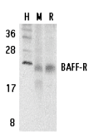
Left: Western blot analysis of BAFF Receptor in human (H), mouse (M), and rat (R) spleen tissue lysates with BAFF Receptor antibody at 5 µg/ml.
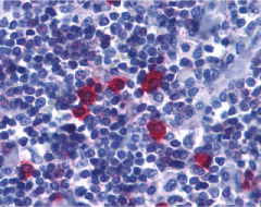
Below: Immunohistochemistry of BAFF Receptor in human tonsil tissue with BAFF Receptor antibody at 5 µg/ml.
Other Product Images
 Source : BAFF Receptor antibody was raised against a synthetic peptide corresponding to amino acids near the carboxy terminus of human BAFF Receptor The peptide sequence is identical between human and mouse origin.
Purification : Affinity chromatography purified via peptide column
Clonality and Clone : This is a polyclonal antibody.
Host : BAFF Receptor antibody was raised in rabbit. Please use anti-rabbit secondary antibodies.
Immunogen : Human BAFF Receptor (C-Terminus) Peptide (Cat. No. 3097P)
Application : BAFF Receptor antibody can be used for detection of BAFF Rexeptor by Western blot at 2 to 5 µg/ml.
Tested Application(s) : E, WB, IHC
Buffer : Antibody is supplied in PBS containing 0.02% sodium azide.
Blocking Peptide : Cat. No. 3097P - BAFF Receptor Peptide
Long-Term Storage : BAFF Receptor antibody can be stored at 4ºC, stable for one year. As with all antibodies care should be taken to avoid repeated freeze thaw cycles. Antibodies should not be exposed to prolonged high temperatures.
Positive Control
1. Cat. No. 1306 - Human Spleen Tissue Lysate
2. Cat. No. 1406 - Mouse Spleen Tissue Lysate
3. Cat. No. 1466 - Rat Spleen Tissue Lysate
4. Cat. No. 1315 - Human Tonsil Tissue Lysate
Species Reactivity :H, M, R
GI Number : 15208475
Accession Number : AAK91826
Short Description : (CT) Receptor for BAFF
References
1. Moore PA, Belvedere O, Orr A, et al. BLyS: member of the tumor necrosis factor family and B lymphocyte stimulator. Science 1999;285:260-3
2. Schneider P, MacKay F, Steiner V, et al. BAFF, a novel ligand of the tumor necrosis factor family, stimulates B cell growth. J Exp Med 1999;189:1747-56
3. Shu HB, Hu WH, Johnson H. TALL-1 is a novel member of the TNF family that is down-regulated by mitogens. J Leukoc Biol 1999;65:680-3
4. Mukhopadhyay A, Ni J, Zhai Y, Yu GL, Aggarwal BB. Identification and characterization of a novel cytokine, THANK, a TNF homologue that activates apoptosis, nuclear factor-kappaB, and c-Jun NH2-terminal kinase. J Biol Chem 1999 ;274:15978-81

 Description
 Source : IL-1RAcP antibody was raised against a peptide corresponding to amino acids near the carboxy terminus of IL-1RAcP, which differs from the sequence of mouse origin by three amino acids.