Tuesday, April 26, 2011

Neuritin Antibody


Catalog# :4101

As the nervous system of a complex organism develops, it establishes functional networks through the growth and retraction of synaptic connections from growing axons and dendrites. This synaptic remodeling involves neuro-transmitter signaling, activation of neurotrophin receptors and alterations in gene expression. One such gene whose expression is increased by neural activity is neuritin, a GPI-anchored protein that is expressed in postmitotic differentiating neurons of the developing nervous system. Its expression is also induced by the neurotrophins BDNF and NT-3. Purified recombinant neuritin promotes neurite outgrowth and arborization in primary embryonic neuronal cultures, suggesting that neuritin may play a role as a downstream effector of activity-induced neurite outgrowth. More recent experiments have shown that neuritin is required for the androgen-induced axonal elongation in motor neurons and is upregulated following spinal cord injury, suggesting that neuritin may also play a role in survival and axonal regeneration.

Additional Names : Neuritin, NRN1, cpg15


Description
Left: Western blot analysis of neuritin in Daudi cell lysate with neuritin antibody at (A) 5 and (B) 10 µg/ml.

Below:Immunohistochemistry of Neuritin in mouse heart tissue with Neuritin antibody at 5 µg/ml.

Other Product Images

Source :Neuritin antibody was raised against a 20 amino acid peptide from near the center of human neuritin.
Purification : Affinity chromatography purified via peptide column
Clonality and Clone : This is a polyclonal antibody.
Host : Neuritin antibody was raised in rabbit.
Please use anti-rabbit secondary antibodies
Application : Neuritin antibody can be used for detection of Neuritin by Western blot at 0.5 – 1 µg/ml.
Tested Application(s) : E, WB, IHC
Buffer : Antibody is supplied in PBS containing 0.02% sodium azide.
Blocking Peptide :Cat.No. 4101P - Neuritin Peptide
Long-Term Storage : Neuritin antibody can be stored at 4ºC, stable for one year. As with all antibodies care should be taken to avoid repeated freeze thaw cycles. Antibodies should not be exposed to prolonged high temperatures.
Positive Control :
1. Cat. No. 1224 - Daudi Cell Lysate
2. Cat. No. 1401 - Mouse Heart Tissue Lysate
Species Reactivity : H, M
GI Number : 7706123
Accession Number : NP_057672
Short Description : a neurite-outgrowth promoting protein
References
1. Li Z and Sheng M. Some assembly required: the development of neuronal synapses. Nat. Rev. Mol. Cell Biol. 2003; 4:833-41.
2. Naeve GS, Ramakrishnan M, Kramer R, et al. Neuritin: A gene induced by neural activity and neurotrophins that promotes neuritogenesis. Proc. Natl. Acad. Sci. USA 1997; 94:2648-53.
3. Marron TU, Guerini V, Rusmini P, et al. Androgen-induced neurite outgrowth is mediated by neuritin in motor neurons. J. Neurochem. 2005; 92:10-20.
4. Di Giovanni S, De Biase A, Yakovlev A, et al. In vivo and in vitro characterization of novel neuronal plasticity factors identified following spinal cord injury. J. Biol. Chem. 2005; 280:2084-91.

Monday, April 25, 2011

RNA Extraction


Biosyn Serving the DNA scientific community since 1984. It provides great resource of Ribosomal RNA services, RNA Service and RNA resources. Our every RNA product is designed and synthesized by professional scientists. Ribosomal RNA (rRNA) is the central component of the ribosome, the protein manufacturing machinery of all living cells.

RNA Separation
Several different possibilities exist to purify RNA from complete cell lysate (see the comparison above). For mammalian cell culture and tissues trizol/tri and silica columns (e.g. RNAeasy) are probably the most common.

RNA Extraction
RNA extraction is the purification of RNA from biological samples. This procedure is complicated by the ubiquitous presence of ribonuclease enzymes in cells and tissues, which can rapidly degrade RNA. Several methods are used in molecular biology to isolate RNA from samples, the most common of these is Guanidinium thiocyanate-phenol-chloroform extraction. This method often uses a proprietary formulation of this reagent called Trizol.

For More Information Visit: www.biosyn.com

NENF Antibody


Catalog# :4753

NENF is a secreted protein that is expressed in neurons but not glial cells of the brain. This protein has neurotrophic activity in primary cultured mouse neurons but not mitogenic activity in primary cultured mouse astrocytes. NENF activated mitogen-activated protein (MAP) and phosphotidylinositol-3 kinase pathways and could be inhibited by pertussis toxin but not receptor tyrosine kinases, suggesting that NENF acts through a Gi/Go-protein-coupled receptor. Despite its predicted molecular weight of ~20kDa, NENF is observed at 37kDa in SDS-PAGE.

Additional Names : NENF, Neuron derived neurotrophic factor, neudesin, CIR2, SPUF, SCIRP10

Description
Left: Western blot analysis of NENF in human kidney tissue lysate with NENF antibody at (A) 1 and (B) 2 µg/ml.

b>Below:Immunohistochemistry of NENF in rat kidney tissue with NENF antibody at 10 μg/ml.


Other Product Images

Source :NENF antibody was raised against a 15 amino acid peptide from near the carboxy terminus of human NENF.
Purification : Affinity chromatography purified via peptide column
Clonality and Clone : This is a polyclonal antibody.
Host : NENF antibody was raised in rabbit.
Please use anti-rabbit secondary antibodies.
Application : NENF antibody can be used for detection of NENF by Western blot at 1 – 2 µg/ml.
Tested Application(s) : E, WB, IHC
Buffer : Antibody is supplied in PBS containing 0.02% sodium azide.
Blocking Peptide :Cat.No. 4753P - NENF Peptide
Long-Term Storage : NENF antibody can be stored at 4ºC, stable for one year. As with all antibodies care should be taken to avoid repeated freeze thaw cycles. Antibodies should not be exposed to prolonged high temperatures.
Positive Control :
1. Cat. No. 1308 - Human Small Intestine Tissue Lysate
Species Reactivity : H, M, R
GI Number : 7019545
Accession Number : NP_037481
Short Description : Neuron derived neurotrophic factor
References
1. Kimura I, Yoshioka M, Konishi M, et al. Neudesin, a novel secreted protein with a unique primary structure and neurotrophic activity. J. Neurosci. Res. 2005; 79:287-94.

MANF Antibody


Catalog# :4349

MANF, also known as ARMET, was initially identified as a protein containing an arginine-rich region that was highly mutated in a variety of tumors. More recently it was identified as a mesencephalic astrocyte-derived neurotrophic factor with selectivity for dopaminergic neurons, similar to glial cell line-derived neurotrophic factor (GDNF) and CDNF. In rat brain slices, MANF enhanced nigral gamma-aminobutyric acid release. Like GDNF and CDNF, MANF has selective neuroprotective activity for dopaminergic neurons suggesting that it may be indicated for the treatment of Parkinson’s disease. Expression of MANF has also been shown to be induced during ER stress, suggesting that it may play a role in protein quality control during ER stress. This antibody does not cross-react with CDNF.

Additional Names : MANF (NT), Mesencephalic astrocyte-derived neurotrophic factor, arginine-rich mutatedin early stage tumor, ARMET, ARP


Description
Left: Western blot analysis of MANF in rat brain tissue lysate with MANF antibody at (A) 1 and (B) 2 µg/ml.

Below:Immunohistochemistry of MANF in human brain tissue with MANF antibody at 2.5 µg/ml.


Other Product Images


Source :MANF antibody was raised against a 12 amino acid peptide from near the amino terminus of human MANF.
Purification : Affinity chromatography purified via peptide column
Clonality and Clone : This is a polyclonal antibody.
Host : MANF antibody was raised in rabbit.
Please use anti-rabbit secondary antibodies.
Application : MANF antibody can be used for detection of MANF by Western blot at 1 – 2 µg/ml.
Tested Application(s) : E, WB, IHC
Buffer : Antibody is supplied in PBS containing 0.02% sodium azide.
Blocking Peptide :Cat.No. 4349P - MANF Peptide
Long-Term Storage : MANF antibody can be stored at 4ºC, stable for one year. As with all antibodies care should be taken to avoid repeated freeze thaw cycles. Antibodies should not be exposed to prolonged high temperatures.
Positive Control :
1. Cat. No. 1463 - Rat Brain Tissue Lysate
Species Reactivity : H, M, R
GI Number : 23503040
Accession Number : P55145
Short Description : (NT) Mesencephalic astrocyte-derived neurotrophic factor
References
1. Shridhar V, Rivard S, Shridhar R, et al. A gene from human chromosomal band 3p21.1 encodes a highly conserved arginine-rich protein and is mutated in renal cell carcinomas. Oncogene 1996; 12:1931-9.
2. Shridhar R, Shridhar V, Rivard S, et al. Mutations in the arginine-rich protein gene, in lung, breast, and prostate cancers, and in squamous cell carcinoma of the head and neck. Cancer Res. 1996; 56:5576-8.
3. Petrova P, Raibekas A, Pevsner J, et al. MANF: a new mesencephalic, astrocyte-derived neurotrophic factor with selectivity for dopaminergic neurons. J. Mol. Neurosci. 2003; 20:173-88.
4. Lindholm P, Voutilainen MH, Lauren J, et al. Novel neurotrophic factor CDNF protects and rescues midbrain dopamine neurons in vivo. Nature 2007; 448:73-7.

MANF Antibody


Catalog# :4347

MANF, also known as ARMET, was initially identified as a protein containing an arginine-rich region that was highly mutated in a variety of tumors. More recently it was identified as a mesencephalic astrocyte-derived neurotrophic factor with selectivity for dopaminergic neurons, similar to glial cell line-derived neurotrophic factor (GDNF) and CDNF. In rat brain slices, MANF enhanced nigral gamma-aminobutyric acid release. Like GDNF and CDNF, MANF has selective neuroprotective activity for dopaminergic neurons suggesting that it may be indicated for the treatment of Parkinson’s disease. Expression of MANF has also been shown to be induced during ER stress, suggesting that it may play a role in protein quality control during ER stress. This antibody does not cross-react with CDNF.

Additional Names : MANF (CT), Mesencephalic astrocyte-derived neurotrophic factor, arginine-rich mutatedin early stage tumor, ARMET, ARP


Description

Left: Western blot analysis of MANF in mouse brain tissue lysate with MANF antibody at (A) 1 and (B) 2 µg/ml.

Below:Immunohistochemistry of MANF in human brain tissue with MANF antibody at 2.5 µg/ml.


Other Product Images


Source :MANF antibody was raised against a 12 amino acid peptide from near the carboxy terminus of human MANF.
Purification : Affinity chromatography purified via peptide column
Clonality and Clone : This is a polyclonal antibody.
Host : MANF antibody was raised in rabbit.
Please use anti-rabbit secondary antibodies.
Application : MANF antibody can be used for detection of MANF by Western blot at 1 – 2 µg/ml.
Tested Application(s) : E, WB, IHC
Buffer : Antibody is supplied in PBS containing 0.02% sodium azide.
Blocking Peptide :Cat.No. 4347P - MANF Peptide
Long-Term Storage : MANF antibody can be stored at 4ºC, stable for one year. As with all antibodies care should be taken to avoid repeated freeze thaw cycles. Antibodies should not be exposed to prolonged high temperatures.
Positive Control :
1. Cat. No. 1403 - Mouse Brain Tissue Lysate
Species Reactivity : H, M, R
GI Number : 23503040
Accession Number : P55145
Short Description : (CT) Mesencephalic astrocyte-derived neurotrophic factor
References
1. Shridhar V, Rivard S, Shridhar R, et al. A gene from human chromosomal band 3p21.1 encodes a highly conserved arginine-rich protein and is mutated in renal cell carcinomas. Oncogene 1996; 12:1931-9.
2. Shridhar R, Shridhar V, Rivard S, et al. Mutations in the arginine-rich protein gene, in lung, breast, and prostate cancers, and in squamous cell carcinoma of the head and neck. Cancer Res. 1996; 56:5576-8.
3. Petrova P, Raibekas A, Pevsner J, et al. MANF: a new mesencephalic, astrocyte-derived neurotrophic factor with selectivity for dopaminergic neurons. J. Mol. Neurosci. 2003; 20:173-88.
4. Lindholm P, Voutilainen MH, Lauren J, et al. Novel neurotrophic factor CDNF protects and rescues midbrain dopamine neurons in vivo. Nature 2007; 448:73-7.

LRFN5 Antibody


Catalog# :5079

LRFN5 is one of a family of five transmembrane glycoproteins that are highly expressed in neuronal tissues. LRFN proteins share leucine-rich repeat (LRR)-immunoglobulin-like (Ig)-fibronectin type III (Fn)-transmembrane domain structure with other members of the LRR-Ig-Fn protein superfamily such as the Slitrk family of proteins. Expression of LRFN1, -3, and -4 mRNA was detected in embryonic neuronal cells, while LRFN2 and LRFN5 expression was primarily restricted to more mature cells. LRFN1, -2, and -4 bound to PDZ domains of postsynaptic PSD95, re-distributing PSD95 to the cell periphery. It has been suggested that the Lrfn proteins play a role in the developing and/or mature vertebrate nervous system. At least two isoforms of LRFN5 are known to exist. This antibody is predicted to not cross-react with other members of the LRFN family.


Additional Names : LRFN5, Leucine-rich repeat and fibronectin typeIII domain-containing protein 5, synaptic adhesion-like molecule 5, SALM5, FIGLER8


Description

Left: Western blot analysis of LRFN5 in EL4 cell lysate with LRFN5 antibody at (A) 0.5 and (B) 1 µg/ml.

Below:Immunohistochemistry of LRFN5 in mouse brain tissue with LRFN5 antibody at 5 µg/ml.


Other Product Images

Source :LRFN5 antibody was raised against a 16 amino acid peptide near the carboxy terminus of human LRFN5.
Purification : Affinity chromatography purified via peptide column
Clonality and Clone : This is a polyclonal antibody.
Host : LRFN5 antibody was raised in rabbit.
Please use anti-rabbit secondary antibodies.
Application : LRFN5 antibody can be used for detection of LRFN5 by Western blot at 0.5 – 1 µg/ml.
Tested Application(s) : E, WB, IHC
Buffer : Antibody is supplied in PBS containing 0.02% sodium azide.
Blocking Peptide :Cat.No. 5079P - LRFN5 Peptide
Long-Term Storage : LRFN5 antibody can be stored at 4ºC, stable for one year. As with all antibodies care should be taken to avoid repeated freeze thaw cycles. Antibodies should not be exposed to prolonged high temperatures.
Positive Control :
1. Cat. No. 1287 - EL4 Cell Lysate
Species Reactivity : H, M, R
GI Number : 116242620
Accession Number : Q96NI6
Short Description : Leucine-rich repeat and fibronectin type III domain-containing protein 5
References
1. Morimura N, Inoue T, Katayama K, et al. Comparative analysis of structure, expression and PSD95-binding capacity of Lrfn, a novel family of neuronal transmembrane proteins. Gene 2006; 380:72-83.
2. Kobe B and Kajava AV. The leucine-rich repeat as a protein recognition motif. Curr. Opin. Struct. Biol. 2001; 11:725-32.
3. Aruga J and Mikoshiba K. Identification and characterization of Slitrk, a novel transmembrane protein family controlling neurite outgrowth. Mol. Cell Neurosci. 2003; 24:117-29.

LRFN4 Antibody

LRFN4 Antibody

Catalog# :5077

LRFN4 is one of a family of five transmembrane glycoproteins that are highly expressed in neuronal tissues. LRFN proteins share leucine-rich repeat (LRR)-immunoglobulin-like (Ig)-fibronectin type III (Fn)-transmembrane domain structure with other members of the LRR-Ig-Fn protein superfamily such as the Slitrk family of proteins. Expression of LRFN1, -3, and -4 mRNA was detected in embryonic neuronal cells, while Lrfn2 and Lrfn5 expression was primarily restricted to more mature cells. LRFN1, -2, and -4 bound to PDZ domains of postsynaptic PSD95, re-distributing PSD95 to the cell periphery. It has been suggested that the Lrfn proteins play a role in the developing and/or mature vertebrate nervous system. At least two isoforms of LRFN4 are known to exist. This antibody is predicted to not cross-react with other members of the LRFN family.

Additional Names : LRFN4, Leucine-rich repeat and fibronectin typeIII domain-containing protein 4, synaptic adhesion-like molecule 3, SALM3, FIGLER6

Description
Left: Western blot analysis of LRFN4 in rat brain lysate with LRFN4 antibody at 1 µg/ml.

Below:Immunohistochemistry of LRFN4 in mouse brain tissue with LRFN4 antibody at 2.5 µg/ml.

Other Product Images


Source :
LRFN4 antibody was raised against a 14amino acid peptide near the carboxy terminus of human LRFN4.
Purification : Affinity chromatography purified via peptide column
Clonality and Clone : This is a polyclonal antibody.
Host : LRFN4 antibody was raised in rabbit.

Please use anti-rabbit secondary antibodies.
Application : LRFN4 antibody can be used for detection of LRFN4 by Western blot at 1 – 2 µg/ml.
Tested Application(s) : E, WB, IHC
Buffer : Antibody is supplied in PBS containing 0.02% sodium azide.
Blocking Peptide :Cat.No. 5077P - LRFN4 Peptide
Long-Term Storage : LRFN4 antibody can be stored at 4ºC, stable for one year. As with all antibodies care should be taken to avoid repeated freeze thaw cycles. Antibodies should not be exposed to prolonged high temperatures.
Positive Control :

1. Cat. No. 1463 - Rat Brain Tissue Lysate
Species Reactivity : H, M, R
GI Number : 62286924
Accession Number : Q6PJG9
Short Description : Leucine-rich repeat and fibronectin type III domain-containing protein 4
References
  1. Morimura N, Inoue T, Katayama K, et al. Comparative analysis of structure, expression and PSD95-binding capacity of Lrfn, a novel family of neuronal transmembrane proteins. Gene 2006; 380:72-83.
  2. Kobe B and Kajava AV. The leucine-rich repeat as a protein recognition motif. Curr. Opin. Struct. Biol. 2001; 11:725-32.
  3. Aruga J and Mikoshiba K. Identification and characterization of Slitrk, a novel transmembrane protein family controlling neurite outgrowth. Mol. Cell Neurosci. 2003; 24:117-29.

LRFN3 Antibody

LRFN3 Antibody

Catalog# :5069

LRFN3 is one of a family of five transmembrane glycoproteins that are highly expressed in neuronal tissues. LRFN proteins share leucine-rich repeat (LRR)-immunoglobulin-like (Ig)-fibronectin type III (Fn)-transmembrane domain structure with other members of the LRR-Ig-Fn protein superfamily such as the Slitrk family of proteins. Expression of LRFN1, -3, and -4 mRNA was detected in embryonic neuronal cells, while Lrfn2 and Lrfn5 expression was primarily restricted to more mature cells. LRFN1, -2, and -4 bound to PDZ domains of postsynaptic PSD95, re-distributing PSD95 to the cell periphery. It has been suggested that the Lrfn proteins play a role in the developing and/or mature vertebrate nervous system. This antibody is predicted to not cross-react with other members of the LRFN family.

Additional Names : LRFN3, Leucine-rich repeat and fibronectin type III domain-containing protein 3, synaptic adhesion-like molecule 4, SALM4, FIGLER1
Description
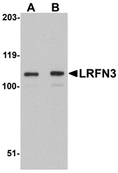
Left: Western blot analysis of LRFN3 in K562 lysate with LRFN3 antibody at (A) 1 and (B) 2 µg/ml.

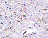
Below:Immunohistochemistry of LRFN3 in mouse brain tissue with LRFN3 antibody at 2.5 µg/ml.

Other Product Images

Source :LRFN3 antibody was raised against a 16 amino acid peptide near the center of human LRFN3.
Purification : Affinity chromatography purified via peptide column
Clonality and Clone : This is a polyclonal antibody.
Host : LRFN3 antibody was raised in rabbit.
Please use anti-rabbit secondary antibodies.
Application : LRFN3 antibody can be used for detection of LRFN3 by Western blot at 1 – 2 µg/ml.
Tested Application(s) : E, WB, IHC
Buffer : Antibody is supplied in PBS containing 0.02% sodium azide.
Blocking Peptide :Cat.No. 5069P - LRFN3 Peptide
Long-Term Storage : LRFN3 antibody can be stored at 4ºC, stable for one year. As with all antibodies care should be taken to avoid repeated freeze thaw cycles. Antibodies should not be exposed to prolonged high temperatures.
Positive Control :
1. Cat. No. 1204 - K562 Cell Lysate
Species Reactivity : H, M, R
GI Number : 13375646
Accession Number : NP_078785
Short Description : Leucine-rich repeat and fibronectin type III domain-containing protein 5
References
1. Morimura N, Inoue T, Katayama K, et al. Comparative analysis of structure, expression and PSD95-binding capacity of Lrfn, a novel family of neuronal transmembrane proteins. Gene 2006; 380:72-83.
2. Kobe B and Kajava AV. The leucine-rich repeat as a protein recognition motif. Curr. Opin. Struct. Biol. 2001; 11:725-32.
3. Aruga J and Mikoshiba K. Identification and characterization of Slitrk, a novel transmembrane protein family controlling neurite outgrowth. Mol. Cell Neurosci. 2003; 24:117-29.

LRFN2 Antibody

LRFN2 Antibody

Catalog# :5067

LRFN2 is one of a family of five transmembrane glycoproteins that are highly expressed in neuronal tissues. LRFN proteins share leucine-rich repeat (LRR)-immunoglobulin-like (Ig)-fibronectin type III (Fn)-transmembrane domain structure with other members of the LRR-Ig-Fn protein superfamily such as the Slitrk family of proteins. Expression of LRFN1, -3, and -4 mRNA was detected in embryonic neuronal cells, while Lrfn2 and Lrfn5 expression was primarily restricted to more mature cells. LRFN1, -2, and -4 bound to PDZ domains of postsynaptic PSD95, re-distributing PSD95 to the cell periphery. It has been suggested that the Lrfn proteins play a role in the developing and/or mature vertebrate nervous system. At least two isoforms of LRFN2 are known to exist. This antibody is predicted to not cross-react with other members of the LRFN family.

Additional Names : LRFN2, Leucine-rich repeat and fibronectin type III domain-containing protein 2, synaptic adhesion-like molecule 1
Description
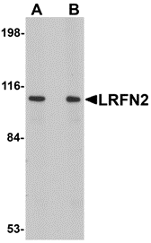
Left:Western blot analysis of LRFN2 in rat brain lysate with LRFN2 antibody at (A) 1 and (B) 2 μg/ml

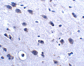
Below:Immunohistochemistry of LRFN2 in mouse brain tissue with LRFN2 antibody at 2.5 μg/ml.

Other Product Images

Source :LRFN2 antibody was raised against a 14 amino acid peptide near the carboxy terminus of human LRFN2.
Purification : Affinity chromatography purified via peptide column
Clonality and Clone : This is a polyclonal antibody.
Host : LRFN2 antibody was raised in rabbit.
Please use anti-rabbit secondary antibodies.
Application : LRFN2 antibody can be used for detection of LRFN2 by Western blot at 1 – 2 µg/ml.
Tested Application(s) : E, WB, IHC
Buffer : Antibody is supplied in PBS containing 0.02% sodium azide.
Blocking Peptide :Cat.No. 5067P - LRFN2 Peptide
Long-Term Storage : LRFN2 antibody can be stored at 4ºC, stable for one year. As with all antibodies care should be taken to avoid repeated freeze thaw cycles. Antibodies should not be exposed to prolonged high temperatures.
Positive Control :
1. Cat. No. 1463 - Rat Brain Tissue Lysate
Species Reactivity : H, M, R
GI Number : 62286951
Accession Number : Q9ULH4
Short Description : Leucine-rich repeat and fibronectin type III domain-containing protein 4
References
1. Morimura N, Inoue T, Katayama K, et al. Comparative analysis of structure, expression and PSD95-binding capacity of Lrfn, a novel family of neuronal transmembrane proteins. Gene 2006; 380:72-83.
2. Kobe B and Kajava AV. The leucine-rich repeat as a protein recognition motif. Curr. Opin. Struct. Biol. 2001; 11:725-32.
3. Aruga J and Mikoshiba K. Identification and characterization of Slitrk, a novel transmembrane protein family controlling neurite outgrowth. Mol. Cell Neurosci. 2003; 24:117-29.

LRFN1 Antibody

LRFN1 Antibody

Catalog# :5065

LRFN1 is one of a family of five transmembrane glycoproteins that are highly expressed in neuronal tissues. LRFN proteins share leucine-rich repeat (LRR)-immunoglobulin-like (Ig)-fibronectin type III (Fn)-transmembrane domain structure with other members of the LRR-Ig-Fn protein superfamily such as the Slitrk family of proteins. Expression of LRFN1, -3, and -4 mRNA was detected in embryonic neuronal cells, while Lrfn2 and Lrfn5 expression was primarily restricted to more mature cells. LRFN1, -2, and -4 bound to PDZ domains of postsynaptic PSD95, re-distributing PSD95 to the cell periphery. It has been suggested that the Lrfn proteins play a role in the developing and/or mature vertebrate nervous system. At least two isoforms of LRFN1 are known to exist. This antibody is predicted to not cross-react with other members of the LRFN family.

Additional Names : LRFN1, Leucine-rich repeat and fibronectin typeIII domain-containing protein 1, synaptic adhesion-like molecule 2
Description
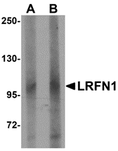
Left: Western blot analysis of LRFN1 in human brain lysate with LRFN1 antibody at (A) 1 and (B) 2 µg/ml.

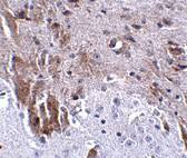
Below:Immunohistochemistry of LRFN1 in mouse brain tissue with LRFN1 antibody at 2.5 µg/ml.

Other Product Images
Source :LRFN1 antibody was raised against an 18 amino acid peptide near the carboxy terminus of human LRFN1.
Purification : Affinity chromatography purified via peptide column
Clonality and Clone : This is a polyclonal antibody.
Host : LRFN1 antibody was raised in rabbit.
Please use anti-rabbit secondary antibodies.
Application : LRFN1 antibody can be used for detection of LRFN1 by Western blot at 1 – 2 µg/ml.
Tested Application(s) : E, WB, IHC
Buffer : Antibody is supplied in PBS containing 0.02% sodium azide.
Blocking Peptide :Cat.No. 5065P - LRFN1 Peptide
Long-Term Storage : LRFN1 antibody can be stored at 4ºC, stable for one year. As with all antibodies care should be taken to avoid repeated freeze thaw cycles. Antibodies should not be exposed to prolonged high temperatures.
Positive Control :
1. Cat. No. 1303 - Human Brain Tissue Lysate
Species Reactivity : H, M, R
GI Number : 189028858
Accession Number : Q9P244
Short Description : Leucine-rich repeat and fibronectin type III domain-containing protein 1
References
1. Morimura N, Inoue T, Katayama K, et al. Comparative analysis of structure, expression and PSD95-binding capacity of Lrfn, a novel family of neuronal transmembrane proteins. Gene 2006; 380:72-83.
2. Kobe B and Kajava AV. The leucine-rich repeat as a protein recognition motif. Curr. Opin. Struct. Biol. 2001; 11:725-32.
3. Aruga J and Mikoshiba K. Identification and characterization of Slitrk, a novel transmembrane protein family controlling neurite outgrowth. Mol. Cell Neurosci. 2003; 24:117-29.