
Tuesday, May 3, 2011
RNA Cell
Biosynthesis can provide RNA, RNA analogs and chimeras in large quantities for your diagnostic and therapeutic research. Biosyn continues the tradition of providing researchers with the highest quality RNA Oligonucleotide synthesis, in quantities ranging from multi-mg to several grams. The quality of our product is controlled in compliance of ISO 9001/2000 and delivered with HPLC profile and MALDI-TOF characterization.
RNA Molecules
RNA molecules play critical and diverse roles in a staggering number of cellular processes. Processes of particular interest in the Department of Cell Biology include the examination of how RNA molecules fold within cells, how RNA-binding proteins recognize their RNA targets, how mRNAs are localized to discrete regions of cells and how small noncoding RNAs influence gene expression and cell function.
RNA Oligonucleotide Synthesis
Oligonucleotide synthesis is the non-biological, chemical synthesis of defined short sequences of nucleic acids. It is extremely useful in laboratory procedures covering a wide range of molecular biology applications. Automated synthesizers allow the synthesis of oligonucleotides up to 200 bases even longer.
RNA Research Analysis
Today RNA biology is becoming important in all areas of biomedical research. This is extremely useful for researchers who design molecules for RNA interference, and need to statistically test complex structure hypotheses.
For More information Visit : www.biosyn.com or call at :972-420-8505 !Now!


PEN2 Antibody
Catalog# :3979
PEN2, in addition to presenilin, nicastrin, and APH-1 forms the gamma-secretase protein complex, a membrane-bound aspartyl protease that can cleave certain proteins at peptide bonds buried within the hydrophobic environment of the lipid bilayer. This cleavage is responsible for a key step in signaling from several cell-surface receptors and is thought to be required for the generation of the neurotoxic amyloid peptides that are central to the pathogenesis of Alzheimer’s disease. Like the tumor necrosis factor-alpha-converting enzyme (TACE) and the beta-site cleavage enzyme (BACE) protease families, gamma-secretase will cleave the amyloid precursor protein (APP), but within the intramembrane region of APP, resulting in either the non-toxic p3 (from the alpha and gamma cleavage site) or the toxic Abeta amyloid peptide (from the beta and gamma cleavage site). It is thought that accumulation of the Abeta peptide is the precursor to Alzheimer’s disease.
Additional Names : PEN2 (CT), Presenilin enhancer protein 2, Gamma-secretase subunit PEN-2

Description
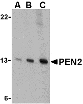
Left: Western blot analysis of PEN2 in K562 cell lysate with PEN2 antibody at (A) 0.5, (B) 1, and (C) 2 µg/ml.
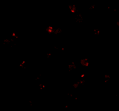
Below: Immunofluorescence of PEN2 in K562 cells with PEN2 antibody at 20 µg/ml.
Other Product Images
Description
Left: Western blot analysis of PEN2 in K562 cell lysate with PEN2 antibody at (A) 0.5, (B) 1, and (C) 2 µg/ml.
Below: Immunofluorescence of PEN2 in K562 cells with PEN2 antibody at 20 µg/ml.
Other Product Images
 Source :PEN2 antibody was raised against a 13 amino acid peptide from near the carboxy terminus of human PEN2.
Purification : Affinity chromatography purified via peptide column
Clonality and Clone : This is a polyclonal antibody.
Host : PEN2 antibody was raised in rabbit.
Please use anti-rabbit secondary antibodies
Application : PEN2 antibody can be used for detection of PEN2 by Western blot at 0.5 – 1 µg/ml.
Tested Application(s) : E, WB, IF
Buffer : Antibody is supplied in PBS containing 0.02% sodium azide.
Blocking Peptide :Cat.No. 3979P - PEN2 Peptide
Long-Term Storage : PEN2 antibody can be stored at 4ºC, stable for one year. As with all antibodies care should be taken to avoid repeated freeze thaw cycles. Antibodies should not be exposed to prolonged high temperatures.
Positive Control :
1.Cat. No. 1204 - K562 Cell Lysate
Species Reactivity : H, M, R
GI Number : 28144920
Accession Number : NP_758844
Short Description : (CT) a member of the gamma-secretase complex
References
1. Weihofen A and Martoglio B. Intramembrane-cleaving proteases: controlled liberation of proteins and bioactive peptides. Trends Cell Biol. 2003; 13:71-8.
2. Periz G and Fortini ME. Functional reconstitution of g-secretase through coordinated expression of presenilin, nicastrin, aph-1, and pen-2. J. Neurosci. Res. 2004; 77:309-22.
3. Selkoe DJ. The cell biology of b-amyloid precursor protein and presenilin in Alzheimer’s disease. Trends Cell Biol. 1998; 8:447-53.
4. Selkoe SJ. Translating cell biology into therapeutic advances in Alzheimer’s disease. Nature 1999; 399:A23-31
PDCD5 Antibody
Catalog# :5019
Programmed cell death 5 (PDCD5), a human apoptosis-related protein, is thought to play an early and universal role in apoptosis. PDCD5 is widely expressed and is upregulated in cells undergoing apoptosis, where it translocates rapidly from the cytoplasm to the nucleus. PDCD5 has a compact core structure of low flexibility with two mobile alpha-helices at N-terminal and a flexible unstructured C-terminal region. The charged residues are crucial for the ability of apoptosis-promoting and cell translocation of the protein. PDCD5 can facilitate apoptosis and enhance TAJ/TROY-induced paraptosis-like cell death. PDCD5 may play a dual role in the Tip60 pathway. It interacts with Tip60 and functions as a Tip60 co-activator to promote apoptosis (5). The nucleotide polymorphisms in the 5′-upstream region of PDCD5 affect promoter activity and the susceptibility of a Chinese population to develop chronic myelogenous leukemia and may represent a novel tumor suppressor gene influencing lung cancer.
Additional Names : PDCD5 (NT), Programmed cell death protein 5, TFAR19, MGC9294

Description
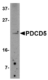
Left: Western blot analysis of PDCD5 in Jurkat cell lysate with PDCD5 antibody at 2.5 µg/ml.
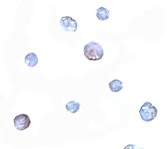
Below: Immunocytochemistry of PDCD5 in Jurkat cells with PDCD5 antibody at 5 μg/ml.
Other Product Images
Description
Left: Western blot analysis of PDCD5 in Jurkat cell lysate with PDCD5 antibody at 2.5 µg/ml.
Below: Immunocytochemistry of PDCD5 in Jurkat cells with PDCD5 antibody at 5 μg/ml.
Other Product Images
Source :PDCD5 antibody was raised against a 13 amino acid peptide from near the amino terminus of human PDCD5.
Purification : Affinity chromatography purified via peptide column
Clonality and Clone : This is a polyclonal antibody.
Host : PDCD5 antibody was raised in rabbit.
Please use anti-rabbit secondary antibodies.
Application : PDCD5 antibody can be used for detection of PDCD5 by Western blot at 1 µg/ml.
Tested Application(s) : E, WB, ICC
Buffer : Antibody is supplied in PBS containing 0.02% sodium azide.
Blocking Peptide :Cat.No. 5019P - PDCD5 Peptide
Long-Term Storage : PDCD5 antibody can be stored at 4ºC, stable for one year. As with all antibodies care should be taken to avoid repeated freeze thaw cycles. Antibodies should not be exposed to prolonged high temperatures.
Positive Control :
1. Cat. No. 1205 - Jurkat Cell Lysate
Species Reactivity : H, M, R
GI Number : 115430252
Accession Number : NP_037365
Short Description : (NT) Programmed cell death protein 5
References
1. Liu H, Wang Y, Zhang Y, et al. TFAR19, a novel apoptosis-related gene cloned from human leukemia cell line TF-1, could enhance apoptosis of some tumor cells induced by growth factor withdrawal. Biochem. Biophys. Res. Commun. 1999; 254:203–10.
2. Chen Y, Sun R, Han W, et al. Nuclear translocation of PDCD5 (TFAR19): an early signal for apoptosis? FEBS Lett. 2001; 509:191–6.
3. Yao H, Xu L, Feng Y, et al. Structure-function correlation of human programmed cell death 5 protein. Arch. Biochem. Biophys. 2009 Apr 7.
4. Wang Y, Li X, Wang L, et al. An alternative form of paraptosis-like cell death triggered by TAJ/TROY and enhanced by PDCD5 overexpression. J. Cell Sci. 2004; 117:1525–32.
PDCD5 Antibody
Catalog# :5011
Programmed cell death 5 (PDCD5), a human apoptosis-related protein, is thought to play an early and universal role in apoptosis. PDCD5 is widely expressed and is upregulated in cells undergoing apoptosis, where it translocates rapidly from the cytoplasm to the nucleus. PDCD5 has a compact core structure of low flexibility with two mobile alpha-helices at N-terminal and a flexible unstructured C-terminal region. The charged residues are crucial for the ability of apoptosis-promoting and cell translocation of the protein. PDCD5 can facilitate apoptosis and enhance TAJ/TROY-induced paraptosis-like cell death. PDCD5 may play a dual role in the Tip60 pathway. It interacts with Tip60 and functions as a Tip60 co-activator to promote apoptosis. The nucleotide polymorphisms in the 5′-upstream region of PDCD5 affect promoter activity and the susceptibility of a Chinese population to develop chronic myelogenous leukemia and may represent a novel tumor suppressor gene influencing lung cancer.
Additional Names : PDCD5 (CT), Programmed cell death protein 5, TFAR19, MGC9294

Description
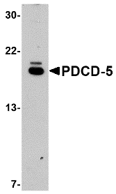
Left: Western blot analysis of PDCD5 in EL4 cell lysate with PDCD5 antibody at 1µg/ml.
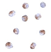
Below: Immunocytochemistry of PDCD5 in Jurkat cells with PDCD5 antibody at 2 µg/ml.
Other Product Images
 Source :PDCD5 antibody was raised against a 21 amino acid peptide from near the carboxy terminus of human PDCD5.
Purification : Affinity chromatography purified via peptide column
Clonality and Clone : This is a polyclonal antibody.
Host : PDCD5 antibody was raised in rabbit.
Please use anti-rabbit secondary antibodies.
Application : PDCD5 antibody can be used for detection of PDCD5 by Western blot at 1 µg/ml.
Tested Application(s) : E, WB, ICC
Buffer : Antibody is supplied in PBS containing 0.02% sodium azide.
Blocking Peptide :Cat.No. 5011P - PDCD5 Peptide
Long-Term Storage : PDCD5 antibody can be stored at 4ºC, stable for one year. As with all antibodies care should be taken to avoid repeated freeze thaw cycles. Antibodies should not be exposed to prolonged high temperatures.
Positive Control :
1.Cat. No. 1287 - EL4 Cell Lysate
Species Reactivity : H, M, R
GI Number : 115430252
Accession Number : NP_037365
Short Description : (CT) Spastic paraplegia 11
References
1. Liu H, Wang Y, Zhang Y, et al. TFAR19, a novel apoptosis-related gene cloned from human leukemia cell line TF-1, could enhance apoptosis of some tumor cells induced by growth factor withdrawal. Biochem. Biophys. Res. Commun. 1999; 254:203–10.
2. Chen Y, Sun R, Han W, et al. Nuclear translocation of PDCD5 (TFAR19): an early signal for apoptosis? FEBS Lett. 2001; 509:191–6.
3. Yao H, Xu L, Feng Y, et al. Structure-function correlation of human programmed cell death 5 protein. Arch. Biochem. Biophys. 2009 Apr 7.
4. Wang Y, Li X, Wang L, et al. An alternative form of paraptosis-like cell death triggered by TAJ/TROY and enhanced by PDCD5 overexpression. J. Cell Sci. 2004; 117:1525–32.
Description
Left: Western blot analysis of PDCD5 in EL4 cell lysate with PDCD5 antibody at 1µg/ml.
Below: Immunocytochemistry of PDCD5 in Jurkat cells with PDCD5 antibody at 2 µg/ml.
Other Product Images
 Source :PDCD5 antibody was raised against a 21 amino acid peptide from near the carboxy terminus of human PDCD5.
Purification : Affinity chromatography purified via peptide column
Clonality and Clone : This is a polyclonal antibody.
Host : PDCD5 antibody was raised in rabbit.
Please use anti-rabbit secondary antibodies.
Application : PDCD5 antibody can be used for detection of PDCD5 by Western blot at 1 µg/ml.
Tested Application(s) : E, WB, ICC
Buffer : Antibody is supplied in PBS containing 0.02% sodium azide.
Blocking Peptide :Cat.No. 5011P - PDCD5 Peptide
Long-Term Storage : PDCD5 antibody can be stored at 4ºC, stable for one year. As with all antibodies care should be taken to avoid repeated freeze thaw cycles. Antibodies should not be exposed to prolonged high temperatures.
Positive Control :
1.Cat. No. 1287 - EL4 Cell Lysate
Species Reactivity : H, M, R
GI Number : 115430252
Accession Number : NP_037365
Short Description : (CT) Spastic paraplegia 11
References
1. Liu H, Wang Y, Zhang Y, et al. TFAR19, a novel apoptosis-related gene cloned from human leukemia cell line TF-1, could enhance apoptosis of some tumor cells induced by growth factor withdrawal. Biochem. Biophys. Res. Commun. 1999; 254:203–10.
2. Chen Y, Sun R, Han W, et al. Nuclear translocation of PDCD5 (TFAR19): an early signal for apoptosis? FEBS Lett. 2001; 509:191–6.
3. Yao H, Xu L, Feng Y, et al. Structure-function correlation of human programmed cell death 5 protein. Arch. Biochem. Biophys. 2009 Apr 7.
4. Wang Y, Li X, Wang L, et al. An alternative form of paraptosis-like cell death triggered by TAJ/TROY and enhanced by PDCD5 overexpression. J. Cell Sci. 2004; 117:1525–32.
PCDH18 Antibody
Catalog# :5081
Protocadherins comprise the largest group within the cadherin family of calcium-dependent cell-cell adhesion molecules. Protocadherin 18 (PCDH18) was initially identified through screening of human and mouse ESTs using conserved cytoplasmic domain motifs. Tissue screening revealed PCDH18 broad expression in tissues such as brain, liver, heart, kidney, lung, and trachea. PCDH18 expression is also temporally and spatially regulated in the mouse embryonic brain and interacts with Disabled-1, an intracellular adapter protein involved in neuronal migration and cell positioning during mammalian brain development. PCDH18 was also found to be expressed in the developing zebrafish neural tube and central nervous system, lending support to the hypothesis that PCDH18 may play a role during brain development. At least three isoforms of PCDH18 are known to exist. This antibody is predicted to not cross-react with PCDH12.
Additional Names : PCDH18, Protocadherin 18
Description
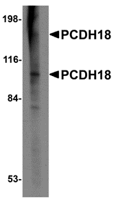
Left: Western blot analysis of PCDH18 in Raji cell lysate with PCDH18 antibody at 2 µg/ml.
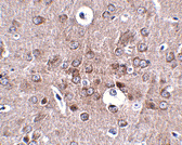
Below:Immunohistochemistry of PCDH18 in mouse brain tissue with PCDH18 antibody at 2.5 µg/ml.
Other Product Images
 Source :PCDH12 antibody was raised against a 17 amino acid peptide near the amino terminus of human PCDH12.
Purification : Affinity chromatography purified via peptide column
Clonality and Clone : This is a polyclonal antibody.
Host : PCDH18 antibody was raised in rabbit.
Application : PCDH18 antibody can be used for detection of PCDH18 by Western blot at 1 – 2 µg/ml.
Tested Application(s) : E, WB, IHC
Buffer : Antibody is supplied in PBS containing 0.02% sodium azide.
Blocking Peptide :Cat.No. 5081P - PCDH18 Peptide
Long-Term Storage : PCDH18 antibody can be stored at 4ºC, stable for one year. As with all antibodies care should be taken to avoid repeated freeze thaw cycles. Antibodies should not be exposed to prolonged high temperatures.
Positive Control :
1. Cat. No. 1207 - Raji Cell Lysate
Species Reactivity : H, M, R
GI Number : 14589929
Accession Number : NP_061908
Short Description : Protocadherin 18
References
1. Frank M and Kemler R. Protocadherins. Curr. Opin. Cell Biol. 2002; 14:557-62.
2. Wolverton T and Lalande M. Identification and characterization of three members of a novel subclass of protocadherins. Genomics 2001; 76:66-72.
3. Homayouni R, Rice DS, and Curran T. Disabled-1 interacts with a novel developmentally regulated protocadherin. Biochem. Biophys. Res. Commun. 2001; 289:539-47.
4. Kubota F, Murakami T, Tajika Y, et al. Expression of protocadherin 18 in the CNS and pharyngeal arches of zebrafish embryos. Int. J. Dev. Biol. 2008; 52:397-405.
Monday, May 2, 2011
Labelling Proteins
Bio-Synthesis offers a complete array of fluorescent reporters and quenchers. Our product offereing includes common dye modifiers such as 6-FAM, HEX and TET, as well as exotic modifictions. When labeling protein/peptide are needed, cross-linking two molecules by using a specific functional group such as an amine, thiol modified oligonucleotide with an certain cross linkers are employed.
Protein Synthesis
The process in which individual amino acids, whether of exogenous or endogenous origin, are connected to each other in peptide linkage in a specific order dictated by the sequence of nucleotides in DNA; this governing sequence is conveyed to the synthesizing apparatus in the ribosomes by mRNA, formed by base-pairing on the DNA template.
DNA Binding Proteins
Structural proteins that bind DNA are well-understood examples of non-specific DNA-protein interactions. Within chromosomes, DNA is held in complexes with structural proteins. These proteins organize the DNA into a compact structure called chromatin. In eukaryotes this structure involves DNA binding to a complex of small basic proteins called his tones, while in prokaryotes multiple types of proteins are involved.
Topoisomerases and Helicases
Topoisomerases are enzymes with both nuclease and ligase activity. These proteins change the amount of supercoiling in DNA. Some of these enzyme work by cutting the DNA helix and allowing one section to rotate, thereby reducing its level of super coiling; the enzyme then seals the DNA break.
PARL Antibody
Catalog# :4761
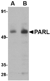
PARL is a mitochondrial integral membrane protein and a member of the highly conserved rhomboid superfamily of membrane proteins. PARL is required for the control of apoptosis during postnatal growth and is required for the processing of OPA1, a protein that prevents the release of cytochrome C from the mitochondria in response to apoptotic signals. In lymphocytes and neurons, PARL’s association with other proteins, such as the Bcl-2 family-related protein Hax1 and Omi can lead to the proteolytic processing of Omi by PARL, preventing the accumulation of Bax, thereby suppressing apoptosis. Variations in this gene have been associated with increased risk for type 2 diabetes. PARL may also play a role in the progression of Alzheimer’s disease through its associations with presenilin-1 and -2. Despite its predicted molecular weight of ~42kDa, PARL is observed at a higher molecular weight in SDS-PAGE. Multiple isoforms of PARL are known to exist.
Additional Names : PARL, Presenilin-associated rhomboid-like protein, PSARL, PSARL1, PSNENIP2

Description
Left: Western blot analysis of PARL in 3T3 cell lysate with PARL antibody at (A) 1 and (B) 2 µg/ml.
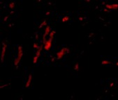
Below:Immunocytochemistry of PARL in 3T3 cells with PARL antibody at 2.5 μg/ml.
Other Product Images
 Source :PARL antibody was raised against a 15 amino acid peptide from near the amino terminus of human PARL.
Purification : Affinity chromatography purified via peptide column
Clonality and Clone : This is a polyclonal antibody.
Host : PARL antibody was raised in rabbit.
Please use anti-rabbit secondary antibodies
Application : PARL antibody can be used for detection of PARL by Western blot at 1 – 2 µg/ml.
Tested Application(s) : E, WB, ICC
Buffer : Antibody is supplied in PBS containing 0.02% sodium azide.
Blocking Peptide :Cat.No. 4761P - PARL Peptide
Long-Term Storage : PARL antibody can be stored at 4ºC, stable for one year. As with all antibodies care should be taken to avoid repeated freeze thaw cycles. Antibodies should not be exposed to prolonged high temperatures.
Positive Control :
1. Cat. No. 1282 - 3T3 (NIH) Cell Lysate
Species Reactivity : H, M, R
GI Number : 143811433
Accession Number : Q9H300
Short Description : Presenilin-associated rhomboid-like protein
References
1. Pellegrini L, Passer BJ, Canelles M, et al. PAMP and PARL, two novel putative metalloproteases interacting with the COOH-terminus of Presenilin-1 and -2. J. Alzheimers Dis. 2001; 3:181-90.
2. Cipolat S, Rudka T, Hartmann D, et al. Mitochondrial rhomboid PARL regulates cytochrome c release during apoptosis via OPA1-dependent cristae remodeling. Cell 2006; 126:163-75.
3. Chao JR, Parganas E, Boyd K, et al. Hax1-mediated processing of HtrA2 by PARL allows survival of lymphocytes and neurons. Nature 2008; 452:98-102.
4. Walder K, Kerr-Bayles L, Civitarese A, et al. The mitochondrial rhomboid protease PSARL is a new candidate gene for type 2 diabetes. Diabetologia 2005; 48:459-68.
Description
Left: Western blot analysis of PARL in 3T3 cell lysate with PARL antibody at (A) 1 and (B) 2 µg/ml.
Below:Immunocytochemistry of PARL in 3T3 cells with PARL antibody at 2.5 μg/ml.
Other Product Images
 Source :PARL antibody was raised against a 15 amino acid peptide from near the amino terminus of human PARL.
Purification : Affinity chromatography purified via peptide column
Clonality and Clone : This is a polyclonal antibody.
Host : PARL antibody was raised in rabbit.
Please use anti-rabbit secondary antibodies
Application : PARL antibody can be used for detection of PARL by Western blot at 1 – 2 µg/ml.
Tested Application(s) : E, WB, ICC
Buffer : Antibody is supplied in PBS containing 0.02% sodium azide.
Blocking Peptide :Cat.No. 4761P - PARL Peptide
Long-Term Storage : PARL antibody can be stored at 4ºC, stable for one year. As with all antibodies care should be taken to avoid repeated freeze thaw cycles. Antibodies should not be exposed to prolonged high temperatures.
Positive Control :
1. Cat. No. 1282 - 3T3 (NIH) Cell Lysate
Species Reactivity : H, M, R
GI Number : 143811433
Accession Number : Q9H300
Short Description : Presenilin-associated rhomboid-like protein
References
1. Pellegrini L, Passer BJ, Canelles M, et al. PAMP and PARL, two novel putative metalloproteases interacting with the COOH-terminus of Presenilin-1 and -2. J. Alzheimers Dis. 2001; 3:181-90.
2. Cipolat S, Rudka T, Hartmann D, et al. Mitochondrial rhomboid PARL regulates cytochrome c release during apoptosis via OPA1-dependent cristae remodeling. Cell 2006; 126:163-75.
3. Chao JR, Parganas E, Boyd K, et al. Hax1-mediated processing of HtrA2 by PARL allows survival of lymphocytes and neurons. Nature 2008; 452:98-102.
4. Walder K, Kerr-Bayles L, Civitarese A, et al. The mitochondrial rhomboid protease PSARL is a new candidate gene for type 2 diabetes. Diabetologia 2005; 48:459-68.
Nudel Antibody
Catalog# :3009
Nudel was initially discovered as a protein homologous to Aspergillus NUDE and that associated with Lis1 and polyprotein complex cytoplasmic dynein, both of which have been linked to neuronal development and migration (1,2). It was later shown to be a substrate of cdk5, a kinase known to be critical during neuronal migration; phosphorylation of Nudel by cdk5 affects its localization in neurons and affects neuritic morphology (1,3). It is thought that together with Lis1, Nudel regulates cytoplasmic dynein, a large polyprotein complex, in neuronal migration and mitosis through direct interactions (4). Similar interactions are thought to also play a role in membrane traffic in other cells as disruption of Nudel expression through RNA interference perturbed normal endomembrane flux and resulted in the fragmentation of the Golgi apparatus (3).
Additional Names : Nudel

Description
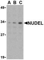
Left: Western blot analysis of Nudel in Jurkat whole cell lysate with Nudel antibody at (A) 0.5, (B) 1, or (C) 2 µg/ml.
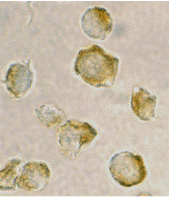
Below: Immunocytochemistry of Nudel in Jurkat cells with Nudel antibody at 2 µg/ml.
Other Product Images
 Source :Nudel antibody was raised against a peptide corresponding to 15 amino acids near the amino terminus of human Nudel.
Purification : Affinity chromatography purified via peptide column
Clonality and Clone : This is a polyclonal antibody.
Host : Nudel antibody was raised in rabbit.
Please use anti-rabbit secondary antibodies
Immunogen : Human Nudel Peptide (Cat. No. 3009P)
Application : Nudel antibody can be used for detection of Nudel by Western blot at 0.5 - 1 µg/ml.
Tested Application(s) : E, WB, ICC
Buffer : Antibody is supplied in PBS containing 0.02% sodium azide.
Blocking Peptide :Cat. No. 3009P - Nudel Peptide
Long-Term Storage : Nudel antibody can be stored at 4ºC, stable for one year. As with all antibodies care should be taken to avoid repeated freeze thaw cycles. Antibodies should not be exposed to prolonged high temperatures.
Positive Control :
1.Cat. No. 1205 - Jurkat Cell Lysate
Species Reactivity : H, M
GI Number : 12043569
Accession Number : AAF97497
Short Description : a protein involved in neuronal development
References
1. Niethammer H, Smith DS, Ayala R, et al. NUDEL is a novel Cdk5 substrate that associates with LIS1 and cytoplasmic dynein. Neuron 2000; 28:697-711.
2. Liu Z, Steward R, and Luo L. Drosophila Lis1 is required for neuroblast proliferation, dendritic elaboration and axonal transport. Nat. Cell Biol. 2000; 2:776-83.
3. Gupta A and Tsai LH. Cyclin-dependent kinase 5 and neuronal migration in the neocortex. Neurosignals 2003; 12:173-9.
4. Liang Y, Yu W, Li Y, et al. Nudel functions in membrane traffic mainly through association with Lis1 and cytoplasmic dynein. J. Cell Biol. 2004; 164:557-66.
NPTX2 Antibody
Catalog# :4573
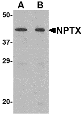
Neuronal pentraxin (NPTX) 2 was initially identified as a member of the pentraxin family having high homology to NPTX1. Unlike NPTX1 however, NPTX2 is expressed in testes, pancreas, liver, heart, skeletal muscle as well as brain. NPTX2 possesses the lectin properties common to the pentraxin family, promotes neurite outgrowth, and is rapidly regulated by neuronal activity. NPTX1 and NPTX2 form heterocomplexes that contribute to both activity-independent and –dependent excitatory synaptogenesis. Recently, NPTX2 has been found to be highly upregulated in Parkinsonian substantia nigra and localizes to Lewy bodies and Lewy neurites in sporadic Parkinson’s disease (PD), suggesting that it is involved in the pathway dysregulation that underlies PD. This NPTX2 antibody is predicted to be specific to NPTX2 and not recognize NPTX1.
Additional Names : NPTX2, Neuronal pentraxin 2, NP2, NP-II, neuronal activity-regulated protein, NARP
Description
Left:Western blot analysis of NPTX2 in mouse brain tissue lysate with NPTX2 antibody at (A) 0.5 and (B) 1 µg/ml.
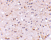
Below: Immunohistochemistry of NPTX2 in human brain with NPTX2 antibody at 5 µg/ml.
Other Product Images
 Source :NPTX2 antibody was raised against a 16 amino acid peptide near the center of the human NPTX2.
Purification : Affinity chromatography purified via peptide column
Clonality and Clone : This is a polyclonal antibody.
Host : NPTX2 antibody was raised in rabbit.
Please use anti-rabbit secondary antibodies.
Application : NPTX2 antibody can be used for detection of NPTX2 by Western blot at 1 – 2 µg/ml.
Tested Application(s) : E, WB, IHC
Buffer : Antibody is supplied in PBS containing 0.02% sodium azide.
Blocking Peptide :Cat.No. 4573P - NPTX2 Peptide
Long-Term Storage : NPTX2 antibody can be stored at 4ºC, stable for one year. As with all antibodies care should be taken to avoid repeated freeze thaw cycles. Antibodies should not be exposed to prolonged high temperatures.
Positive Control :
1. Cat. No. 1403 - Mouse Brain Tissue Lysate
Species Reactivity : H, M, R
GI Number : 28195384
Accession Number : NP_002514
Short Description : Neuronal pentraxin 2
References
1. Hsu YC and Perin MS. Human neuronal pentraxin II (NPTX2): conservation, genomic structure, and chromosomal localization. Genomics 1995; 28:220-7.
2. Tsu CC, Copeland GG, Gilbert DJ, et al. Narp, a novel member of the pentraxin family, promotes neurite outgrowth and is dynamically regulated by neuronal activity. J. Neurosci. 1996; 16:2463-78.
3. Xu D, Hopf C, Reddy R, et al. Narp and NP1 form heterocomplexes that function in developmental and activity-dependent synaptic plasticity. Neuron 2003; 39:513-28.
4. Moran LB, Hickey L, Michael GJ, et al. Neuronal pentraxin II is highly upregulated in Parkinson’s disease and a novel component of Lewy bodies. Acta Neuropathol. 2008; 115:471-8
NPAS3 Antibody
Catalog# :4109
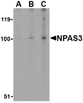
Neuronal PAS domain protein 3 (NPAS3) is a brain-enriched basic helix-loop-helix PAS domain transcription factor and is broadly expressed in the developing neuroepithelium and has recently found to be disrupted by genetic translocation in a family affected with schizophrenia. It was recently shown to be involved in the regulation of FGF signaling in the dentate gyrus by controlling the expression of the FGF receptor subtype 1 and in turn neurogenesis emanating from this region. NPAS3-null mice were growth-retarded and displayed brain defects that included reduced size of the anterior hippocampus, hypoplasia of the corpus callosum, and enlargement of the ventricles, as well as several behavioral abnormalities. Furthermore, these NPAS3-null mice also exhibited disruptions in several neurosignaling pathways involving glutamate, dopamine, and serotonin. These results demonstrate the essential role played by NPAS3 during structural and functional brain development. At least three isoforms of NPAS3 are known to exist.
Additional Names : NPAS3 (IN), Neuronal PAS domain-containing protein 3, member of Pas protein 6, MOP6
Description
Left: Western blot analysis of NPAS3 in rat brain tissue lysate with NPAS3 antibody at (A) 0.5, (B) 1 and (C) 2 µg/ml.
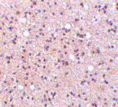
Below:Immunohistochemistry of NPAS3 in human brain tissue with NPAS3 antibody at 5 µg/ml.
Other Product Images
 Source :NPAS3 antibody was raised against a 28 amino acid peptide from near the center of human NPAS3.
Purification : Affinity chromatography purified via peptide column
Clonality and Clone : This is a polyclonal antibody.
Host : NPAS3 antibody was raised in rabbit.
Please use anti-rabbit secondary antibodies.
Application : NPAS3 antibody can be used for detection of NPAS3 by Western blot at 1 – 2 µg/ml.
Tested Application(s) : E, WB, IHC
Buffer : Antibody is supplied in PBS containing 0.02% sodium azide.
Blocking Peptide :Cat.No. 4109P - NPAS3 Peptide
Long-Term Storage : NPAS3 antibody can be stored at 4ºC, stable for one year. As with all antibodies care should be taken to avoid repeated freeze thaw cycles. Antibodies should not be exposed to prolonged high temperatures.
Positive Control :
1. Cat. No. 1463 - Rat Brain Tissue Lysate
2. Cat. No. 1303 - Human Brain Tissue Lysate
Species Reactivity : H, M, R
GI Number : 38604888
Accession Number : Q8IXF0
Short Description : (IN) Neuronal PAS domain-containing protein 3
References
1. Brunskill EW, Witte DP, Shreiner AB et al. Characterization of npas3, a novel basic helix-loop-helix PAS gene expressed in the developing mouse nervous system. Mech. Dev. 1999; 88:237-41.
2. Kamnasaran D, Muir WJ, Ferguson-Smith MA, et al. Disruption of the neuronal PAS3 gene in a family affected with schizophrenia. J. Med. Genet. 40:325-32.
3. Brunskill EW, Ehrman LA, Williams MT, et al. Abnormal neurodevelopment, neurosignaling and behaviour in Npas3-deficient mice. Euro. J. Neurosci. 2005; 22:1265-76.
Subscribe to:
Posts (Atom)