Tuesday, November 30, 2010

MATN3 Antibody

MATN3 Antibody

Catalog# :5127

Matrilins (MATNs) are a family of non-collagenous extra-cellular matrix (ECM) proteins consisting of four known members that have been proposed to play key roles in modulating cellular phenotypes during chondrogenesis of mesenchymal stem cells (MSCs). MATN1 and MATN3 are expressed specifically in cartilage and are among the most up-regulated ECM proteins during chondrogenesis. MATN3 is composed of a single N-terminal von Willebrand Factor A (vWFA) domain followed by four epidermal growth factor (EGF) repeats and a coiled-coil domain whereas MATN1 is composed of two vWFA domains separated by one EGF-like domain. MATN1 or MATN3 may play a role in modulating chondrogenesis during the chondrocyte differentiation process. Mutations of this gene have been associated with variety of inherited chondrodysplasias. Recent studies show that aberrant expression and processing of MATN3 are hallmarks of conventional cartilaginous neoplasms.

Additional Names : MATN3 (CT), Matrilin 3,HOA, OS2, EDM5


Description
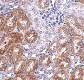
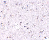
Left: Western blot analysis of MATN3 in 3T3 cell lysate with MATN3 antibody at (A) 1 and (B) 2 µg/ml.

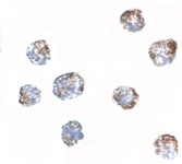
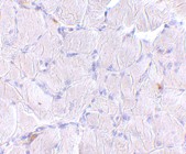
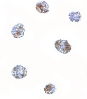
Below: Immunocytochemistry of MATN3 in 3T3 cells with MATN3 antibody at 5 μg/ml.

Other Product Images
Source :MATN3 antibody was raised against a 13 amino acid peptide from near the carboxy terminus of human MATN3.
Purification : Affinity chromatography purified via peptide column
Clonality and Clone : This is a polyclonal antibody.
Host : MATN3 antibody was raised in rabbit. Please use anti-rabbit secondary antibodies.
Application : MATN3 antibody can be used for detection of Matn3 by Western blot at 1 - 2 µg/ml.
Tested Application(s) : E, WB
Buffer : Antibody is supplied in PBS containing 0.02% sodium azide.
Blocking Peptide :Cat.No. 5127P - MATN3 Peptide
Long-Term Storage : MATN3 antibody can be stored at 4ºC, stable for one year. As with all antibodies care should be taken to avoid repeated freeze thaw cycles. Antibodies should not be exposed to prolonged high temperatures.
Positive Control :
1. Cat. No. 1282 - 3T3 (NIH) Cell Lysate
Species Reactivity : H, M, R
GI Number : 146218451
Accession Number : AAI39908
Short Description : (CT) Matrilin 3
References
1. Pei M, Luo J, and Chen Q. Enhancing and maintaining matrilins. Osteoarthritis Cartilage 2008; 16:1110-7.
2. Frank S, Schulthess T, Landwehr R, et al. Characterization of the matrilin coiled-coil domains reveals seven novel isoforms. J. Biol. Chem. 2002; 277:19071-9.
3. Chen Q, Johnson DM, Haudenschild DR, et al. Progression and recapitulation of the chondrocyte differentiation program: cartilage matrix protein is a marker for cartilage maturation. Dev. Biol. 1995; 172:293–306.
4. Stokes DG, Liu G, Coimbra IB, et al. Assessment of the gene expression profile of differentiated and dedifferentiated human fetal chondrocytes by microarray analysis. Arthritis Rheum 2002; 46:404–19.

Hydrolysis Synthesis


Hydrolysis is a chemical reaction during which one or more water molecules are split into hydrogen and hydroxide ions which may go on to participate in further reactions. It is the type of reaction that is used to break down certain polymers, especially those made by step-growth polymerization. Such polymer degradation is usually catalysed by either acid or alkali attack, often increasing with their strength or pH.

Hydrolysis of Amide Links
In the hydrolysis of an amide link into a carboxylic acid and an amine or ammonia, the carboxylic acid has a hydroxyl group derived from a water molecule and the amine (or ammonia) gains the hydrogen ion. A specific case of the hydrolysis of an amide link is the hydrolysis of peptides to smaller fragments or amino acids. Other polymers made by step-growth polymerization are susceptible to similar polymer degradation reactions. The problem is known as stress corrosion cracking.

Hydrolysis of polysaccharides

In polysaccharides, monosaccharide molecules are linked together by a Glycosidic bond. This bond can be cleaved by hydrolysis to yield monosaccharide. The best known disaccharide is sucrose, commonly known a sugar. Hydrolysis of sucrose yields glucose and fructose. There are many enzymes which speed up the hydrolysis of polysaccharides. Invertase is used industrially to hydrolyze sucrose to so-called invert sugar.

Monday, November 29, 2010

MATN3 Antibody

MATN3 Antibody

Catalog# :5141

Matrilins (MATNs) are a family of non-collagenous extra-cellular matrix (ECM) proteins consisting of four known members that have been proposed to play key roles in modulating cellular phenotypes during chondrogenesis of mesenchymal stem cells (MSCs). MATN1 and MATN3 are expressed specifically in cartilage and are among the most up-regulated ECM proteins during chondrogenesis. MATN3 is composed of a single N-terminal von Willebrand Factor A (vWFA) domain followed by four epidermal growth factor (EGF) repeats and a coiled-coil domain whereas MATN1 is composed of two vWFA domains separated by one EGF-like domain. MATN1 or MATN3 may play a role in modulating chondrogenesis during the chondrocyte differentiation process. Mutations of this gene have been associated with variety of inherited chondrodysplasias. Recent studies show that aberrant expression and processing of MATN3 are hallmarks of conventional cartilaginous neoplasms.

Additional Names : MATN3 (IN), Matrilin 3,HOA, OS2, EDM5

Description
Left: Western blot analysis of MATN3 in rat thymus tissue lysate with MATN3 antibody at (A) 1 and (B) 2 µg/ml.

Below: Immunocytochemistry of MATN3 in 3T3 cells tissue with MATN3 antibody at 2.5 μg/ml.

Other Product Images


Source :
MATN3 antibody was raised against a 13 amino acid peptide from near the center of human MATN3.
Purification : Affinity chromatography purified via peptide column
Clonality and Clone : This is a polyclonal antibody.
Host : MATN3 antibody was raised in rabbit. Please use anti-rabbit secondary antibodies.
Application : MATN3 antibody can be used for detection of MATN3 by Western blot at 1 - 2 µg/ml.
Tested Application(s) : E, WB
Buffer : Antibody is supplied in PBS containing 0.02% sodium azide.
Blocking Peptide :Cat.No. 5141P - MATN3 Peptide
Long-Term Storage : MATN3 antibody can be stored at 4ºC, stable for one year. As with all antibodies care should be taken to avoid repeated freeze thaw cycles. Antibodies should not be exposed to prolonged high temperatures.
Positive Control :
1. Cat. No. 1471 - Rat Thymus Tissue Lysate
Species Reactivity : H, M, R
GI Number : 146218451
Accession Number : AAI39908
Short Description : (IN) Matrilin 3
References

1. Pei M, Luo J, and Chen Q. Enhancing and maintaining matrilins. Osteoarthritis Cartilage 2008; 16:1110-7.
2. Frank S, Schulthess T, Landwehr R, et al. Characterization of the matrilin coiled-coil domains reveals seven novel isoforms. J. Biol. Chem. 2002; 277:19071-9.
3. Chen Q, Johnson DM, Haudenschild DR, et al. Progression and recapitulation of the chondrocyte differentiation program: cartilage matrix protein is a marker for cartilage maturation. Dev. Biol. 1995; 172:293–306.
4. Stokes DG, Liu G, Coimbra IB, et al. Assessment of the gene expression profile of differentiated and dedifferentiated human fetal chondrocytes by microarray analysis. Arthritis Rheum 2002; 46:404–19.

MATN2 Antibody

MATN2 Antibody

Catalog# :5105

Matrilin (MATNs) are a family of non-collagenous extracellular matrix proteins consisting of four known members that have been proposed to play key roles in the formation of both collagen-dependent and collagen-independent filamentous networks. The matrilin family all share a structure made up of von Willebrand factor A domains, epidermal growth factor-like domains and a coiled coil alpha-helical module. MATN1 and MATN3 are expressed mainly in cartilage, while MATN2 and MATN4 occur in a wide variety of extracellular matrices. The matrilin genes are strictly and differently regulated and their expression may serve as markers for cellular differentiation and diseases such as astrocytoma and liver carcinoma. Recent studies show that MATN2 is a permissive substrate for axonal growth and cell migration, and it is required for successful nerve regeneration.

Additional Names : MATN2, Matrilin 2


Description
Left: Western blot analysis of MATN2 in human liver tissue lysate with MATN2 antibody at (A) 1 and (B) 2 µg/ml.


Source :
MATN2 antibody was raised against a 15 amino acid peptide from near the carboxy terminus of human MATN2.
Purification : Affinity chromatography purified via peptide column
Clonality and Clone : This is a polyclonal antibody.
Host : MATN2 antibody was raised in rabbit. Please use anti-rabbit secondary antibodies.
Application : MATN2 antibody can be used for detection of MATN2 by Western blot at 1 - 2 µg/ml.
Tested Application(s) : E, WB
Buffer : Antibody is supplied in PBS containing 0.02% sodium azide.
Blocking Peptide :Cat.No. 5105P - MATN2 Peptide
Long-Term Storage : MATN2 antibody can be stored at 4ºC, stable for one year. As with all antibodies care should be taken to avoid repeated freeze thaw cycles. Antibodies should not be exposed to prolonged high temperatures.
Positive Control :
1. Cat. No. 1304 - Human Liver Tissue Lysate
Species Reactivity : H, M, R
GI Number : 62548862
Accession Number : NP_085072
Short Description : Matrilin 2
References

1. Pei M, Luo J, and Chen Q. Enhancing and maintaining chondrogenesis of synovial fibroblasts by cartilage extracellular matrix protein matrilins. Osteoarthritis Cartilage 2008; 16:1110-7.
2. Frank S, Schulthess T, Landwehr R, et al. Characterization of the Matrilin Coiled-coil Domains Reveals Seven Novel Isoforms. J. Biol. Chem. 2002; 277:19071-9.
3. Deak F, Wagener R, Kiss I, et al. The matrilins: a novel family of oligomeric extracellular matrix proteins. Matrix Biol. 1999; 18:55-64.
4. Szabo E, Korpos E, Batmunkh E, et al. Expression of matrilin-2 in liver cirrhosis and hepatocellular carcinoma. Pathol. Oncol. Res. 2008; 14:15-22.


MATN1 Antibody

MATN1 Antibody

Catalog# :5125

Matrilins (MATNs) are a family of non-collagenous extra-cellular matrix (ECM) proteins consisting of four known members that have been proposed to play key roles in modulating cellular phenotypes during chondrogenesis of mesenchymal stem cells (MSCs). MATN1 and MATN3 are expressed specifically in cartilage and are among the most up-regulated ECM proteins during chondrogenesis. MATN1 is composed of two Willebrand Factor A (vWFA) domains separated by one EGF-like domain, whereas MATN3 is composed of a single N-terminal vWFA domain followed by four epidermal growth factor (EGF) repeats and a coiled-coil domain. MATN1 or MATN3 may play a role in modulating chondrogenesis during the chondrocyte differentiation process. Mutations of this gene have been associated with variety of inherited chondrodysplasias. Recent studies show that the MATN1 promoter region was associated with both susceptibility and disease progression in Adolescent idiopathic scoliosis.

Additional Names : MATN1 (CT), Matrilin 1, Cartilage matrix protein, CMP, CRTM


Description
Left: Western blot analysis of MATN1 in rat liver tissue lysate with MATN1 antibody at (A) 1 and (B) 2 µg/ml.

Below: Immunohistochemistry of MATN1 in human liver tissue with MATN1 antibody at 5 µg/ml.

Other Product Images


Source :
MATN1 antibody was raised against a 13 amino acid peptide from near the carboxy terminus of human MATN1.
Purification : Affinity chromatography purified via peptide column
Clonality and Clone : This is a polyclonal antibody.
Host : MATN1 antibody was raised in rabbit. Please use anti-rabbit secondary antibodies.
Application : MATN1 antibody can be used for detection of MATN1 by Western blot at 1 - 2 µg/ml.
Tested Application(s) : E, WB, IHC
Buffer : Antibody is supplied in PBS containing 0.02% sodium azide.
Blocking Peptide :Cat.No. 5125P - MATN1 Peptide
Long-Term Storage : MATN1 antibody can be stored at 4ºC, stable for one year. As with all antibodies care should be taken to avoid repeated freeze thaw cycles. Antibodies should not be exposed to prolonged high temperatures.
Positive Control :
1. Cat. No. 1464 - Rat Liver Tissue Lysate
Species Reactivity : H, M, R
GI Number : 4505111
Accession Number : NP_002370
Short Description : (CT) Matrilin 1
References
1. Pei M, Luo J, and Chen Q. Enhancing and maintaining matrilins. Osteoarthritis Cartilage 2008; 16:1110-7.
2. Frank S, Schulthess T, Landwehr R, et al. Characterization of the matrilin coiled-coil domains reveals seven novel isoforms. J. Biol. Chem. 2002; 277:19071-9.
3. Chen Q, Johnson DM, Haudenschild DR, et al. Progression and recapitulation of the chondrocyte differentiation program: cartilage matrix protein is a marker for cartilage maturation. Dev. Biol. 1995; 172:293–306.
4. Stokes DG, Liu G, Coimbra IB, et al. Assessment of the gene expression profile of differentiated and dedifferentiated human fetal chondrocytes by microarray analysis. Arthritis Rheum 2002; 46:404–19.


Peptide Drug Development


Bio-Synthesis, founded in 1984, is an international provider of genomic and proteomic services established around the core business lines oligonucleotide and peptide synthesis. We offer wide variety of peptide modifications and labelings from small discovery research scale to multi-gram ASR/cGMP for clinical diagnostic production. As always, quality is guaranteed!

Protein-Protein Interactions

The interactions between proteins are important for many biological functions. For example, signals from the exterior of a cell are mediated to the inside of that cell by protein-protein interactions of the signaling molecules. This process, called signal transduction, plays a fundamental role in many biological processes and in many diseases (e.g. cancer). Proteins might interact for a long time to form part of a protein complex, a protein may be carrying another protein (for example, from cytoplasm to nucleus or vice versa in the case of the nuclear pore importins), or a protein may interact briefly with another protein just to modify it (for example, a protein kinase will add a phosphate to a target protein).

Protein Sequencing

The first step is to determine the amino acid composition of the peptide. We provide high throughput automation with more precise and powerful protein analysis. The protein of interest having been purified and its mass determined, the next analysis usually performed is to determine the protein's amino acid sequence, or primary structure.

Genetic Testing


There are about 900 genetic tests available at present times, these are tests on blood or other tissue to find genetic disorders. Bio-Synthesis offer DNA microarray printing and analysis for genetidc disease detection and analysis. Microarray technology makes it feasible to test for the presence of thousands of specific genetic abnormalities or variations simultaneously.

Genetic Testing

Genetic testing allows the genetic diagnosis of vulnerabilities to inherited diseases, and can also be used to determine a person's ancestry. Normally, every person carries two copies of every gene, one inherited from their mother, one inherited from their father. The human genome is believed to contain around 20,000 - 25,000 genes. In addition to studying chromosomes to the level of individual genes, genetic testing in a broader sense includes biochemical tests for the possible presence of genetic diseases, or mutant forms of genes associated with increased risk of developing genetic disorders.

Newborn Screening

Newborn screening is used just after birth to identify genetic disorders that can be treated early in life. The routine testing of infants for certain disorders is the most widespread use of genetic testing—millions of babies is tested each year in the United States. All states currently test infants for phenylketonuria (a genetic disorder that causes mental retardation if left untreated) and congenital hypothyroidism.

Friday, November 26, 2010

DNA RNA Sequence

DNA RNA Sequence

Bio-Synthesis, founded in 1984, is an international provider of genomic services established around the core business lines oligonucleotide and gene synthesis. Base, ribose and backbone modifications and reporter groups like dyes and haptens are routinely incorporated in DNA/RNA hybrid synthesis. Through innovative production process and synthesis know-how, our services are the best in their class, providing you with innovative and trustworthy solutions to accelerate your research.

DNA Synthesis

When DNA is synthesized the free 3´ hydroxyl (OH) group from the growing strand of DNA attacks the phosphate on the next base to be added (see the red arrows below). Pyrophosphate is released and the new base forms a phosphodiester bond with the growing strand of DNA. The free 3´ hydroxyl group is then free to attack the next base to be added. This reaction is catalyzed by DNA polymerases.

RNA Synthesis

RNA synthesis is said to proceed in the 5′ to 3′ direction, reflecting the fact that the attachment of new nucleotides always occurs at the 3′ hydroxyl group of the growing RNA chain. RNA synthesis goes through phases that are typical of polymerization processes: initiation, elongation, and termination, yielding an RNA product of defined size and sequence.

Read More : DNA RNA Sequence

Wednesday, November 24, 2010

DNA RNA Proteins


Biosynthesis has studied that the structure of DNA is actually a double helix each molecule of DNA is actually made up of 2 strands of DNA cross-linked together. Each nucleotide base in the DNA strand will cross-link (via hydrogen bonds) with a nucleotide base in a second strand of DNA forming a structure that resembles a ladder. Protein synthesis is a 2 part process that involves a second type of nucleic acid along with DNA. Biosynthesis provides custom oligonucleotide-peptide synthesis and conjugation services.

DNA Structure

DNA is actually a double helix each molecule of DNA is actually made up of 2 strands of DNA cross-linked together. Each nucleotide base in the DNA strand will cross-link (via hydrogen bonds) with a nucleotide base in a second strand of DNA forming a structure that resembles a ladder.

Protein Synthesis

The process in which individual amino acids, whether of exogenous or endogenous origin, are connected to each other in peptide linkage in a specific order dictated by the sequence of nucleotides in DNA; this governing sequence is conveyed to the synthesizing apparatus in the ribosomes by mRNA, formed by base-pairing on the DNA template. A process where information is taken from DNA to acts as a blue print for creating a particular protein that is in demand by the body. This blueprint will allow the construction of the protein with the various materials required in its production.

DNA Label

DNA Label
Bio-Synthesis, founded in 1984, is an international provider of genomic services established around the core business lines oligonucleotide and gene synthesis. Base, ribose and backbone modifications and reporter groups like dyes and haptens are routinely incorporated in DNA, RNA, LNA, and BNA. Through innovative production process and synthesis know-how, our services are the best in their class, providing you with innovative and trustworthy solutions to accelerate your research.

DNA Binding Proteins
Structural proteins that bind DNA are well-understood examples of non-specific DNA-protein interactions. Within chromosomes, DNA is held in complexes with structural proteins. These proteins organize the DNA into a compact structure called chromatin. In eukaryotes this structure involves DNA binding to a complex of small basic proteins called his tones, while in prokaryotes multiple types of proteins are involved.

DNA Modifying Enzymes
Nucleases are enzymes that cut DNA strands by catalyzing the hydrolysis of the phosphodiester bonds. Nucleases that hydrolyse nucleotides from the ends of DNA strands are called exonucleases, while endonucleases cut within strands.

Topoisomerases and Helicases
Topoisomerases are enzymes with both nuclease and ligase activity. These proteins change the amount of super coiling in DNA. Some of this enzyme work by cutting the DNA helix and allowing one section to rotate, thereby reducing its level of super coiling; the enzyme then seals the DNA break.

Monday, November 22, 2010

DNA Sequencing Companies


Bio-Synthesis, founded in 1984 , is an international provider of genomic and proteomic provider established around the core business lines Oligonucleotide, peptide synthesis, antibody productions and analytical services such as DNA, peptide, protein sequencing, microarray printing, screening and analysis.

DNA Sequencing

The term DNA sequencing encompasses biochemical methods for determining the order of the nucleotide bases, adenine, guanine, cytosine, and thymine, in a DNA oligonucleotide. The sequence of DNA constitutes the heritable genetic information in nuclei, plasmids, mitochondria, and chloroplasts that forms the basis for the developmental programs of all living organisms. Determining the DNA sequence is therefore useful in basic research studying fundamental biological processes, as well as in applied fields such as diagnostic or forensic research. The advent of DNA sequencing has significantly accelerated biological research and discovery.

Dye-terminator Sequencing

An alternative to primer labelling is labelling of the chain terminators, a method commonly called 'dye-terminator sequencing'. The major advantage of this method is that the sequencing can be performed in a single reaction, rather than four reactions as in the labelled-primer method. In dye-terminator sequencing, each of the four dideoxynucleotide chain terminators is labelled with a different fluorescent dye, each fluorescing at a different wavelength.

Gene Hybridization


Hybridization is the process, discovered by Alexander Rich, of combining complementary, single-stranded nucleic acids into a single molecule. Nucleotides will bind to their complement under normal conditions, so two perfectly complementary strands will bind to each other readily. This is called annealing. However, due to the different molecular geometries of the nucleotides, a single inconsistency between the two strands will make binding between them more energetically unfavorable.

Hybridization Probe

In molecular biology, a hybridization probe is a fragment of DNA of variable length (usually 100-1000 bases long), which is used to detect in DNA or RNA samples the presence of nucleotide sequences (the DNA target) that are complementary to the sequence in the probe. The probe thereby hybridizes to single-stranded nucleic acid (DNA or RNA) whose base sequence allows probe-target base pairing due to complementarily between the probe and target. The labeled probe is first denatured (by heating or under alkaline conditions) into single DNA strands and then hybridized to the target DNA (Southern blotting) or RNA (northern blotting) immobilized on a membrane or in situi.

Immunohistochemistry

Immunohistochemistry is a technique for identifying cellular or tissue constituents (antigens) by means of antigen-antibody interactions, the site of antibody binding being identify Either by direct labelling of the antibody, or by use of a secondary labelling method.

Thursday, November 18, 2010

DNA Oligo


For over 20 years, BioSynthesis has provided custom oligonucleotide production services worldwide, including researcher at university, biotechnology and pharmaceutical institutions. We offer wide variety of oligo modifications and labelings from small discovery research scale to multi-gram ASR/cGMP for clinical diagnostic production. As always, quality is guaranteed!

Deoxyribonucleic Acid

A deoxyribonucleic acid (DNA) or ribonucleic acid (RNA) sequence composed of two or more covalently linked nucleotides. Oligonucleotides are classified as deoxyribooligonucleotides or ribooligonucleotides. Fragments containing up to 50 nucleotides are generally termed oligonucleotides, and longer fragments are called polynucleotides. Gene synthesis from synthetic deoxyoligonucleotides is now routinely used to prepare genes and modified genes for proteins having potential clinical applications. Oligonucleotides have also been used to diagnose genetic disorders and bacterial or viral infections. Oligonucleotides are sometimes referred to as oligos.

Deoxyribooligonucleotide

A deoxyribooligonucleotide consists of a 5-carbon sugar called deoxyribose joined covalently to phosphate at the 5′ and 3′ carbons of this sugar to form an alternating, unbranched polymer. A ribooligonucleotide consists of a similar repeating structure where the 5-carbon sugar is ribose. Chemically synthesized oligonucleotides of predetermined sequence have proven to be very useful for studying a large number of biochemical processes. In the 1960s, these compounds were used to decipher the genetic code. Later, chemically prepared deoxyoligonucleotides were joined to form genes for transfer RNAs.

MATN1 Antibody

MATN1 Antibody

Catalog# : 5139

Matrilins (MATNs) are a family of non-collagenous extra-cellular matrix (ECM) proteins consisting of four known members that have been proposed to play key roles in modulating cellular phenotypes during chondrogenesis of mesenchymal stem cells (MSCs). MATN1 and MATN3 are expressed specifically in cartilage and are among the most up-regulated ECM proteins during chondrogenesis. MATN1 is composed of two Willebrand Factor A (vWFA) domains separated by one EGF-like domain, whereas MATN3 is composed of a single N-terminal vWFA domain followed by four epidermal growth factor (EGF) repeats and a coiled-coil domain. MATN1 or MATN3 may play a role in modulating chondrogenesis during the chondrocyte differentiation process. Mutations of this gene have been associated with variety of inherited chondrodysplasias. Recent studies show that the MATN1 promoter region was associated with both susceptibility and disease progression in Adolescent idiopathic scoliosis.

Additional Names : MATN1 (NT), Matrilin 1, Cartilage matrix protein, CMP, CRTM

Description
Left: Western blot analysis of MATN1 in mouse liver tissue lysate with MATN1 antibody at (A) 1 and (B) 2 µg/ml.


Source :
MATN1 antibody was raised against a 12 amino acid peptide from near the amino terminus of human MATN1.
Purification : Affinity chromatography purified via peptide column
Clonality and Clone : This is a polyclonal antibody.
Host : MATN1 antibody was raised in rabbit. Please use anti-rabbit secondary antibodies.
Application : MATN1 antibody can be used for detection of MATN1 by Western blot at 1 - 2 µg/ml.
Tested Application(s) : E, WB
Buffer : Antibody is supplied in PBS containing 0.02% sodium azide.
Blocking Peptide :Cat.No. 5139P - MATN1 Peptide
Long-Term Storage : MATN1 antibody can be stored at 4ºC, stable for one year. As with all antibodies care should be taken to avoid repeated freeze thaw cycles. Antibodies should not be exposed to prolonged high temperatures.
Positive Control :
1. Cat. No. 1404 - Mouse Liver Tissue Lysate
Species Reactivity : H, M, R
GI Number : 4505111
Accession Number : NP_002370
Short Description : (NT) Matrilin 1
References
1. Pei M, Luo J, and Chen Q. Enhancing and maintaining matrilins. Osteoarthritis Cartilage 2008; 16:1110-7.
2. Frank S, Schulthess T, Landwehr R, et al. Characterization of the matrilin coiled-coil domains reveals seven novel isoforms. J. Biol. Chem. 2002; 277:19071-9.
3. Chen Q, Johnson DM, Haudenschild DR, et al. Progression and recapitulation of the chondrocyte differentiation program: cartilage matrix protein is a marker for cartilage maturation. Dev. Biol. 1995; 172:293–306.
4. Stokes DG, Liu G, Coimbra IB, et al. Assessment of the gene expression profile of differentiated and dedifferentiated human fetal chondrocytes by microarray analysis. Arthritis Rheum 2002; 46:404–19.

LSD1 Antibody

LSD1 Antibody

Catalog# :3761

Histone modifications mediate changes in gene expression by altering chromatin structure or by serving as a platform to recruit other proteins. LSD1 is a recently discovered amine oxidase that catalyzes the lysine-specific demethylation of histone proteins via an FAD-dependent oxidative reaction (1). Methylation on histone H3-K9 is thought to play an important role in heterochromatin formation, while methylation on arginine and some lysine residues (such as H3-K4) is associated with active transcription (2). LSD1 associates with various proteins, including HDAC1/2, CoREST, and BHC80, that act to regulate LSD1 activity in vivo, and in a histone H3-K4-specific methylase complex that is involved in transcriptional regulation (3,4). Experiments have shown that CoREST, a SANT domain-containing corepressor (5) acts to enhance LSD1 activity, while BHC80, a PHD domain-containing protein (6), inhibits CoREST/LSD1 activity in vitro (3). LSD1-mediated histone demethylation thus may have significant effects on gene expression.

Additional Names : LSD1 (NT), Lysine-specific histone demethylase, amine oxidase flavin containing domain protein 2, AOF2

Description
Left: Western blot analysis of LSD1 in P815 cell lysate with LSD1 antibody at (A) 1 and (B) 2 µg/ml.


Source :
LSD1 antibody was raised against a 17 amino acid peptide from near the amino terminus of human LSD1.
Purification : Affinity chromatography purified via peptide column
Clonality and Clone : This is a polyclonal antibody.
Host : LSD1 antibody was raised in rabbit.Please use anti-rabbit secondary antibodies.
Immunogen : Human LSD1 (N-Terminus) Peptide (Cat. No. 3761P)
Application : LSD1 antibody can be used for the detection of LSD1 by Western blot at 1 – 2 µg/ml.
Tested Application(s) : E, WB
Buffer : Antibody is supplied in PBS containing 0.02% sodium azide.
Blocking Peptide :Cat. No. 3761P - LSD1 Peptide
Long-Term Storage : LSD1 antibody can be stored at 4ºC, stable for one year. As with all antibodies care should be taken to avoid repeated freeze thaw cycles. Antibodies should not be exposed to prolonged high temperatures.
Positive Control :
1. Cat. No. 1286 - P815 Cell Lysate
Species Reactivity : H, M, R
GI Number : 58761544
Accession Number : NP_001009999
Short Description : (NT) A histone demethylase
References
1. Shi Y, Lan F, Matson C, et al. Histone demethylation mediated by the nuclear amine oxidase homolog LSD1. Cell 2004; 119:941-53.
2. Kouzarides T. Histone methylation in transcriptional control. Curr. Opin. Genet. Dev. 2002; 12:198-209.
3. Shi YJ, Matson C, Lan F, et al. Regulation of LSD1 histone demethylase activity by its associated factors. Mol. Cell 2005; 19:857-64.
4. Nakamura T, Mori T, Tada S, et al. ALL-1 is a histone methyltransferase that assembles a supercomplex of proteins involved in transcriptional regulation. Mol. Cell 2002; 10:1119-28.

LSD1 Antibody

LSD1 Antibody

Catalog# :3763

Histone modifications mediate changes in gene expression by altering chromatin structure or by serving as a platform to recruit other proteins. LSD1 is a recently discovered amine oxidase that catalyzes the lysine-specific demethylation of histone proteins via an FAD-dependent oxidative reaction (1). Methylation on histone H3-K9 is thought to play an important role in heterochromatin formation, while methylation on arginine and some lysine residues (such as H3-K4) is associated with active transcription (2). LSD1 associates with various proteins, including HDAC1/2, CoREST, and BHC80, that act to regulate LSD1 activity in vivo, and in a histone H3-K4-specific methylase complex that is involved in transcriptional regulation (3,4). Experiments have shown that CoREST, a SANT domain-containing corepressor (5) acts to enhance LSD1 activity, while BHC80, a PHD domain-containing protein (6), inhibits CoREST/LSD1 activity in vitro (3). LSD1-mediated histone demethylation thus may have significant effects on gene expression.

Additional Names : LSD1 (IN), Lysine-specific histone demethylase, amine oxidase flavin containing domain protein 2, AOF2
Description
Left: Western blot analysis of LSD1 in P815 cell lysate with LSD1 antibody at (A) 1 and (B) 2 µg/ml.

Below: Immunohistochemistry of LSD1 in human small intestine tissue with LSD1 antibody at 2 µg/ml.

Other Product Images
Source :
LSD1 antibody was raised against a 16 amino acid peptide from near the center of human LSD1.
Purification : Affinity chromatography purified via peptide column
Clonality and Clone : This is a polyclonal antibody.
Host : LSD1 antibody was raised in rabbit. Please use anti-rabbit secondary antibodies.
Immunogen : Human LSD1 (Intermediate Domain) Peptide (Cat. No. 3763P)
Application : LSD1 antibody can be used for the detection of LSD1 by Western blot at 1 – 2 µg/ml.
Tested Application(s) : E, WB, IHC
Buffer : Antibody is supplied in PBS containing 0.02% sodium azide.
Blocking Peptide :Cat. No. 3763P - LSD1 Peptide
Long-Term Storage : LSD1 antibody can be stored at 4ºC, stable for one year. As with all antibodies care should be taken to avoid repeated freeze thaw cycles. Antibodies should not be exposed to prolonged high temperatures.
Positive Control :
1. Cat. No. 1286 - P815 Cell Lysate
2. Cat. No. 1308 - Human Small Intestine Tissue Lysate
Species Reactivity : H, M, R
GI Number : 58761544
Accession Number : NP_001009999
Short Description : (IN) A histone demethylase
References
1. Shi Y, Lan F, Matson C, et al. Histone demethylation mediated by the nuclear amine oxidase homolog LSD1. Cell 2004; 119:941-53.
2. Kouzarides T. Histone methylation in transcriptional control. Curr. Opin. Genet. Dev. 2002; 12:198-209.
3. Shi YJ, Matson C, Lan F, et al. Regulation of LSD1 histone demethylase activity by its associated factors. Mol. Cell 2005; 19:857-64.
4. Nakamura T, Mori T, Tada S, et al. ALL-1 is a histone methyltransferase that assembles a supercomplex of proteins involved in transcriptional regulation. Mol. Cell 2002; 10:1119-28.

LIMP2 Antibody

LIMP2 Antibody

Catalog# :4621

The lysosomal integral membrane protein 2 (LIMP2) is a heavily glycosylated type III transmembrane protein, the majority of which exists in the lumen of the lysosome and a cytoplasmic domain of approximately 20 amino acids. A deficiency of LIMP2 in mice causes uretic pelvic junction obstruction, deafness, and peripheral neuropathy associated with impaired vesicular trafficking and distribution of apically expressed proteins. More recently, LIMP2 was shown to act as a receptor to bind b-glucocerebrosidase, the enzyme defective in Gaucher disease, a lysosomal storage disorder. LIMP2-deficient mice showed missorted as well as secreted b-glucocerebrosidase, suggesting that LIMP2 also functions as the mannose-6-phosphate-independent trafficking receptor. Despite its predicted molecular weight, LIMP2 runs at approximately 80 – 85 kDa in SDS-PAGE.

Additional Names : LIMP2, Lysosomal integral membrane protein 2, CD36L2, Scavenger receptor class B member 2, SR-B2, SCARB2

Description
Left: Western blot analysis of LIMP2 in human skeletal muscle tissue lysate with LIMP2 antibody at 1 µg/ml in (A) the absence and (B) presence of blocking peptide.

Below: Immunohistochemistry of LIMP2 in human skeletal muscle with LIMP2 antibody at 2.5 µg/ml.

Other Product Images
Source :LIMP2 antibody was raised against a 16 amino acid peptide from near the center of human LIMP2.
Purification : Affinity chromatography purified via peptide column
Clonality and Clone : This is a polyclonal antibody.
Host : LIMP2 antibody was raised in rabbit. Please use anti-rabbit secondary antibodies.
Application : LIMP2 antibody can be used for detection of LIMP2 by Western blot at 1 – 2 µg/ml.
Tested Application(s) : E, WB, IHC
Buffer : Antibody is supplied in PBS containing 0.02% sodium azide.
Blocking Peptide :Cat.No. 4621P - LIMP2 Peptide
Long-Term Storage : LIMP2 antibody can be stored at 4ºC, stable for one year. As with all antibodies care should be taken to avoid repeated freeze thaw cycles. Antibodies should not be exposed to prolonged high temperatures.
Positive Control :
1. Cat. No. 1375 - Human Skeletal Muscle Tissue Lysate
Species Reactivity : H, M, R
GI Number : 18257312
Accession Number : AAH21892
Short Description : Lysosomal integral membrane protein 2
References
1. Fujita H, Saeki M, Yasunaga K, et al. Isolation and sequencing of a cDNA clone encoding 85kDa sialoglycoprotein in rat liver lysosomal membranes. Biochem. Biophys. Res. Commun. 1991; 178:444-52.
2. Gamp A, Tanaka Y, Lullmann-Rauch R, et al. LIMP-2/LGP85 deficiency causes uretic pelvic junction obstruction, deafness and peripheral neuropathy in mice. Hum. Mol. Genet. 2003; 12:631-46.
3. Knipper M, Claussen C, Ruttiger L, et al. Deafness in LIMP2-deficient mice due to early loss of the potassium channel KCNQ1/KCNE1 in marginal cells of the stria vascularis. J. Physiol. 2006; 576:73-86.
4. Reczek D, Schwake M, Schroder J, et al. LIMP-2 is a receptor for lysosomal mannose-6-phosphate-independent targeting of b-glucocerebrosidase. Cell 2007; 131:770-83.

A peptomimetic inhibitor of BCL6 with potent antilymphoma effects in vitro and in vivo

A peptomimetic inhibitor of BCL6 with potent antilymphoma effects in vitro and in vivo

The BCL6 transcriptional repressor is the most commonly involved oncogene in diffuse large B-cell lymphomas (DLBCLs). BCL6 lymphomagenic activity is dependent on its ability to recruit corepressor proteins to a unique binding site on its N-terminal BTB domain. A recombinant peptide fragment of the SMRT (silencing mediator for retinoid and thyroid hormone receptor) corepressor that blocks this site can inhibit BCL6 biologic functions. Shortening and conversion of this peptide to D-amino acid and retro configuration as well as the addition of a fusogenic motif yielded a far more potent and stable BCL6 inhibitor that still retained the specificity of the original SMRT fragment. Like the L-peptide, retroinverso BCL6 peptide inhibitor (RI-BPI) selectively killed BCR rather than OxPhos-type DLBCL cells. The RI-BPI could recapitulate the failure to form germinal centers seen in BCL6 null mice yet was nontoxic and nonimmunogenic even when administered for up to 52 weeks. RI-BPI showed superior duration of tissue penetration and could accordingly powerfully suppress the growth of human DLBCLs xenografts in a dose-dependent manner. Finally, RI-BPI could kill primary human DLBCL cells but had no effect on normal lymphoid tissue or other tumors.

SAFETY AND IMMUNOGENICITY OF A BIVALENT CMV DNA VACCINE IN HEALTHY ADULT SUBJECTS

Background

VCL-CB01, a candidate CMV DNA vaccine containing plasmids encoding CMV phosphoprotein 65 (pp65) and glycoprotein B (gB) to induce cellular and humoral immune responses and formulated with poloxamer CRL1005 and benzalkonium chloride to enhance immune responses, was evaluated in a Phase 1 clinical trial.

Methods

VCL-CB01 was evaluated in 44 healthy adult subjects (22 CMV-seronegative, 22 CMV-seropositive) ages 18-43. Thirty-two subjects received 1 mg or 5 mg doses of vaccine on a 0-, 2-, and 8-week schedule, and 12 subjects received 5 mg doses of vaccine on a 0-, 3-, 7-, and 28-day schedule.

Results

Overall, the vaccine was well tolerated with no serious adverse events. Local reactions included mild to moderate injection site pain and tenderness, induration, and erythema. Systemic reactions included mild to moderate malaise and myalgia. All reactions resolved without sequelae. Through Week 16 of the study, immunogenicity, as measured by ELISA and/or ex vivo IFN-γ ELISPOT assay, was documented in 45% of CMV-seronegative subjects and 25% of CMV-seropositive subjects who received the full vaccine series and 68% of CMV-seronegative subjects had memory IFN-γ T-cell responses at Week 32.

Conclusion

The safety and immunogenicity data from this trial support further evaluation of VCL-CB01. Keywords: DNA vaccine, cytomegalovirus, clinical trial, hematopoietic cell transplant, congenital CMV

LIMP2 Antibody

LIMP2 Antibody

Catalog# :4621

The lysosomal integral membrane protein 2 (LIMP2) is a heavily glycosylated type III transmembrane protein, the majority of which exists in the lumen of the lysosome and a cytoplasmic domain of approximately 20 amino acids. A deficiency of LIMP2 in mice causes uretic pelvic junction obstruction, deafness, and peripheral neuropathy associated with impaired vesicular trafficking and distribution of apically expressed proteins. More recently, LIMP2 was shown to act as a receptor to bind b-glucocerebrosidase, the enzyme defective in Gaucher disease, a lysosomal storage disorder. LIMP2-deficient mice showed missorted as well as secreted b-glucocerebrosidase, suggesting that LIMP2 also functions as the mannose-6-phosphate-independent trafficking receptor. Despite its predicted molecular weight, LIMP2 runs at approximately 80 – 85 kDa in SDS-PAGE.
Additional Names : LIMP2, Lysosomal integral membrane protein 2, CD36L2, Scavenger receptor class B member 2, SR-B2, SCARB2
Description
Left: Western blot analysis of LIMP2 in human skeletal muscle tissue lysate with LIMP2 antibody at 1 µg/ml in (A) the absence and (B) presence of blocking peptide.

Below: Immunohistochemistry of LIMP2 in human skeletal muscle with LIMP2 antibody at 2.5 µg/ml.

Other Product Images

Source :LIMP2 antibody was raised against a 16 amino acid peptide from near the center of human LIMP2.
Purification : Affinity chromatography purified via peptide column
Clonality and Clone : This is a polyclonal antibody.
Host : LIMP2 antibody was raised in rabbit. Please use anti-rabbit secondary antibodies.
Application : LIMP2 antibody can be used for detection of LIMP2 by Western blot at 1 – 2 µg/ml.
Tested Application(s) : E, WB, IHC
Buffer : Antibody is supplied in PBS containing 0.02% sodium azide.
Blocking Peptide :Cat.No. 4621P - LIMP2 Peptide
Long-Term Storage : LIMP2 antibody can be stored at 4ºC, stable for one year. As with all antibodies care should be taken to avoid repeated freeze thaw cycles. Antibodies should not be exposed to prolonged high temperatures.
Positive Control :
1. Cat. No. 1375 - Human Skeletal Muscle Tissue Lysate
Species Reactivity : H, M, R
GI Number : 18257312
Accession Number : AAH21892
Short Description : Lysosomal integral membrane protein 2
References
1. Fujita H, Saeki M, Yasunaga K, et al. Isolation and sequencing of a cDNA clone encoding 85kDa sialoglycoprotein in rat liver lysosomal membranes. Biochem. Biophys. Res. Commun. 1991; 178:444-52.
2. Gamp A, Tanaka Y, Lullmann-Rauch R, et al. LIMP-2/LGP85 deficiency causes uretic pelvic junction obstruction, deafness and peripheral neuropathy in mice. Hum. Mol. Genet. 2003; 12:631-46.
3. Knipper M, Claussen C, Ruttiger L, et al. Deafness in LIMP2-deficient mice due to early loss of the potassium channel KCNQ1/KCNE1 in marginal cells of the stria vascularis. J. Physiol. 2006; 576:73-86.
4. Reczek D, Schwake M, Schroder J, et al. LIMP-2 is a receptor for lysosomal mannose-6-phosphate-independent targeting of b-glucocerebrosidase. Cell 2007; 131:770-83.

LIMP2 Antibody

LIMP2 Antibody

Catalog# :4655

The lysosomal integral membrane protein 2 (LIMP2) is a heavily glycosylated type III transmembrane protein, the majority of which exists in the lumen of the lysosome and a cytoplasmic domain of approximately 20 amino acids. A deficiency of LIMP2 in mice causes uretic pelvic junction obstruction, deafness, and peripheral neuropathy associated with impaired vesicular trafficking and distribution of apically expressed proteins. More recently, LIMP2 was shown to act as a receptor to bind b-glucocerebrosidase, the enzyme defective in Gaucher disease, a lysosomal storage disorder. LIMP2-deficient mice showed missorted as well as secreted b-glucocerebrosidase, suggesting that LIMP2 also functions as the mannose-6-phosphate-independent trafficking receptor. Despite its predicted molecular weight, LIMP2 runs at approximately 80 – 85 kDa in SDS-PAGE.
Additional Names : LIMP2 (CT), Lysosomal integral membrane protein 2, CD36L2, Scavenger receptor class B member 2, SR-B2, SCARB2
Description
Left: Western blot analysis of LIMP2 in human skeletal muscle tissue lysate with LIMP2 antibody at (A) 1 and (B) 2 µg/ml.

Below: Immunohistochemistry of LIMP2 in human skeletal muscle tissue with LIMP2 antibody at 10 μg/ml.

Other Product Images

Source :LIMP2 antibody was raised against a 18 amino acid peptide from near the carboxy terminus of human LIMP2.
Purification : Affinity chromatography purified via peptide column
Clonality and Clone : This is a polyclonal antibody.
Host : LIMP2 antibody was raised in rabbit. Please use anti-rabbit secondary antibodies.
Application : LIMP2 antibody can be used for detection of LIMP2 by Western blot at 2 – 4 µg/ml.
Tested Application(s) : E, WB
Buffer : Antibody is supplied in PBS containing 0.02% sodium azide.
Blocking Peptide :Cat.No. 4655P - LIMP2 Peptide
Long-Term Storage : LIMP2 antibody can be stored at 4ºC, stable for one year. As with all antibodies care should be taken to avoid repeated freeze thaw cycles. Antibodies should not be exposed to prolonged high temperatures.
Positive Control :
1. Cat. No. 1375 - Human Skeletal Muscle Tissue Lysate
Species Reactivity : H, M
GI Number : 18257312
Accession Number : AAH21892
Short Description : (CT) Lysosomal integral membrane protein 2
References
1. Fujita H, Saeki M, Yasunaga K, et al. Isolation and sequencing of a cDNA clone encoding 85kDa sialoglycoprotein in rat liver lysosomal membranes. Biochem. Biophys. Res. Commun. 1991; 178:444-52.
2. Gamp A, Tanaka Y, Lullmann-Rauch R, et al. LIMP-2/LGP85 deficiency causes uretic pelvic junction obstruction, deafness and peripheral neuropathy in mice. Hum. Mol. Genet. 2003; 12:631-46.
3. Knipper M, Claussen C, Ruttiger L, et al. Deafness in LIMP2-deficient mice due to early loss of the potassium channel KCNQ1/KCNE1 in marginal cells of the stria vascularis. J. Physiol. 2006; 576:73-86.
4. Reczek D, Schwake M, Schroder J, et al. LIMP-2 is a receptor for lysosomal mannose-6-phosphate-independent targeting of b-glucocerebrosidase. Cell 2007; 131:770-83.

LIAR Antibody

LIAR Antibody

Catalog# :5261

Ankyrin (ANK) repeats mediate protein-protein interactions in diverse families of proteins. The number of ANK repeats in a protein can range from 2 to over 20. ANK repeats may occur in combinations with other types of domains. The structural repeat unit contains two anti-parallel helices and a beta-hairpin, with repeats stacked in a superhelical arrangement. LIAR, also known as ANKRD54, is a recently identified ANK repeat-containing protein that is predominantly expressed in tissues rich in cilliated cells, such as olfactory sensory neurons and is predicted to be important to cilia. At least three isoforms of LIAR are known to exist.
Additional Names : LIAR, Lyn-interacting ankyrin repeat protein, ankyrin repeat domain 54, ANKRD54

Description
Left: Western blot analysis of LIAR in mouse kidney tissue lysate with LIAR antibody at 1 µg/ml in (A) the absence and (B) the presence of blocking peptide.

Below: Immunohistochemistry of LIAR in mouse kidney tissue with LIAR antibody at 2.5 μg/ml.

Other Product Images
Source :LIAR antibody was raised against a 16 amino acid peptide near the carboxy terminus of human LIAR.
Purification : Affinity chromatography purified via peptide column
Clonality and Clone : This is a polyclonal antibody.
Host : LIAR antibody was raised in rabbit. Please use anti-rabbit secondary antibodies.
Application : LIAR antibody can be used for detection of LIAR by Western blot at 1 - 2 µg/ml.
Tested Application(s) : E, WB
Buffer : Antibody is supplied in PBS containing 0.02% sodium azide.
Blocking Peptide :Cat.No. 5261P - LIAR Peptide
Long-Term Storage : LIAR antibody can be stored at 4ºC, stable for one year. As with all antibodies care should be taken to avoid repeated freeze thaw cycles. Antibodies should not be exposed to prolonged high temperatures.
Positive Control :
1. Cat. No. 1405 - Mouse Kidney Tissue Lysate
Species Reactivity : H, M, R
GI Number : 20270347
Accession Number : NP_620152
Short Description : Ankyrin repeat domain 54
References
1. Li J, Mahajan A, and Tsai MD. Ankyrin repeat: a unique motif mediating protein-protein interactions. Biochemistry 2006; 45:15168-78.

LASS6 Antibody

LASS6 Antibody

Catalog# :4941

The LASS (longevity assurance homolog) family members represent a subgroup of the homeobox gene family and are highly conserved from yeasts to mammals. Six members of this family of proteins have been characterized (LASS1-6) and all are involved in ceramide synthesis during cell growth regulation and cancer differentiation. Like the highly homologous LASS5, LASS6 is also an endoplasmic reticulum, multi-pass membrane protein. LASS6 is also involved in the synthesis of C14, C16 and C18-ceramide, but shows a preference for unsaturated fatty acids. LASS6 is broadly expressed in a wide range of tissues and microarray data suggests that it may play a role in cancer differentiation and early embryonic development. At least two isoforms of LASS6 are known to exist. This antibody is predicted not to cross-react with LASS5.

Description
Left: Western blot analysis of LASS6 in rat brain tissue lysate with LASS6 antibody at (A) 1 and (B) 2 µg/ml.

Below: Immunohistochemistry of LASS6 in mouse brain tissue with LASS6 antibody at 2.5 µg/ml.

Other Product Images
Source :LASS6 antibody was raised against a 13 amino acid peptide near the amino terminus of the human LASS6.
Purification : Affinity chromatography purified via peptide column
Clonality and Clone : This is a polyclonal antibody.
Host : LASS6 antibody was raised in rabbit. Please use anti-rabbit secondary antibodies.
Application : LASS6 antibody can be used for detection of LASS6 by Western blot at 1 – 2 µg/ml.
Tested Application(s) : E, WB, IHC
Buffer : Antibody is supplied in PBS containing 0.02% sodium azide.
Blocking Peptide :Cat.No. 4941P - LASS6 Peptide
Long-Term Storage : LASS6 antibody can be stored at 4ºC, stable for one year. As with all antibodies care should be taken to avoid repeated freeze thaw cycles. Antibodies should not be exposed to prolonged high temperatures.
Positive Control :
1. Cat. No. 1463 - Rat Brain Tissue Lysate
Species Reactivity : H, M, R
GI Number : 80478334
Accession Number : AAI09285
Short Description : longetivity, sphingolipids, ceramide synthesis
References
1. Riebeling C, Allegood JC, Wang E, et al. Two mammalian longevity assurance gene (LAG1) family members, Trh1 and Trh, regulate dihydroceramide synthesis using different fatty acyl-CoA donors. J. Biol. Chem. 2003; 278:43452-9.
2. Mizutani Y, Kihara A and Igarashi Y. Mammalian Lass6 and its related family members regulate synthesis of specific ceramides. Biochem. J. 2005; 390:263-71.
3. Lahiri S and Futerman AH. LASS5 is a bona fide dihydroceramide synthase that selectively utilizes palmitoyl-CoA as acyl donor. J. Biol Chem. 2005; 280:33735-8.
4. Weinmann A, Galle PR, and Teufel A. LASS6, an additional member of the longevity assurance gene family. Int. J. Mol. Med. 2005; 16:905-10.

DNA Molecular Biology


Molecular biology is the study of biology at a molecular level. The field overlaps with other areas of biology and chemistry, particularly genetics and biochemistry. Molecular biology chiefly concerns itself with understanding the interactions between the various systems of a cell, including the interactions between DNA, RNA and protein biosynthesis and learning how these interactions are regulated.

Expression Cloning

One of the most basic techniques of molecular biology to study protein function is expression cloning. In this technique, DNA coding for a protein of interest is cloned (using PCR and/or restriction enzymes) into a plasmid (known as an expression vector). This plasmid may have special promoter elements to drive production of the protein of interest, and may also have antibiotic resistance markers to help follow the plasmid. This plasmid can be inserted into either bacterial or animal cells. Introducing DNA into bacterial cells can be done by transformation (via uptake of naked DNA), conjugation (via cell-cell contact) or by transduction (via viral vector).

Polymerase Chain Reaction

The polymerase chain reaction is an extremely versatile technique for copying DNA. In brief, PCR allows a single DNA sequence to be copied (millions of times), or altered in predetermined ways. For example, PCR can be used to introduce restriction enzyme sites, or to mutate (change) particular bases of DNA, the latter is a method referred to as "Quick change". PCR can also be used to determine whether a particular DNA fragment is found in a cDNA library. PCR has many variations, like reverse transcription PCR (RT-PCR) for amplification of RNA, and, more recently, real-time PCR (QPCR) which allow for quantitative measurement of DNA or RNA molecules.

Wednesday, November 17, 2010

DNA MRNA


Bio-Synthesis continued to provide quality mRNA products for your gene knockdown studies. At Bio-Synthesis we offer disovery scale synthesis to mid-scale multi gram quantities of antisense oligos for clinical diagnostic applications, in addition to the synthesis of these modified oligos, we routinely assist customers in the design of the oligos that are particularly suited to their applications.

MRNA Display

MRNA display is a display technique used for in vitro protein, and/or peptide evolution to create molecules that can bind to a desired target. The process results in translated peptides or proteins that are associated with their mRNA progenitor via a puromycin linkage. The complex then binds to an immobilized target in a selection step (affinity chromatography). The mRNA-protein fusions that bind well are then reverse transcribed to cDNA and their sequence amplified via a polymerase chain reaction. The end result is a nucleotide sequence that encodes a peptide with high affinity for the molecule of interest.

Complementary DNA

In genetics, complementary DNA (cDNA) is DNA synthesized from a mature mRNA template in a reaction catalyzed by the enzyme reverse transcriptase.[ cDNA is often used to clone eukaryotic genes in prokaryotes. CDNA is also produced by retrovirus (such as HIV-1, HIV-2, Simian Immunodeficiency Virus, etc.) which is integrated into its host to create a provirus.

Tuesday, November 16, 2010

DNA Oligos


For over 20 years, BioSynthesis has provided custom oligonucleotide production services worldwide, including researcher at university, biotechnology and pharmaceutical institutions. We offer wide variety of oligo modifications and labels from small discovery research scale to multi-gram ASR/cGMP for clinical diagnostic production. As always, quality is guaranteed!

Deoxyribooligonucleotide

A deoxyribooligonucleotide consists of a 5-carbon sugar called deoxyribose joined covalently to phosphate at the 5′ and 3′ carbons of this sugar to form an alternating, unbranched polymer. A ribooligonucleotide consists of a similar repeating structure where the 5-carbon sugar is ribose. Chemically synthesized oligonucleotides of predetermined sequence have proven to be very useful for studying a large number of biochemical processes. In the 1960s, these compounds were used to decipher the genetic code. Later, chemically prepared deoxyoligonucleotides were joined to form genes for transfer RNAs.

Deoxyribonucleic Acid

A deoxyribonucleic acid (DNA) or ribonucleic acid (RNA) sequence composed of two or more covalently linked nucleotides. Oligonucleotides are classified as deoxyribooligonucleotides or ribooligonucleotides. Fragments containing up to 50 nucleotides are generally termed oligonucleotides, and longer fragments are called polynucleotides. Gene synthesis from synthetic deoxyoligonucleotides is now routinely used to prepare genes and modified genes for proteins having potential clinical applications. Oligonucleotides have also been used to diagnose genetic disorders and bacterial or viral infections. Oligonucleotides are sometimes referred to as oligos.

Monday, November 15, 2010

LASS5 Antibody

LASS5 Antibody

Catalog# : 4939

The LASS (longevity assurance homolog) family members represent a subgroup of the homeobox gene family and are highly conserved from yeasts to mammals. Six members of this family of proteins have been characterized (LASS1-6) and all are involved in ceramide synthesis during cell growth regulation and cancer differentiation. LASS5, also called Trh4, is a 392 amino acid endoplasmic reticulum, multi-pass membrane protein. Functioning as a dihydro-ceramide synthase, LASS5 is involved in the production of sphingolipids containing mainly one fatty acid donor (N-linked palmitoyl-ceramide) in a fumonisin B1-independent manner. It uses palmitoyl-CoA as an acyl donor and is involved in the synthesis of C14, C16 and C18-ceramide. LASS5 is the most abundantly expressed and predominant ceramide synthase isoform in lung epithelia. Recent studies show that LASS5 partially correct growth and apoptosis. Multiple isoforms of LASS5 are known to exist. This antibody is predicted not to cross-react with the highly homologous LASS6.
Additional Names : LASS5 (NT2), Longevity Assurance Homolog 5, LAG1, TRH4
Description
Left: Western blot analysis of LASS5 in SK-N-SH lysate with LASS5 antibody at (A) 1 and (B) 2 µg/ml.

Below: Immunohistochemistry of LASS5 in mouse brain tissue with LASS5 antibody at 2.5 µg/ml.

Other Product Images

Source : LASS5 antibody was raised against a 14 amino acid peptide near the amino terminus of the human LASS5.
Purification : Affinity chromatography purified via peptide column
Clonality and Clone : This is a polyclonal antibody.
Host : LASS5 antibody was raised in rabbit. Please use anti-rabbit secondary antibodies.
Application : LASS5 antibody can be used for detection of LASS5 by Western blot at 1 – 2 µg/ml.
Tested Application(s) : E, WB, IHC
Buffer : Antibody is supplied in PBS containing 0.02% sodium azide.
Blocking Peptide : Cat.No. 4939P - LASS5 Peptide
Long-Term Storage : LASS5 antibody can be stored at 4ºC, stable for one year. As with all antibodies care should be taken to avoid repeated freeze thaw cycles. Antibodies should not be exposed to prolonged high temperatures.
Positive Control
1. Cat. No. 1220 - SK-N-SH Cell Lysate
Species Reactivity :H, M, R
GI Number : 22218345
Accession Number : NP_671723
Short Description : (NT2) longetivity, sphingolipids, ceramide synthesis
References
1. Riebeling C, Allegood JC, Wang E, et al. Two mammalian longevity assurance gene (LAG1) family members, Trh1 and Trh, regulate dihydroceramide synthesis using different fatty acyl-CoA donors. J. Biol. Chem. 2003; 278:43452-9.
2. Lahiri S and Futerman AH. LASS5 is a bona fide dihydroceramide synthase that selectively utilizes palmitoyl-CoA as acyl donor. J. Biol Chem. 2005; 280:33735-8.
3. Nishi M, Sakagami H, Komazaki S, et al. Coexpression of junctophilin type 3 and type 4 in brain. Brain Res. Mol. Brain Res. 2003; 118:102-10.
4. Xu Z, Zhou J, McCoy DM, et al. LASS5 is the predominant ceramide synthase isoform involved in de novo sphingolipid synthesis in lung epithelia. J. Lipid Res. 2005; 46:1229-38.

LASS5 Antibody

LASS5 Antibody

Catalog# : 4697

The LASS (longevity assurance homolog) family members represent a subgroup of the homeobox gene family and are highly conserved from yeasts to mammals. Six members of this family of proteins have been characterized (LASS1-6) and all are involved in ceramide synthesis during cell growth regulation and cancer differentiation. LASS5, also called Trh4, is a 392 amino acid endoplasmic reticulum, multi-pass membrane protein. Functioning as a dihydro-ceramide synthase, LASS5 is involved in the production of sphingolipids containing mainly one fatty acid donor (N-linked palmitoyl-ceramide) in a fumonisin B1-independent manner. It uses palmitoyl-CoA as an acyl donor and is involved in the synthesis of C14, C16 and C18-ceramide. LASS5 is the most abundantly expressed and predominant ceramide synthase isoform in lung epithelia. Recent studies show that LASS5 partially correct growth and apoptosis. Multiple isoforms of LASS5 are known to exist. This antibody may cross-react with the highly homologous LASS6.
Additional Names : LASS5 (NT), Longevity Assurance Homolog 5, LAG1, TRH4

Description
Left: Western blot analysis of LASS5 in rat brain tissue lysate with LASS5 antibody at (A) 1 and (B) 2 µg/ml.

Below: Immunohistochemistry of LASS5 in human brain tissue with LASS5 antibody at 2.5 μg/ml.

Other Product Images
Source : LASS5 antibody was raised against a 18 amino acid peptide near the amino terminus of the human LASS5.
Purification : Affinity chromatography purified via peptide column
Clonality and Clone : This is a polyclonal antibody.
Host : LASS5 antibody was raised in rabbit. Please use anti-rabbit secondary antibodies.
Application : LASS5 antibody can be used for detection of LASS5 by Western blot at 1 – 2 µg/ml.
Tested Application(s) : E, WB
Buffer : Antibody is supplied in PBS containing 0.02% sodium azide.
Blocking Peptide : Cat.No. 4697P - LASS5 Peptide
Long-Term Storage : LASS5 antibody can be stored at 4ºC, stable for one year. As with all antibodies care should be taken to avoid repeated freeze thaw cycles. Antibodies should not be exposed to prolonged high temperatures.
Positive Control
1. Cat. No. 1463 - Rat Brain Tissue Lysate
Species Reactivity :H, M, R
GI Number : 22218345
Accession Number : NP_671723
Short Description : (NT) Longevity assurance homolog 5
References
1. Riebeling C, Allegood JC, Wang E, et al. Two mammalian longevity assurance gene (LAG1) family members, Trh1 and Trh, regulate dihydroceramide synthesis using different fatty acyl-CoA donors. J. Biol. Chem. 2003; 278:43452-9.
2. Lahiri S and Futerman AH. LASS5 is a bona fide dihydroceramide synthase that selectively utilizes palmitoyl-CoA as acyl donor. J. Biol Chem. 2005; 280:33735-8.
3. Spassieva S, Seo JG, Jiang JC, et al. 2006. Necessary role for the LAG1p motif in (dihydro)ceramide synthase activity. J. Biol. Chem. 2006; 281:33931-8.
4. Xu Z, Zhou J, McCoy DM, et al. LASS5 is the predominant ceramide synthase isoform involved in de novo sphingolipid synthesis in lung epithelia. J. Lipid Res. 2005; 46:1229-38.