Leukocyte RNA Purification Kit
Catalog# : NB-21200
Price/Unit : $295.00
Unit : 50 preps
For the rapid extraction and purification of total RNA from leukocytes
* Fractionate leukocytes from whole blood in minutes
* Isolate total RNA, including microRNA, without phenol
* Rapid and convenient spin-column format
Bio-Synthesis's Leukocyte RNA Purification Kit provides a rapid method for the isolation and purification of total leukocyte RNA from mammalian blood samples in 40 minutes. Selective isolation of leukocyte RNA results in improved expression profiling and other downstream applications by removing the masking effects of some RNAs which are very abundant in whole blood, such as globin mRNAs. The kit is able to isolate total leukocyte RNA, including both large mRNA and all small RNA species containing microRNA (miRNA) and small silencing RNA (siRNA). The purified RNA is of the highest quality and can be used in a number of downstream applications.
Purification is based on spin column chromatography using Bio-Synthesis's proprietary resin as the separation matrix. Briefly, the red blood cells are first removed from the sample through differential red blood cell lysis, and the leukocytes are recovered through centrifugation. The leukocytes are lysed, and the leukocyte RNA is bound to Bio-Synthesis's column (BIND). Under these conditions only the RNA will bind to Bio-Synthesis's resin, while the DNA, proteins and other contaminants will be removed in the flowthrough. The bound RNA is then washed to remove any remaining impurities (WASH). Lastly, the purified RNA will be eluted into 50 µL of the provided Elution Buffer or water (ELUTE). Please see the procedure flowchart to the right.
 FEATURES AND BENEFITS
* Fast and easy processing - Rapid spin-column format allows for the processing of 10 samples in 40 minutes.
* No phenol:chloroform extractions - Bio-Synthesis's Leukocyte RNA Purification Kit isolates RNA without the use of harmful chemicals such as phenol or chloroform.
* Recovered RNA is suitable for downstream applications - Purified RNA can be used in a number of downstream applications including real-time PCR, reverse transcription PCR, Northern blotting, RNase protection and primer extension, and expression array analysis requiring the use of intact RNA.
* Isolate total leukocyte RNA - All leukocyte RNA species are isolated, from large mRNA down to microRNA.
* Fractionate leukocytes from whole blood in minutes - Rapid removal of red blood cells from whole blood samples using differential red blood cell lysis.
APPLICATIONS
The purified RNA is of the highest quality and can be used in a number of downstream applications including:
* Bioanalyzer
* Quantitative, real-time RT-PCR for large RNA and small RNA including miRNA
* RT-PCR for large RNA and small RNA including miRNA
* Northern blotting
* RNase protection
* Primer extension
* Expression array assays
* Next Generation Sequencing
* microRNA Cloning
 Figure 1. Consistent Isolation of High Quality Leukocyte RNA. Bio-Synthesis's Leukocyte RNA Purification Kit isolates leukocyte RNA of high quality with great consistency. Total leukocyte RNA was isolated from 100 µL of hamster blood using Bio-Synthesis's Leukocyte RNA Purification Kit. A total of 6 replicates were performed, and 7 µL of the 50 µL purified RNA was then resolved on a 1.2% formaldehyde-agarose gel. As it can be seen, Bio-Synthesis's kit not only isolated high and consistent yields of total RNA, but the RNA was also of high quality as evidenced by intactness of the major 28S and 18S rRNA.

Figure 2. Higher Yield and Quality of Leukocyte RNA Isolated by Bio-Synthesis's Leukocyte RNA Purification Kit. Bio-Synthesis's Leukocyte RNA Purification Kit isolates Leukocyte RNA that exceeds the yield and quality of competitors. Total RNA was isolated from 200 µL of hamster blood using Bio-Synthesis's Leukocyte RNA Purification Kit and a leading competitor’s kit. One microliter of the 50 µL purified RNA was resolved on an Agilent RNA Nano 6000 chip. The gel diagram (Upper panel) and the electropherogram (Lower panel) showed better quality of RNA isolated by Bio-Synthesis's kit. In particular, RNA isolated by Bio-Synthesis's Leukocyte RNA Purification Kit did not have any evidence of the RNA degradation that was present in RNA isolated by the competitor’s kit (Red Circle). In addition, Bio-Synthesis's kit isolated higher amounts of RNA (Blue Line) with the additional recovery of small RNAs including miRNA (Black Circle) which were not present in RNA isolated by the competitor’s kit. The above observation was further confirmed in RT-qPCR reactions as illustrated Figure 3.
 Figure 3. High Yield of a Diversity of RNA Species. Bio-Synthesis's Leukocyte RNA Purification Kit effectively recovers all sizes of RNA from large mRNA to small RNA including microRNAs. Total RNA was isolated from 200 µL of hamster blood sample using Bio-Synthesis's Leukocyte RNA Purification Kit and a leading competitor’s kit as illustrated in Figure 2. Two hundred nanograms of the purified RNA (both 50 µL elution volumes) was then used as the template in a RT-qPCR for detecting the beta-Actin gene (Lower Panel) and for detecting miR-21(Upper Panel). In both graphs the blue lines correspond to Bio-Synthesis's isolated-RNA and the red lines correspond to competitor-isolated RNA. As it can be seen, Bio-Synthesis's kit isolated higher yields of microRNA, as indicated by the lower Ct values of the blue lines (Upper Panel). Also, Bio-Synthesis's kit successfully isolated a similar amount of the large RNA compared to the competitor’s kit (Lower Panel) indicating the full diversity of RNA species isolated.
Leukocyte RNA Purification Kit Contents
1.RBC Lysis Solution
2.Binding Solution
3.Wash Solution
4.Elution Buffer
5.Micro Spin Columns
6.Collection Tubes
7.Elution Tubes
8.Product Insert
Long-Term Storage : All solutions should be kept tightly sealed and stored at room temperature. The RBC Lysis Buffer should be stored at 4°C upon arrival. These reagents should remain stable for at least 1 year in their unopened containers.
Product Usage
For laboratory research use only, not for diagnostic use! Not for use in humans.
ProteoSpin Inclusion Body Isolation Micro Kit
Catalog# : NB-10300
Price/Unit : $322.00
Unit : 20 preps
For the rapid isolation of inclusion body proteins - cells to gels in 60 minutes
* All-in-one solution for inclusion body protein isolation and purification
* Fast and convenient spin column protocol
* Complete kit with Cell Lysis Reagent, Inclusion Body Solubilization Reagent, buffers and spin columns to purify proteins
The ProteospinTM Inclusion Body Isolation Micro Kit is designed for the purification of recombinant proteins expressed in bacteria as inclusion bodies. Bacteria are widely used for the expression of different recombinant protein. However, 70-80% of the proteins expressed in E. coli by recombinant techniques are contained in inclusion bodies (i.e., protein aggregates). Procedures for the purification of the expressed proteins from inclusion bodies are often labour-intensive, time consuming and not cost effective. Bio-Synthesis's ProteospinTM Inclusion Body Isolation Micro Kit offers many advantages over these other procedures. The kit provides the essential reagents for cell disruption, inclusion body solubilisation and purification using spin column chromatography - all optimized to work together thereby simplifying the process and saving a tremendous amount of time and cost.
The ProteospinTM Inclusion Body Isolation Micro Kit uses a proprietary Cell Lysis Reagent to selectively lyse the cells and release inclusion bodies in their solid form. Next, inclusion bodies are dissolved and their contents released using the IB Solubilization Reagent. Inclusion body proteins are then further purified by loading onto spin columns containing Bio-Synthesis's proprietary resin (BIND). Non-specifically bound components in the solution can then be washed from the column, leaving the inclusion body protein bound to the resin (WASH). These specific proteins can then be eluted into 50 µL of the supplied Elution Buffer (ELUTE). Please see the procedure flowchart to the right.
The process is efficient and steamlined. The kit can process up to 12 samples in only 60 minutes. Each spin column is able to recover up to 50 µg of acidic or basic protein. This kit is often used to screen recombinants prior to scale up. Purified recombinant proteins are then ready for SDS-PAGE, 2D gels, Western blots, mass spectrometry analysis, and other applications
 FEATURES AND BENEFITS
* Three kits in one for acidic and basic proteins - The kit provides essential reagents for cell disruption, inclusion body solubilization and purification using spin-column chromatography
* Gentle disruption procedure - Cell lysis is accomplished through non-ionic detergent chemical disruption. Passing cells through a needle eliminates the need for cell sonication or other forms of mechanical disruption, producing proteins that are >95% pure.
* Long shelf life - Enzyme-free process with stable solutions for longer shelf life.
* Fast and easy processing - Process up to 12 samples in only 60 minutes using a rapid spin-column procedure
* Ready-to-use columns - No column preparations required.
* Proteins suitable for downstream applications - The purified proteins are suitable for a number of downstream applications such as SDS-PAGE, 2D gels, mass spectrometry analysis or other procedures.
APPLICATIONS
Purified proteins can be used in a number of downstream applications including:
* SDS-PAGE analysis
* Western blot
* ELISA
* Refolding experiments
* Mass spectrometry
* Further purification and scale up
 Figure 1. Isolation of Acidic Recombinant Protein. Small (1.5 mL) cultures of BL21 (DE3) pLysS bacteria expressing a 50 kDa chimeric protien (acidic) were pelleted. Cells were lysed and inclusion bodies separated and dissolved using the ProteoSpin™ Inclusion Body Protein Isolation Micro Kit. The resulting protein was bound to the ProteoSpinTM column, washed and eluted in 50 µL of the provided elution buffer. The eluted protein samples were analyzed in 12.5% polyacrylamide gels, which were run for 45 minutes at 200 V/6.5 cm. The protein bands were made visible by staining with Coomassie Blue R-250.
 Figure 2. Isolation of Basic Recombinant Protein (RNase A). Small (1.5 mL) cultures of BL21 (DE3) pLysS bacteria expressing RNaseA (basic) were pelleted. Cells were lysed and inclusion bodies separated and dissolved using the ProteoSpin™ Inclusion Body Protein Isolation Micro Kit. The resulting protein was bound to the ProteoSpinTM column, washed and eluted in 50 µL of the provided elution buffer. The eluted protein samples were analyzed in 12.5% polyacrylamide gels, which were run for 45 minutes at 200 V/6.5 cm. The protein bands were made visible by staining with Coomassie Blue R-250.

Figure 3. Efficient Isolation of Inclusion Body Proteins Using ProteoSpin Inclusion Body Protein Isolation Kits. Following gene expression,1.5 mL cultures of BL21 (DE3) pLysS bacteria expressing proteins of various molecular weights were pelleted by centrifugation. Cells were lysed and inclusion bodies separated and dissolved using the ProteoSpin™ Inclusion Body Protein Isolation Micro Kit. The resulting proteins were bound to the ProteoSpinTM columns, washed and eluted in 50 µL of the provided elution buffer. The eluted protein samples were analyzed in 12.5% polyacrylamide gels, which were run for 45 minutes at 200 V/6.5 cm. The protein bands were made visible by staining with Coomassie Blue R-250. Lane labels indicate the size of each protein in kDa.
ProteoSpin Inclusion Body Protein Isolation Micro Kit Contents
1.Cell Lysis Reagent
2.Inclusion Body Solubilization Reagent
3.Column Activation and Wash Buffer for Acidic Proteins
4.pH Binding Buffer for Acidic Proteins
5.Column Activation and Wash Buffer for Basic Proteins
6.pH Binding Buffer for Basic Proteins
7.Elution Buffer
8.Neutralizer
9.Maxi Spin Columns (20)
10.Collection Tubes (20)
11.Elution Tubes (20)
12.Syringes (20)
13.Needles (20)
14.Product Insert
Long-Term Storage : The Cell Lysis Reagent and the IB Solubilization Reagent should be stored at 4°C upon receipt of the kit. All other unopened solutions should be stored at room temperature. Once opened, the solutions should be stored at 4°C when not in use, except for the Basic and Acidic Binding Buffers, which should be stored at room temperature.
Product Usage
For laboratory research use only, not for diagnostic use! Not for use in humans.
ProteoSpin Inclusion Body Isolation Maxi Kit
Catalog# : NB-17700
Price/Unit : $175.00
Unit : 4 preps
For the rapid isolation of inclusion body proteins
All-in-one solution for inclusion body protein isolation and purifcation
Complete kit with Cell Lysis Reagent, Inclusion Body Solubilization Reagent, buffers and spin columns to purify proteins
Fast and convenient spin column protocol
The ProteospinTM Inclusion Body Isolation Maxi Kit is a complete kit that is designed for the purification of recombinant proteins expressed in bacteria as inclusion bodies. Bacteria are widely used for the expression of different recombinant protein, however 70-80% of the proteins expressed in E. coli by recombinant techniques are contained in inclusion bodies (i.e., protein aggregates). This kit is stream- lined for rapid and efficient cell lysis, protein solubilisation and on-column protein purification of up to 12 mg of proteins from 100 mL of bacterial culture. No other kit on the market offers all of these processes in one kit. The purified proteins can be run directly gels or further characterized without any additional steps such as dialysis or extra desalting.
Procedures for the purification of inclusion body proteins are often labour-intensive, time consuming and not cost effective. The ProteospinTM Inclusion Body Isolation Maxi Kit offers many advantages over these other procedures. The kit provides the essential reagents for cell disruption, inclusion body solubilisation and purification using spin column chromatography - all optimized to work together thereby simplifying the procees, and saving tremendous amounts of time and money.
The ProteospinTM Inclusion Body Isolation Maxi Kit uses a proprietary Cell Lysis Reagent to selectively lyse the cells and release inclusion bodies in their solid form. Next, inclusion bodies are dissolved and their contents released using the IB Solubilization Reagent. Inclusion body proteins are then further purified by loading onto spin columns containing Bio-Synthesis's proprietary resin (BIND). Non-specifically bound components in the solution can then be washed from the column, leaving the inclusion body protein bound to the resin (WASH). These specific proteins can then be eluted into 6 mL of the supplied Elution Buffer (ELUTE). Please see the procedure flowchart to the right.
The process is efficient and steamlined. Purified recombinant proteins are ready for SDS-PAGE, 2D gels, Western blots, mass spectrometry analysis, and other applications.
 FEATURES AND BENEFITS
* Three kits in one for acidic and basic proteins - The kit provides essential reagents for cell disruption, inclusion body solubilization and purification using spin column chromatography. The kit includes solutions and protocols for use with either acidic or basic proteins.
* Gentle disruption procedure provides high purity final product - Cell lysis is accomplished through non-ionic detergent chemical disruption. The use of a needle and syringe reduces viscosity and facilitates purification, producing protein often >95% pure. Proteins are then ready for SDS-PAGE, 2D gels, mass spectrometry analysis or other procedures.
* Convenient process volume - Analyze 100 mL of microbial cell culture yielding 12 mg of protein.
* Long shelf life - The kit uses an enzyme-free process with stable solutions for longer shelf life.
* Fast process time - Process time is about 2 hours for a single sample.
* Ready-to-use columns - No column preparation is required.
APPLICATIONS
Purified proteins can be used directly in a number of downstream applications including:
* Mass Spectrometry
* SDS-PAGE analysis
* Refolding Experiments
* Western blot
* ELISA
 Figure 1: No Loss of Proteins when Isolating a Basic Protein. 100 mL of induced bacterial culture expressing a recombinant 30 kD BASIC protein were pelleted and processed using the ProteoSpin™ Inclusion Body Protein Isolation Maxi Kit. Briefly, pelleted cells were lysed, inclusion bodies were separated and subsequently dissolved using the provided Inclusion Body Solubilization Reagent. Fractions of input, flowthrough, wash and elution were loaded on a 12.5% acrylamide gel. Lane 1 is the input, Lane 2 is the flowthrough from the input, Lane 3 is the wash, Lane 4 is the first elution and Lane 5 is the second elution. As can be seen, proteins are not lost in the flowthrough or wash. Recombinant proteins were efficiently bound to the column and eluted.
 Figure 2: No Loss of Proteins when Isolating An Acidic Protein. 100 mL of induced bacterial culture expressing a recombinant 70 kD ACIDIC protein were pelleted and processed using the ProteoSpin™ Inclusion Body Protein Isolation Maxi Kit. Briefly, pelleted cells were lysed, inclusion bodies were separate and subsequently dissolved using the provided Inclusion Body Solubilization Reagent. Fractions of input, flowthrough, wash and elution were loaded on a 12.5% acrylamide gel. Lane 1 is the input, Lane 2 is the flowthrough from the input, Lane 3 is the wash, Lane 4 is the first elution and Lane 5 is the second elution. As can be seen, proteins are not lost in the flowthrough or wash. Recombinant proteins were efficiently bound to the column and eluted.
Figure 3: Efficient Isolation of Inclusion Body Proteins Using ProteoSpin Inclusion Body Protein Isolation Kits. Following gene expression, inclusion bodies were extracted, solubilized and proteins were purified using Bio-Synthesis's Inclusion Body Protein Isolation Kits. Briefly, cells were pelleted and lysed using the provided Cell Lysis Reagent. The inclusion bodies were separated via centrifiguation and washing and dissolved using the Inclusion Body Solubilization Reagent. Recovered proteins were analyzed on 12% SDS-PAGE and stained with Coomassie Brilliant Blue R-250. Numbers represent kDa sizes of protein bands. As can be seen, proteins of a wide range of masses are efficiently purified. Lane numbers represent size of protein purified in kDa.
ProteoSpin Inclusion Body Protein Isolation Maxi Kit Contents
1.Cell Lysis Reagent
2.Inclusion Body Solubilization Reagent
3.Column Activation and Wash Buffer for Acidic Proteins
4.pH Binding Buffer for Acidic Proteins
5.Column Activation and Wash Buffer for Basic Proteins
6.pH Binding Buffer for Basic Proteins
7.Elution Buffer
8.Neutralizer
9.Maxi Spin Columns (4)
10.Collection Tubes (4)
11.Elution Tubes (4)
12.Syringes (4)
13.Needles (4)
14.Product Insert
Long-Term Storage : The Cell Lysis Reagent and the IB Solubilization Reagent should be stored at 4°C upon receipt of the kit. All other unopened solutions should be stored at room temperature. Once opened, the solutions should be stored at 4°C when not in use, except for the Basic and Acidic Binding Buffers, which should be stored at room temperature.
Product Usage
For laboratory research use only, not for diagnostic use! Not for use in humans.
Bio-Synthesis assists client in developing DNA-based test kits for diseases by using custom PCR (Polymerase Chain Reaction) assays which are designed, manufactured, quality controlled, and tested on biological materials at our facilities in Lewisville, Texas. This Molecular Test is also known as an Nucleic Acid Amplification Test. It is based on the amplification of the presence of target DNA in the specimen, rather than the conventional method of bacteria or cell cultures. The group of molecular genetic research scientists at Bio-Synthesis, Inc. has successfully developed the DNA PCR –SSP Fastype™ System Detection Kits for human infectious diseases for clients in need of custom formulation. We also provide fresh prepared DNA-Based Human Infectious Detection kits designed to detect Conjunctivitis, Adenovirus and Chlamydia, useful for molecular testing labs of infectious diseases.
As a one-stop-shop company, we put all of our products to work more efficiently through our Custom Assay Development. Bio-Synthesis has provided industrial and academic scientists with a unique service platform dedicated to the design, development and testing of the DNA-Based assays that you have been waiting for.
Bio-Synthesis can guide you through all steps, starting from your basic needs to finishing with your DNA-based PCR detection kits.
The primary features of the unique Fastype™ System Kits are listed as follows:
All primers, dNTPs and PCR buffer (with red dye as an indicator) are pre-aliquot and lyophilized in high-quality PCR thermal cycle strip tubes or plates. Positive Control Kits are also prepared with the same procedure by just adding genomic DNA template into the wells.
Lyophilized primers, dNTPs and PCR buffer are more stable than in the liquid form under ambient condition, and it is easier to transport.
The final lyophilized kits contain a red dot (reagent) in the bottom of the PCR test tubes, and one can distinguish clearly the kits from the regular PCR tubes. The PCR is performed with the special red color gel loading dye, and you can simply apply the PCR product onto the agarose gel without adding loading buffer.
The Fastype™ PCR-SSP Kits reduce the preparation time to less than 10 minutes. All you have to do is add DNA template, DDH2O and TAQ, and start the PCR program.
We can custom develop testing kits of your research interest, whether you prefer a volume of 10µl or 50µl per PCR experiment. And, we have strong technical support to guide you along the way.
See products on Fastype™ PCR-SSP Detection Kits for Human Infectious Disease
Contact:
Before going into technical steps, your project will be fully analyzed in order to evaluate its feasibility and to build its strategy, providing the highest chance of success. You will be provided with a detailed quotation, listing all steps and associated costs in order to facilitate your decision process.
Please contact us to consult with our specialists and start designing a Custom PCR-SSP Molecular Detection Kit.
FLIP Antibody
Catalog# : 2055
Apoptosis is related to many diseases and induced by a family of cell death receptors and their ligands. Cell death signals are transduced by death domain (DD)-containing adapter molecules and members of the ICE/CED-3 protease family. Caspases-8 (FLICE) and -10 (FLICE2) are two pivotal members in the ICE/CED-3 protease family. FLICE-inhibitory proteins were identified in virus and human and designated v-FLIPs and c-FLIPs, respectively1,2. The human FLIPs were also cloned by several labs independently and termed Casper , I-FLICE, FLAME-1, CASH, CLARP and usurpin3-7. FLIP contains two death effector domains (DEDs) and a caspase-like domain. FLIP interacts with adapter protein FADD and caspase-8 and 10, and potently inhibits apoptosis induced by all known death receptors CD95, DR3, TRAIL-R and TNFR1. Four splice variants of c-FLIPs have been identified and termed FLIPalpha, beta, gamma, and delta, respectively (8).
Additional Names : FLIP (CT), FLIPs, FLIPg/d
 Description
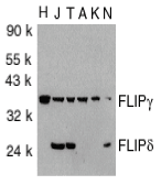
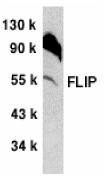
Left: Western blot analysis of FLIP in total cell lysates from HeLa (H), Jurkat (J), THP-1 (T), A431 (A), K562 (K) and NIH3T3 (N) cells with FLIP antibody at 1:1000 dilution.
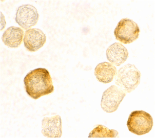
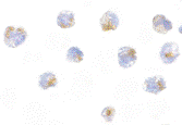
Below: Immunocytochemistry staining of HeLa cells using FLIP antibody at 2 µg/ml.
Other Product Images
 Source : FLIP antibody was raised against a peptide corresponding to amino acids 191 to 209 at C-terminus of human FLIP form2.
Purification : Affinity chromatography purified via peptide column
Clonality and Clone : This is a polyclonal antibody.
Host : FLIP antibody was raised in rabbit. Please use anti-rabbit secondary antibodies.
Immunogen : Human FLIP (C-Terminus) Peptide (Cat. No. 2055P)
Application : FLIP can be used for detection of FLIP by Western blot at 1:1000 dilution. 35 kDa and 25 kDa bands can be detected. antibody recognizes human and murine FLIPy and FLIPd but not FLIPa and FLIPd.
Tested Application(s) : E, WB, ICC
Buffer : Antibody is supplied in PBS containing 0.02% sodium azide.
Blocking Peptide : Cat. No. 2055P - FLIP Peptide
Long-Term Storage : FLIP antibody can be stored at 4ºC, stable for one year. As with all antibodies care should be taken to avoid repeated freeze thaw cycles. Antibodies should not be exposed to prolonged high temperatures.
Positive Control
1. Cat. No. 1205 - Jurkat Cell Lysate
Species Reactivity :H
GI Number : 2253681
Accession Number : AAC51623
Short Description : (CT) FLICE inhibitory protein
References
1. Thome M, Schneider P, Hofmann K, Fickenscher H, Meinl E, Neipel F, Mattmann C, Burns K, Bodmer JL, Schroter M, Scaffidi C, Krammer PH, Peter ME, Tschopp J. Viral FLICE-inhibitory proteins (FLIPs) prevent apoptosis induced by death receptors. Nature 1997;386:517-521
2. Irmler M, Thome M, Hahne M, Schneider P, Hofmann K, Steiner V, Bodmer JL, Schroter M, Burns K, Mattmann C, Rimoldi D, French LE, Tschopp J. Inhibition of death receptor signals by cellular FLIP. Nature 1997;388:190-195
3. Shu HB, Halpin DR , Goeddel DV. Casper is a FADD- and caspase-related inducer of apoptosis. Immunity 1997;6:751-763
4. Hu S, Vincenz C, Ni J, Gentz R, Dixit VM. I-FLICE, a novel inhibitor of tumor necrosis factor receptor-1- and CD-95-induced apoptosis. J Biol Chem 1997;272:17255-17257
FLIP Antibody
Catalog# : 1161
Apoptosis is related to many diseases and induced by a family of cell death receptors and their ligands. Cell death signals are transduced by death domain (DD)- containing adapter molecules and members of the ICE/CED-3 protease family. Caspases-8 (FLICE) and -10 (FLICE2) are two pivotal members in the ICE/CED-3 protease family. FLICE-inhibitory proteins were identified in virus and human and designated v-FLIPs and FLIP, respectively1,2. The human FLIP was also cloned by several labs independently and termed Casper , I-FLICE, FLAME-1, CASH and CLARP3-7. FLIP contains two death effector domains (DEDs) and a caspase-like domain. FLIP interacts with adapter protein FADD and caspase-8 and 10, and potently inhibits apoptosis induced by all known death receptors. Four splice variants of c-FLIPs have been identified and termed FLIPalpha, beta, gamma, and delta, respectively (8).
Additional Names : FLIP (CT), I-FLICE
 Description
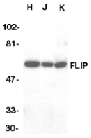
Left: Western blot analysis of FLIP in HeLa (H), Jurkat (J), and K562 (K) whole cell lysate with FLIP antibody at 1:1000 dilution.
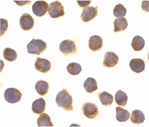
Below: Immunocytochemistry of FLIP in Jurkat cells with FLIP antibody at 10 µg/ml.
Other Product Images

Source : FLIP antibody was raised against a peptide corresponding to amino acids near the C-terminus of human FLIPaFLIPl form.
Purification : Antibody is DEAE purified
Clonality and Clone : This is a polyclonal antibody.
Host : FLIP antibody was raised in rabbit. Please use anti-rabbit secondary antibodies.
Immunogen : Bacteria FLIP (C-Terminus) Peptide (Cat. No. 1161P)
Application : Flip antibody can be used for detection of FLIP by Western blot at 1:500 to 1:2000 dilution. A 55 kDa band can be detected. antibody recognizes the FLIPa only.
Tested Application(s) : E, WB, ICC
Buffer : Antibody is supplied in PBS containing 0.02% sodium azide.
Blocking Peptide : Cat. No. 1161P - FLIP Peptide
Long-Term Storage : FLIP antibody can be stored at 4ºC, stable for one year. As with all antibodies care should be taken to avoid repeated freeze thaw cycles. Antibodies should not be exposed to prolonged high temperatures.
Positive Control
1. Cat. No. 1201 - HeLa Cell Lysate
Species Reactivity :H, M, R
GI Number : 2253679
Accession Number : AAC51622
Short Description : (CT) FLICE inhibitory protein
References
1. Thome M, Schneider P, Hofmann K, et al. Viral FLICE-inhibitory proteins (FLIPs) prevent apoptosis induced by death receptors. Nature 1997;386:517-521
2. Irmler M, Thome M, Hahne M, et al. Inhibition of death receptor signals by cellular FLIP. Nature 1997;388:190-195
3. Shu HB, Halpin DR , Goeddel DV. Casper is a FADD- and caspase-related inducer of apoptosis. Immunity 1997;6:751-763
4. Hu S, Vincenz C, Ni J, Gentz R, Dixit VM. I-FLICE, a novel inhibitor of tumor necrosis factor receptor-1- and CD-95-induced apoptosis. J Biol Chem 1997;272:17255-17257
FLIP Antibody
Catalog# : 1156
Apoptosis is related to many diseases and induced by a family of cell death receptors and their ligands. Cell death signals are transduced by death domain (DD) containing adapter molecules and members of the ICE/CED-3 protease family.Caspases-8 (FLICE) and -10 (FLICE2) are two pivotal members in the ICE/CED-3 protease family. FLICE-inhibitory proteins were identified in virus and human and designated v-FLIPs and FLIP respectively. The human FLIP was also cloned by several labs independently and termed Casper , I-FLICE, FLAME-1, CASH, CLARP and MRIT. FLIP contains two death effector domains (DEDs) and a caspase-like domain. FLIP interacts with adapter protein FADD and caspase-8 and 10, and potently inhibits apoptosis induced by all known death receptors CD95, DR3, TRAIL-R and TNFR1.
Additional Names : FLIP (CT), I-FLICE
 Description
Left: Western blot analysis of FLIP in NIH/3T3 whole cell lysate with FLIP antibody at 1:500 dilution.
Below: Immunocytochemistry of FLIP in 3T3 cells with FLIP antibody at 5 µg/ml.
Other Product Images
 Source : FLIP antibody was raised against a peptide corresponding to amino acids 449 to 465 of mouse FLIPL/CASHa.
Purification : Antibody is DEAE purified
Clonality and Clone : This is a polyclonal antibody.
Host : FLIP antibody was raised in rabbit. Please use anti-rabbit secondary antibodies.
Immunogen : Bacteria FLIP (C-Terminus) Peptide (Cat. No. 1156P)
Application : FLIP antibody can be used for the detection of FLIP by Western blot at 1:500 to 1:1000 dilution. 55 kDa band can be detected. FLIP has short form (FLIPS) and long form (FLIPL) and antibody recognizes the FLIPL only.
Tested Application(s) : E, WB, ICC
Buffer : Antibody is supplied in PBS containing 0.02% sodium azide.
Blocking Peptide : Cat. No. 1156P - FLIP Peptide
Long-Term Storage : FLIP antibody can be stored at 4ºC, stable for one year. As with all antibodies care should be taken to avoid repeated freeze thaw cycles. Antibodies should not be exposed to prolonged high temperatures.
Positive Control
1. Cat. No. 1212 - 3T3 Cell Lysate
Species Reactivity :M
GI Number : 46559393
Accession Number : NP_997536
Short Description : (CT) FLICE inhibitory protein
References
1. Thome M, Schneider P, Hofmann K, Fickenscher H, Meinl E, Neipel F, Mattmann C, Burns K, Bodmer JL, Schroter M, Scaffidi C, Krammer PH, Peter ME, Tschopp J. Viral FLICE-inhibitory proteins (FLIPs) prevent apoptosis induced by death receptors. Nature 1997;386:517-521.
2. Irmler M, Thome M, Hahne M, Schneider P, Hofmann K, Steiner V, Bodmer JL, Schroter M, Burns K, Mattmann C, Rimoldi D, French LE, Tschopp J. Inhibition of death receptor signals by cellular FLIP. Nature 1997;388:190-195.
3. Shu HB, Halpin DR , Goeddel DV. Casper is a FADD- and caspase-related inducer of apoptosis. Immunity 1997;6:751-763.
4. Hu S, Vincenz C, Ni J, Gentz R, Dixit VM. I-FLICE, a novel inhibitor of tumor necrosis factor receptor-1- and CD-95-induced apoptosis. J Biol Chem 1997;272:17255-17257.
FLIP Antibody
Catalog# : 1159
Apoptosis is related to many diseases and induced by a family of cell death receptors and their ligands. Cell death signals are transduced by death domain (DD)- containing adapter molecules and members of the ICE/CED-3 protease family. Caspases-8 (FLICE) and -10 (FLICE2) are two pivotal members in the ICE/CED-3 protease family. FLICE-inhibitory proteins were identified in virus and human and designated v-FLIPs and FLIP, respectively. The human FLIP was also cloned by several labs independently and termed Casper , I-FLICE, FLAME-1, CASH and CLARP3-7. FLIP contains two death effector domains (DEDs) and a caspase-like domain. FLIP interacts with adapter protein FADD and caspase-8 and 10, and potently inhibits apoptosis induced by all known death receptors. Four splice variants of c-FLIPs have been identified and termed FLIPalpha, beta, gamma, and delta, respectively.
Additional Names : FLIP (NT), I-FLICE
 Description
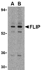
Left: Western blot analysis of FLIP in K562 cell lysate with FLIP antibody at (A) 1 and (B) 2 µg/ml.
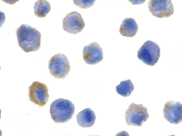
Below: Immunocytochemistry of FLIP in HeLa cells with FLIP antibody at 5 µg/ml.
Other Product Images

Source : FLIP antibody was raised against a peptide corresponding to amino acids near the amino terminus of human FLIP The sequence is identical in all FLIP splice variants.
Purification : Affinity chromatography purified via peptide column
Clonality and Clone : This is a polyclonal antibody.
Host : FLIP antibody was raised in rabbit. Please use anti-rabbit secondary antibodies.
Immunogen : Bacteria FLIP (N-Terminus) Peptide (Cat. No. 1159P)
Application : FLIP can be used for Western blot at 1:500 to 1:1000 dilution.FLIP recognizes all FLIP splice variants including FLIPa, b, and y.
Tested Application(s) : E, WB, ICC
Buffer : Antibody is supplied in PBS containing 0.02% sodium azide.
Blocking Peptide : Cat. No. 1159P - FLIP Peptide
Long-Term Storage : FLIP antibody can be stored at 4ºC, stable for one year. As with all antibodies care should be taken to avoid repeated freeze thaw cycles. Antibodies should not be exposed to prolonged high temperatures.
Positive Control
1. Cat. No. 1205 - Jurkat Cell Lysate
Species Reactivity :H, M, R
GI Number : 2253679
Accession Number : AAC51622
Short Description : (NT) FLICE inhibitory protein
References
1. Thome M, Schneider P, Hofmann K, Fickenscher H, Meinl E, Neipel F, Mattmann C, Burns K, Bodmer JL, Schroter M, Scaffidi C, Krammer PH, Peter ME, Tschopp J. Viral FLICE-inhibitory proteins (FLIPs) prevent apoptosis induced by death receptors. Nature 1997;386:517-521
2. Irmler M, Thome M, Hahne M, Schneider P, Hofmann K, Steiner V, Bodmer JL, Schroter M, Burns K, Mattmann C, Rimoldi D, French LE, Tschopp J. Inhibition of death receptor signals by cellular FLIP. Nature 1997;388:190-195
3. Shu HB, Halpin DR , Goeddel DV. Casper is a FADD- and caspase-related inducer of apoptosis. Immunity 1997;6:751-763
4. Hu S, Vincenz C, Ni J, Gentz R, Dixit VM. I-FLICE, a novel inhibitor of tumor necrosis factor receptor-1- and CD-95-induced apoptosis. J Biol Chem 1997;272:17255-17257
FLASH Antibody
Catalog# : 2271
A novel mammalian CED-4 homologous was recently identified and cloned in mouse (1) and human (2) and designated FLASH (for FLICE-associated huge protein). FLASH is involved in Fas induced apoptosis. It is recruited to Fas after the receptor cross-linking. Overexpression of wild type of FLASH facilitates and its dominant negative form inhibits Fas induced apoptosis. FLASH interacts with the DEDs of caspase-8 and FADD through the DED-like domain of FLASH and mediates activation of caspase-8 (1). There are parallels between FLASH and Apaf-1/CED-4 although there are arguments against their structural similarity (1,2). FLASH is widely expressed.
Additional Names : FLASH (CT), FLASH
 Description
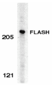
Left: Western blot analysis of FLASH in HeLa whole cell lysate with FLASH antibody at 0.5 µg/ml.
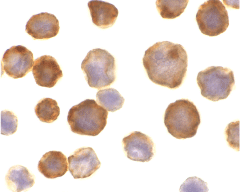
Below: Immunocytochemistry of FLASH in HeLa cells with FLASH antibody at 10 µg/ml.
Other Product Images
 Source : FLASH antibody was raised against a synthetic peptide corresponding to amino acids near the carboxy-terminus of human FLASH which differ from those of mouse by one amino acid.
Purification : Affinity chromatography purified via peptide column
Clonality and Clone : This is a polyclonal antibody.
Host : FLASH antibody was raised in rabbit. Please use anti-rabbit secondary antibodies.
Immunogen : Human FLASH (C-Terminus) Peptide (Cat. No. 2271P)
Application : FLASH antibody can be used for detection of FLASH by Western blot at 0.25 to 0.5 µg/ml.An approximate 220 kDa band can be detected.
Tested Application(s) : E, WB, ICC
Buffer : Antibody is supplied in PBS containing 0.02% sodium azide.
Blocking Peptide : Cat. No. 2271P - FLASH Peptide
Long-Term Storage : FLASH antibody can be stored at 4ºC, stable for one year. As with all antibodies care should be taken to avoid repeated freeze thaw cycles. Antibodies should not be exposed to prolonged high temperatures.
Positive Control
1.Cat. No. 1201 - HeLa Cell Lysate
Species Reactivity :H
GI Number : 5533376
Accession Number : AF165161
Short Description : (CT) Homolog of CED4
References
1.Imai Y, Kimura T, Murakami A, Yajima N, Sakamaki K, Yonehara S. The CED-4-homologous protein FLASH is involved in Fas-mediated activation of caspase-8 during apoptosis. Nature 1999;398:777-85
2.Koonin EV, Aravind L, Hofmann K, Tschopp J, Dixit VM Apoptosis. Searching for FLASH domains. Nature 1999;401:662-3
3.Medema JP. Apoptosis. Life and death in a FLASH. Nature 1999;398:756-7 (WD0800)
BSI offers amino acid analysis because an exact knowledge of protein or peptide quantities is required for further protein studies. Amino acid analysis is not only a suitable tool for precise determination of protein quantities, but also provides detailed information regarding the relative amino acid composition. The relative amino acid composition gives a characteristic profile for proteins, which is often sufficient for identification of a protein. We perform amino acid analysis using Water's ACQ-Tag method to derivatize amino acids, followed by separating the derivatives by reverse-phase HPLC and quantitifying the derivatives by fluorescent detection. Other analytical services also available are: sequencing and mass spectrometry.
For further details on Amino Acid Analysis, call 800-227-0627, or write to us at biosyn@biosyn.com.
Features:
Hydrolysis: hydrolysis is typically achieved by acid conditions. A standard procedure is hydrolysis with 6 M hydrochloric acid (24 hours, 110 ºC). This standard procedure is a compromise between time requirement and temperature. Sensitive amino acids such as tryptophan and cysteine will be partially destroyed. Gas phase hydrolysis and addition of other acids such as propionic acid, TFA, methansulfonic acid can be applied to shorten the hydrolysis time and improve the yield of sensitive amino acids.
Seperation, Detection and Quantification: Internal and external standards is used during the analyses. The hydrolyzed amino acid samples are derivatized and separated by HPLC.
Fast and Economical: low cost results in 2 days
Quantity Required: 0.1 to 0.5 mgs
Resources:
1. FAQ about analytical services
2. Technical and educational warehouse on Analytical Services

 FEATURES AND BENEFITS
 Figure 1. Consistent Isolation of High Quality Leukocyte RNA. Bio-Synthesis's Leukocyte RNA Purification Kit isolates leukocyte RNA of high quality with great consistency. Total leukocyte RNA was isolated from 100 µL of hamster blood using Bio-Synthesis's Leukocyte RNA Purification Kit. A total of 6 replicates were performed, and 7 µL of the 50 µL purified RNA was then resolved on a 1.2% formaldehyde-agarose gel. As it can be seen, Bio-Synthesis's kit not only isolated high and consistent yields of total RNA, but the RNA was also of high quality as evidenced by intactness of the major 28S and 18S rRNA.

 Figure 3. High Yield of a Diversity of RNA Species. Bio-Synthesis's Leukocyte RNA Purification Kit effectively recovers all sizes of RNA from large mRNA to small RNA including microRNAs. Total RNA was isolated from 200 µL of hamster blood sample using Bio-Synthesis's Leukocyte RNA Purification Kit and a leading competitor’s kit as illustrated in Figure 2. Two hundred nanograms of the purified RNA (both 50 µL elution volumes) was then used as the template in a RT-qPCR for detecting the beta-Actin gene (Lower Panel) and for detecting miR-21(Upper Panel). In both graphs the blue lines correspond to Bio-Synthesis's isolated-RNA and the red lines correspond to competitor-isolated RNA. As it can be seen, Bio-Synthesis's kit isolated higher yields of microRNA, as indicated by the lower Ct values of the blue lines (Upper Panel). Also, Bio-Synthesis's kit successfully isolated a similar amount of the large RNA compared to the competitor’s kit (Lower Panel) indicating the full diversity of RNA species isolated.