LASS5 Antibody
Catalog# : 4695
The LASS (longevity assurance homolog) family members represent a subgroup of the homeobox gene family and are highly conserved from yeasts to mammals. Six members of this family of proteins have been characterized (LASS1-6) and all are involved in ceramide synthesis during cell growth regulation and cancer differentiation. LASS5, also called Trh4, is a 392 amino acid endoplasmic reticulum, multi-pass membrane protein. Functioning as a dihydro-ceramide synthase, LASS5 is involved in the production of sphingolipids containing mainly one fatty acid donor (N-linked palmitoyl-ceramide) in a fumonisin B1-independent manner. It uses palmitoyl-CoA as an acyl donor and is involved in the synthesis of C14, C16 and C18-ceramide. LASS5 is the most abundantly expressed and predominant ceramide synthase isoform in lung epithelia. Recent studies show that LASS5 partially correct growth and apoptosis. Multiple isoforms of LASS5 are known to exist. This antibody may cross-react with the highly homologous LASS6.
Additional Names : LASS5 (CT), Longevity Assurance Homolog 5, LAG1, TRH4
 Description
Left: Western blot analysis of LASS5 in SK-N-SH lysate with LASS5 antibody at 1 µg/ml in the (A) absence and (B) presence of blocking peptide.
Below: Immunofluorescence of LASS5 in human brain tissue with LASS5 antibody at 20 μg/ml.
Other Product Images

Source : LASS5 antibody was raised against a 17 amino acid peptide near the carboxy terminus of the human LASS5.
Purification : Affinity chromatography purified via peptide column
Clonality and Clone : This is a polyclonal antibody.
Host : LASS5 antibody was raised in rabbit. Please use anti-rabbit secondary antibodies.
Application : LASS5 antibody can be used for detection of LASS5 by Western blot at 1 – 2 µg/ml.
Tested Application(s) : E, WB
Buffer : Antibody is supplied in PBS containing 0.02% sodium azide.
Blocking Peptide : Cat.No. 4695P - LASS5 Peptide
Long-Term Storage : LASS5 antibody can be stored at 4ºC, stable for one year. As with all antibodies care should be taken to avoid repeated freeze thaw cycles. Antibodies should not be exposed to prolonged high temperatures.
Positive Control
1. Cat. No. 1220 - SK-N-SH Cell Lysate
Species Reactivity :H, M, R
GI Number : 22218345
Accession Number : NP_671723
Short Description : (CT) Longevity assurance homolog 5
References
1. Riebeling C, Allegood JC, Wang E, et al. Two mammalian longevity assurance gene (LAG1) family members, Trh1 and Trh, regulate dihydroceramide synthesis using different fatty acyl-CoA donors. J. Biol. Chem. 2003; 278:43452-9.
2. Lahiri S and Futerman AH. LASS5 is a bona fide dihydroceramide synthase that selectively utilizes palmitoyl-CoA as acyl donor. J. Biol Chem. 2005; 280:33735-8.
3. Spassieva S, Seo JG, Jiang JC, et al. 2006. Necessary role for the LAG1p motif in (dihydro)ceramide synthase activity. J. Biol. Chem. 2006; 281:33931-8.
4. Xu Z, Zhou J, McCoy DM, et al. LASS5 is the predominant ceramide synthase isoform involved in de novo sphingolipid synthesis in lung epithelia. J. Lipid Res. 2005; 46:1229-38.
DNA Protocol
Deoxyribonucleic acid (DNA) is a nucleic acid that contains the genetic instructions used in the development and functioning of all known living organisms and some viruses. The main role of DNA molecules is the long-term storage of information. DNA is often compared to a set of blueprints or a recipe, since it contains the instructions needed to construct other components of cells, such as proteins and RNA molecules.
Base Pairing
Each type of base on one strand forms a bond with just one type of base on the other strand. This is called complementary base pairing. Here, purines form hydrogen bonds to pyrimidines, with A bonding only to T, and C bonding only to G. This arrangement of two nucleotides binding together across the double helix is called a base pair. The double helix is also stabilized by the hydrophobic effect and pi stacking, which are not influenced by the sequence of the DNA.
DNA Extraction
DNA extraction is a routine procedure to collect DNA for subsequent molecular or forensic analysis. There are three basic steps in a DNA extraction:a) Breaking the cells open to expose the DNA within, such as by grinding or sonicating the sample b) Removing membrane lipids by adding a detergent. c) Precipitating the DNA with an alcohol — usually ethanol or isopropanol. Since DNA is insoluble in these alcohols, it will aggregate together, giving a pellet on centrifugation. This step also removes alcohol-soluble salt.
Small nucleolar RNAs (snoRNAs) are a class of small RNA molecules that guide chemical modifications (methylation or pseudouridylation) of ribosomal RNAs (rRNAs) and other RNA genes (tRNAs and other small nuclear RNAs (snRNAs). They are classified under snRNA in MeSH. snoRNAs are commonly referred to as guide RNAs but should not be confused with the guide RNAs (gRNA) that direct RNA editing in trypanosomes.
5' cap
The 5' cap is a specially altered nucleotide end to the 5' end of precursor messenger RNA and some other primary RNA transcripts as found in eukaryotes and, as a special exception, caliciviruses, of which norovirus is one. The process of 5' capping is vital to creating mature messenger RNA which is then able to undergo translation. Capping ensures the messenger RNA's stability while it undergoes translation in the process of protein synthesis, and is a highly regulated process which occurs in the cell nucleus.
5' cap Structure
The 5' cap is found on the 5' end of an mRNA molecule and consists of a guanine nucleotide connected to the mRNA via an unusual 5' to 5' triphosphate linkage. This guanosine is methylated on the 7 position directly after capping in vitro by a methyl transferase. It is referred to as a 7-methylguanosine cap, abbreviated m7G.
SnoRNA Guided Modifications
After transcription, nascent rRNA molecules (termed pre-rRNA) are required to undergo a series of processing steps in order to generate the mature rRNA molecule. Prior to cleavage by exo- and endonucleases the pre-rRNA undergoes a complex pattern of nucleoside modifications. These include methylations and pseudouridylations, guided by snoRNAs. Methylation is the attachment or substitution of a methyl group onto various substrates.
Bio-Synthesis, founded in 1984, is an international provider of genomic services established around the core business lines oligonucleotide and gene synthesis. Base, ribose and backbone modifications and reporter groups like dyes and haptens are routinely incorporated in DNA/RNA hybrid synthesis. Through innovative production process and synthesis know-how, our services are the best in their class, providing you with innovative and trustworthy solutions to accelerate your research.
DNA Methylation
The universal mechanism that regulates genetic activity is DNA methylation, where a methyl group is joined to a section of a molecule. Special methyl-DNA binding proteins spring into action, bond with a section of the methylated DNA and suppress gene activity.
DNA Methylation Analysis
DNA methylation analysis and propose that MeDIP assays may constitute a key method for elucidating the hypermethylome of cancer.
Read More DNA Methylation Protocol
DNA Paternity Lab
Biosyn is a leading Private Investigation company. Biosyn has team of professional scientist. Biosyn is work for lot of Legal attorneys, Civil Cases and Insurance companies. Biosyn also helps in immigration process. Biosyn is also work for searching evidence of adultery or other illegal conduct within marriage to establish grounds for a divorce. Biosyn is an active member of the AABB Parentage Testing Accreditation Program and also participates in proficiency testing by the College of American Pathologists.
Paternity Testing
A maternity or paternity identification test is conducted to establish whether a person is the biological parent of another person. A test to prove paternity (whether a man is someone's father) is known as a paternity test; a test to prove whether a woman is someone's mother) is called a maternity test.
Maternity Testing
Maternity DNA testing determines whether a woman could be the biological mother of a child. Like a DNA paternity test, it compares a child’s DNA pattern with that of the alleged mother to determine how likely it is that the child has inherited the DNA from the alleged mother.
JMJD8 Antibody
Catalog# : 5387
The jumonji domain-containing protein (JMJD) family is defined by the presence of the JmjC domain that is observed in several diverse species. While several JMJD proteins have been identified as being involved in chromatin regulation, histone demethylation and development, the function of JMJD8 has not been identified.
Additional Names : JMJD8, Jumonji domain-containing protein 8, JmjC domain-containing protein 8
 Description
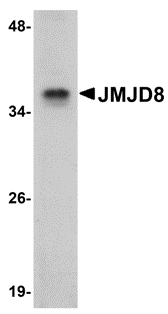
Left: Western blot analysis of JMJD8 in rat kidney tissue lysate with JMJD8 antibody at 1µg/ml.
Source : JMJD8 antibody was raised against an 18 amino acid peptide from near the carboxy terminus of human JMJD8.
Purification : Affinity chromatography purified via peptide column
Clonality and Clone : Polyclonal
Host : JMJD8 antibody was raised in rabbit. Please use anti-rabbit secondary antibodies.
Application : JMJD8 antibody can be used for detection of JMJD8 by Western blot at 1 - 2 µg/ml.
Tested Application(s) : E, WB
Buffer : Antibody is supplied in PBS containing 0.02% sodium azide.
Blocking Peptide : Cat.No. 5387P - JMJD8 Peptide
Long-Term Storage : JMJD8 antibody can be stored at 4ºC, stable for one year. As with all antibodies care should be taken to avoid repeated freeze thaw cycles. Antibodies should not be exposed to prolonged high temperatures.
Positive Control
1. Cat. No. 1465 - Rat Kidney Tissue Lysate
Species Reactivity :H, M, R
GI Number : 74717267
Accession Number : Q96S16
Short Description : Jumonji domain-containing protein 8
References
1. Takeuchi T, Watanabe Y, Takano-Shimizu T, et al. Roles of jumonji and jumonji family genes in chromatin regulation and development. Dev. Dyn. 2006; 235:2449-59.
JMJD7 Antibody
Catalog# : 5385
The jumonji domain-containing protein (JMJD) family is defined by the presence of the JmjC domain that is observed in several diverse species. While several JMJD proteins have been identified as being involved in chromatin regulation, histone demethylation and development, the function of JMJD7 has not been identified. JMJD7 was initially thought to be a novel splice form of the phospholipase PLA2G4B which is located downstream of JMJD7 as most tissues express a read-through transcript of the two genes.
Additional Names : JMJD7, Jumonji domain-containing protein 7, JmjC domain-containing protein 7
 Description
Left: Western blot analysis of JMJD7 in 3T3 cell lysate with JMJD7 antibody at 1µg/ml.
Source : JMJD7 antibody was raised against an 18 amino acid peptide from near the carboxy terminus of human JMJD7.
Purification : Affinity chromatography purified via peptide column
Clonality and Clone : Polyclonal
Host : JMJD7 antibody was raised in rabbit. Please use anti-rabbit secondary antibodies.
Application : JMJD7 antibody can be used for detection of JMJD7 by Western blot at 1 - 2 µg/ml.
Tested Application(s) : E, WB
Buffer : Antibody is supplied in PBS containing 0.02% sodium azide.
Blocking Peptide : Cat.No. 5385P - JMJD7 Peptide
Long-Term Storage : JMJD7 antibody can be stored at 4ºC, stable for one year. As with all antibodies care should be taken to avoid repeated freeze thaw cycles. Antibodies should not be exposed to prolonged high temperatures.
Positive Control
1. Cat. No. 1282 - 3T3 (NIH) Cell Lysate
Species Reactivity :H, M, R
GI Number : 205783894
Accession Number : P0C870
Short Description : Jumonji domain-containing protein 7
References
1. Takeuchi T, Watanabe Y, Takano-Shimizu T, et al. Roles of jumonji and jumonji family genes in chromatin regulation and development. Dev. Dyn. 2006; 235:2449-59.
2. Clissold PM and Ponting CP. JmjC: cupin metalloenzyme-like domains in jumonji, hairless and phospholipase A2beta. Trends Biochem. Sci. 2001; 26:7-9.
JMJD6 Antibody
Catalog# : 5383
The jumonji domain-containing protein (JMJD) family is defined by the presence of the JmjC domain that is observed in several diverse species. JMJD6 was initially identified as a membrane protein that participates in phagocytosis but recent studies have shown that it has other functions when it is expressed in the cytosol and nucleus. JMJD6 is thought to play important roles in regulation of development and differentiation as knockdown experiments in mice resulted in neonatal lethality with severe defects in the morphology of numerous organs. JMJD6 also can catalyze the lysyl-hydroxylation of U2AF65, a protein involved with RNA splicing, suggesting that some of the functions attributed to JMJD6 may be due to its regulatory activity of RNA splicing.
Additional Names : JMJD6, Jumonji domain-containing protein 6, phosphatidylserine receptor, PTDSR, PSR
 Description
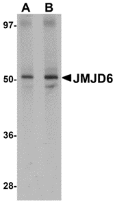
Left: Western blot analysis of JMJD6 in human brain tissue lysate with JMJD6 antibody at (A) 1 and (B) 2 µg/ml.
Source : JMJD6 antibody was raised against a 15 amino acid peptide from near the amino terminus of human JMJD6.
Purification : Affinity chromatography purified via peptide column
Clonality and Clone : Polyclonal
Host : JMJD6 antibody was raised in rabbit. Please use anti-rabbit secondary antibodies.
Application : JMJD6 antibody can be used for detection of JMJD6 by Western blot at 1 - 2 µg/ml.
Tested Application(s) : E, WB
Buffer : Antibody is supplied in PBS containing 0.02% sodium azide.
Blocking Peptide : Cat.No. 5383P - JMJD6 Peptide
Long-Term Storage : JMJD6 antibody can be stored at 4ºC, stable for one year. As with all antibodies care should be taken to avoid repeated freeze thaw cycles. Antibodies should not be exposed to prolonged high temperatures.
Positive Control
1. Cat. No. 1303 - Human Brain Tissue Lysate
Species Reactivity :H, M, R
GI Number : 67461014
Accession Number : Q6NYC1
Short Description : Jumonji domain-containing protein 6
References
1. Takeuchi T, Watanabe Y, Takano-Shimizu T, et al. Roles of jumonji and jumonji family genes in chromatin regulation and development. Dev. Dyn. 2006; 235:2449-59.
2. Fadok VA, Bratton DL, Rose DM, et al. A receptor for phosphatidylserine-specific clearance of apoptotic cells. Nature 2000; 405:85-90.
3. Zakharova L, Dadsetan S, and Fomina AF. Endogenous JMJD6 gene product is expressed at the cell surface and regulates phagocytosis in immature monocyte-like activated THP-1 cells. J. Cell. Phys. 2009; 221:84-91.
4. Bose J, Gruber AD, Helming L, et al. The phosphatidylserine receptor has essential functions during embryogenesis but not in apoptotic cell removal. J. Biol. 2004; 3:15.
JMJD5 Antibody
Catalog# : 5381
The jumonji domain-containing protein (JMJD) family is defined by the presence of the JmjC domain that is observed in several diverse species. While several JMJD proteins have been identified as being involved in chromatin regulation, histone demethylation and development, the function of JMJD5 has not been identified. JMJD5 is expressed in multiple tissues.
Additional Names : JMJD5, Jumonji domain-containing protein 5
 Description
Left: Western blot analysis of JMJD5 in human liver tissue lysate with JMJD5 antibody at (A) 1 and (B) 2 µg/ml.
Source : JMJD5 antibody was raised against an 18 amino acid peptide from near the carboxy terminus of human JMJD5.
Purification : Affinity chromatography purified via peptide column
Clonality and Clone : Polyclonal
Host : JMJD5 antibody was raised in rabbit. Please use anti-rabbit secondary antibodies.
Application : JMJD5 antibody can be used for detection of JMJD5 by Western blot at 1 - 2 µg/ml.
Tested Application(s) : E, WB
Buffer : Antibody is supplied in PBS containing 0.02% sodium azide.
Blocking Peptide : Cat.No. 5381P - JMJD5 Peptide
Long-Term Storage : JMJD5 antibody can be stored at 4ºC, stable for one year. As with all antibodies care should be taken to avoid repeated freeze thaw cycles. Antibodies should not be exposed to prolonged high temperatures.
Positive Control
1. Cat. No. 1304 - Human Liver Tissue Lysate
Species Reactivity :H, M, R
GI Number : 223942018
Accession Number : NP_001138820
Short Description : Jumonji domain-containing protein 5
References
1. Takeuchi T, Watanabe Y, Takano-Shimizu T, et al. Roles of jumonji and jumonji family genes in chromatin regulation and development. Dev. Dyn. 2006; 235:2449-59.

 Description