
Showing posts with label Peptide Library Synthesis. Show all posts
Showing posts with label Peptide Library Synthesis. Show all posts
Tuesday, March 1, 2011
RNA Synthesis
With over 20 years experience in custom synthesis for the biomedical research communities, BSI has developed the expertise to deliver custom synthesized RNA with quality that meets all your RNAi, siRNA, shRNA and other RNA projects.
Unmodified RNA
Over 100 different chemical types of modifications have been identified in thousands of sites in tRNAs, rRNAs, mRNAs, small nuclear RNAs, and other RNAs. Some modifications are highly conserved, while others are more specialized.
Modified RNA
We are committed to long-term continuous improvement and developing the incorporation of modifications and minor bases. It may be available as a product under development or we may be able to add it to our list of R&D projects.
Labeled Probes
BioSynthesis offer design and synthesis of labeled oligos where a fluorescent reporter and a fluorescence quencher are placed on the same oligo, enable closed-tube system which permit rapid and sensitive high-throughput assays.
RNA Conjugations
The BNA molecules can be chemically linked to a variety of other chemical groups, as we detail below. These bioconjugates can be used in a variety of applications including but not limited to, Control of Gene Expression, Antisense Therapy, Cellular Uptake/Delivery, Drug/Effectors Delivery and many others.


Thursday, December 16, 2010
OVGP1 Antibody
OVGP1 Antibody
Catalog# :4763
Oviductins belong to a family of glycoproteins that have been suggested to play several roles during the early processes of reproduction. OVGP1 is a large carbohydrate-rich, epithelial glycoprotein with numerous O-glycosylation sites within threonine, serine, and proline-rich tandem repeats. The protein is secreted from non-ciliated oviductal epithelial cells and associates with ovulated oocytes, blastomeres, and spermatozoan acrosomal regions. Despite its predicted molecular weight, OVGP1 will often run at higher molecular weight in SDS-PAGE. At least two isoforms of OVGP are known to exist.
Additional Names : OVGP1, OVGP, Oviductin, oviduct-specific glycoprotein, muc9
 Description
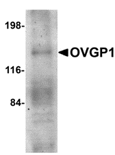
Left: Western blot analysis of OVGP1 in human placenta tissue lysate with OVGP1 antibody at 1 µg/ml.
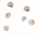
Below: Immunocytochemistry of OVGP1 in 293 cells with OVGP1L antibody at 2.5 μg/ml.
Other Product Images
 Source :
OVGP1 antibody was raised against a 18 amino acid peptide from near the amino terminus of human OVGP1.
Purification : Affinity chromatography purified via peptide column
Clonality and Clone : This is a polyclonal antibody.
Host : OVGP1 antibody was raised in rabbit. Please use anti-rabbit secondary antibodies.
Application : OVGP1 antibody can be used for the detection of OVGP1 by Western blot at 1 µg/ml.
Tested Application(s) : E, WB
Buffer : Antibody is supplied in PBS containing 0.02% sodium azide.
Blocking Peptide :Cat.No. 4763P - OVGP1 Peptide
Long-Term Storage : OVGP1 antibody can be stored at 4ºC, stable for one year. As with all antibodies care should be taken to avoid repeated freeze thaw cycles. Antibodies should not be exposed to prolonged high temperatures.
Positive Control :
1. Cat. No. 1309 - Human Placenta Lysate
Species Reactivity : H, M, R
GI Number : 58386720
Accession Number : NP_002548
Short Description : an oviduct-specific protein
References
1. Buhi WC. Characterization and biological roles of oviduct-specific, oestrogen-dependent glycoprotein. Reproduction 123:355-62.
2. Arias EB, Verhage HG and Jaffe RC. Complementary deoxyribonucleic acid cloning and molecular characterization of an estrogen-dependent human oviductal glycoprotein. Biol. Reprod. 1994; 51:685-94.
Catalog# :4763
Oviductins belong to a family of glycoproteins that have been suggested to play several roles during the early processes of reproduction. OVGP1 is a large carbohydrate-rich, epithelial glycoprotein with numerous O-glycosylation sites within threonine, serine, and proline-rich tandem repeats. The protein is secreted from non-ciliated oviductal epithelial cells and associates with ovulated oocytes, blastomeres, and spermatozoan acrosomal regions. Despite its predicted molecular weight, OVGP1 will often run at higher molecular weight in SDS-PAGE. At least two isoforms of OVGP are known to exist.
Additional Names : OVGP1, OVGP, Oviductin, oviduct-specific glycoprotein, muc9
 Description
Left: Western blot analysis of OVGP1 in human placenta tissue lysate with OVGP1 antibody at 1 µg/ml.
Below: Immunocytochemistry of OVGP1 in 293 cells with OVGP1L antibody at 2.5 μg/ml.
Other Product Images
 Source :
OVGP1 antibody was raised against a 18 amino acid peptide from near the amino terminus of human OVGP1.
Purification : Affinity chromatography purified via peptide column
Clonality and Clone : This is a polyclonal antibody.
Host : OVGP1 antibody was raised in rabbit. Please use anti-rabbit secondary antibodies.
Application : OVGP1 antibody can be used for the detection of OVGP1 by Western blot at 1 µg/ml.
Tested Application(s) : E, WB
Buffer : Antibody is supplied in PBS containing 0.02% sodium azide.
Blocking Peptide :Cat.No. 4763P - OVGP1 Peptide
Long-Term Storage : OVGP1 antibody can be stored at 4ºC, stable for one year. As with all antibodies care should be taken to avoid repeated freeze thaw cycles. Antibodies should not be exposed to prolonged high temperatures.
Positive Control :
1. Cat. No. 1309 - Human Placenta Lysate
Species Reactivity : H, M, R
GI Number : 58386720
Accession Number : NP_002548
Short Description : an oviduct-specific protein
References
1. Buhi WC. Characterization and biological roles of oviduct-specific, oestrogen-dependent glycoprotein. Reproduction 123:355-62.
2. Arias EB, Verhage HG and Jaffe RC. Complementary deoxyribonucleic acid cloning and molecular characterization of an estrogen-dependent human oviductal glycoprotein. Biol. Reprod. 1994; 51:685-94.
Thursday, November 18, 2010
LSD1 Antibody
LSD1 Antibody
Catalog# :3761
Histone modifications mediate changes in gene expression by altering chromatin structure or by serving as a platform to recruit other proteins. LSD1 is a recently discovered amine oxidase that catalyzes the lysine-specific demethylation of histone proteins via an FAD-dependent oxidative reaction (1). Methylation on histone H3-K9 is thought to play an important role in heterochromatin formation, while methylation on arginine and some lysine residues (such as H3-K4) is associated with active transcription (2). LSD1 associates with various proteins, including HDAC1/2, CoREST, and BHC80, that act to regulate LSD1 activity in vivo, and in a histone H3-K4-specific methylase complex that is involved in transcriptional regulation (3,4). Experiments have shown that CoREST, a SANT domain-containing corepressor (5) acts to enhance LSD1 activity, while BHC80, a PHD domain-containing protein (6), inhibits CoREST/LSD1 activity in vitro (3). LSD1-mediated histone demethylation thus may have significant effects on gene expression.
Additional Names : LSD1 (NT), Lysine-specific histone demethylase, amine oxidase flavin containing domain protein 2, AOF2
 Description
Left: Western blot analysis of LSD1 in P815 cell lysate with LSD1 antibody at (A) 1 and (B) 2 µg/ml.
Source :
LSD1 antibody was raised against a 17 amino acid peptide from near the amino terminus of human LSD1.
Purification : Affinity chromatography purified via peptide column
Clonality and Clone : This is a polyclonal antibody.
Host : LSD1 antibody was raised in rabbit.Please use anti-rabbit secondary antibodies.
Immunogen : Human LSD1 (N-Terminus) Peptide (Cat. No. 3761P)
Application : LSD1 antibody can be used for the detection of LSD1 by Western blot at 1 – 2 µg/ml.
Tested Application(s) : E, WB
Buffer : Antibody is supplied in PBS containing 0.02% sodium azide.
Blocking Peptide :Cat. No. 3761P - LSD1 Peptide
Long-Term Storage : LSD1 antibody can be stored at 4ºC, stable for one year. As with all antibodies care should be taken to avoid repeated freeze thaw cycles. Antibodies should not be exposed to prolonged high temperatures.
Positive Control :
1. Cat. No. 1286 - P815 Cell Lysate
Species Reactivity : H, M, R
GI Number : 58761544
Accession Number : NP_001009999
Short Description : (NT) A histone demethylase
References
1. Shi Y, Lan F, Matson C, et al. Histone demethylation mediated by the nuclear amine oxidase homolog LSD1. Cell 2004; 119:941-53.
2. Kouzarides T. Histone methylation in transcriptional control. Curr. Opin. Genet. Dev. 2002; 12:198-209.
3. Shi YJ, Matson C, Lan F, et al. Regulation of LSD1 histone demethylase activity by its associated factors. Mol. Cell 2005; 19:857-64.
4. Nakamura T, Mori T, Tada S, et al. ALL-1 is a histone methyltransferase that assembles a supercomplex of proteins involved in transcriptional regulation. Mol. Cell 2002; 10:1119-28.
Monday, November 8, 2010
JMJD7 Antibody
JMJD7 Antibody
Catalog# : 5385
The jumonji domain-containing protein (JMJD) family is defined by the presence of the JmjC domain that is observed in several diverse species. While several JMJD proteins have been identified as being involved in chromatin regulation, histone demethylation and development, the function of JMJD7 has not been identified. JMJD7 was initially thought to be a novel splice form of the phospholipase PLA2G4B which is located downstream of JMJD7 as most tissues express a read-through transcript of the two genes.
Additional Names : JMJD7, Jumonji domain-containing protein 7, JmjC domain-containing protein 7
 Description
Left: Western blot analysis of JMJD7 in 3T3 cell lysate with JMJD7 antibody at 1µg/ml.
Source : JMJD7 antibody was raised against an 18 amino acid peptide from near the carboxy terminus of human JMJD7.
Purification : Affinity chromatography purified via peptide column
Clonality and Clone : Polyclonal
Host : JMJD7 antibody was raised in rabbit. Please use anti-rabbit secondary antibodies.
Application : JMJD7 antibody can be used for detection of JMJD7 by Western blot at 1 - 2 µg/ml.
Tested Application(s) : E, WB
Buffer : Antibody is supplied in PBS containing 0.02% sodium azide.
Blocking Peptide : Cat.No. 5385P - JMJD7 Peptide
Long-Term Storage : JMJD7 antibody can be stored at 4ºC, stable for one year. As with all antibodies care should be taken to avoid repeated freeze thaw cycles. Antibodies should not be exposed to prolonged high temperatures.
Positive Control
1. Cat. No. 1282 - 3T3 (NIH) Cell Lysate
Species Reactivity :H, M, R
GI Number : 205783894
Accession Number : P0C870
Short Description : Jumonji domain-containing protein 7
References
1. Takeuchi T, Watanabe Y, Takano-Shimizu T, et al. Roles of jumonji and jumonji family genes in chromatin regulation and development. Dev. Dyn. 2006; 235:2449-59.
2. Clissold PM and Ponting CP. JmjC: cupin metalloenzyme-like domains in jumonji, hairless and phospholipase A2beta. Trends Biochem. Sci. 2001; 26:7-9.
Wednesday, November 3, 2010
GRTP1 Antibody
GRTP1 Antibody
Catalog# : 4591
Growth hormone regulated TBC protein 1 (GRTP1) was identified through microarray analysis as a gene whose expression is regulated by growth hormone (GH). It contains the TBC signature motif of GTPase activator proteins of Rab-like small GTPases. GRTP1 mRNA is expressed at highest levels in testes, and is also present in kidney, lung, liver, and intestine. Administration of GH to mice resulted in an increase in GRTP1 mRNA in testes, but a decrease of GRTP1 mRNA in kidney and liver (1). GRTP1 was identified as an HIV dependency factor (HDF), suggesting that GRTP1 may be an important drug target in HIV treatment. At least three isoforms of GRTP1 are known to exist; this antibody should recognize all isoforms.
Additional Names : GRTP1 (IN), Growth hormone regulated TBC protein 1, TBC1 domain family member 6
 Description
Left: Western blot analysis of GRTP1 in SK-N-SH cell lysate with GRTP1 antibody at 1 µg/ml in the (A) absence and (B) presence of blocking peptide.
Source : GRTP1 antibody was raised against a 16 amino acid peptide from near the center of human GRTP1.
Purification : Affinity chromatography purified via peptide column
Clonality and Clone : This is a polyclonal antibody.
Host : GRTP1 antibody was raised in rabbit. Please use anti-rabbit secondary antibodies.
Application : GRTP1 antibody can be used for detection of GRTP1 by Western blot at 1 – 2 µg/ml.
Tested Application(s) : E, WB
Buffer : Antibody is supplied in PBS containing 0.02% sodium azide.
Blocking Peptide : Cat.No. 4591P - GRTP1 Peptide
Long-Term Storage : GRTP1 antibody can be stored at 4ºC, stable for one year. As with all antibodies care should be taken to avoid repeated freeze thaw cycles. Antibodies should not be exposed to prolonged high temperatures.
Positive Control
1. Cat. No. 1220 - SK-N-SH Cell Lysate
Species Reactivity :H, M, R
GI Number : 119629611
Accession Number : EAX09206
Short Description : (IN) Growth hormone regulated TBC protein 1
References
1. Lu C, Kasik J, Stephan DA, et al. Grtp1, a novel gene regulated by growth hormone. Endocriniology 2001; 142:4568-71.
2. Brass AL, Dykxhoorn DM, Benita Y, et al. Identification of host proteins required for HIV infection through a functional genomic screen. Science 2008; 319:921-6.
Wednesday, October 27, 2010
EVER1 Antibody
EVER1 Antibody
Catalog# : 4549
Epidermodysplasia verruciformis (EV) is an autosomal recessive dermatosis characterized by abnormal susceptibility to human papillomaviruses (HPVs) and a high rate of progression to squamous cell carcinoma on sun-exposed skin. EV is caused by mutations in either of two adjacent genes, EVER1 and EVER2, located on chromosome 17q25.3. Both of these genes encode integral membrane proteins that localize to the endoplasmic reticulum and are predicted to form transmembrane channels. Both EVER1 and EVER2 are members of the transmembrane channel-like (TMC) protein family. EVER1 possesses eight trans-membrane domains and two leucine zipper motifs. EVER1 and EVER2 form a complex and interact with the zinc transporter 1 (ZnT-1), suggesting that EVER1 and EVER2 act to regulate cellular zinc balance. At least four isoforms of EVER1 are known to exist. This EVER1 antibody does not cross-react with EVER2.
Additional Names : EVER1 (IN), EV1, EVIN1, Transmembrane protein channel-like protein 6, TMC6
 Description
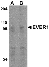
Left: Western blot analysis of EVER1 in human spleen tissue lysate with EVER1 antibody at (A) 1 and (B) 2 µg/ml.
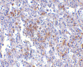
Below: Immunohistochemistry of EVER1 in human spleen with EVER1 antibody at 2.5 µg/ml.
Other Product Images.
 Source : EVER1 antibody was raised against a 15 amino acid peptide from near the center of human EVER1.
Purification : Affinity chromatography purified via peptide column
Clonality and Clone : This is a polyclonal antibody.
Host : EVER1 antibody was raised in rabbit. Please use anti-rabbit secondary antibodies.
Application : EVER1 antibody can be used for the detection of EVER1 by Western blot at 1 – 2 µg/ml.
Tested Application(s) : E, WB, IHC
Buffer : Antibody is supplied in PBS containing 0.02% sodium azide.
Blocking Peptide : Cat.No. 4549P - EVER1 Peptide
Long-Term Storage : EVER1 antibody can be stored at 4ºC, stable for one year. As with all antibodies care should be taken to avoid repeated freeze thaw cycles. Antibodies should not be exposed to prolonged high temperatures.
Positive Control
1. Cat. No. 1306 - Human Spleen Tissue Lysate
Species Reactivity :H, M
GI Number : 25527208
Accession Number : AAM44452
Short Description : (IN) Transmembrane protein channel-like protein 6
References
1. Majewski S, Jablonska J and Orth G. Epidermodysplasia verruciformis. Immunological and nonimmunological surveillance mechanisms: role in tumor progression. Clin. Dermatol. 1997; 15:321-34.
2. Ramoz N, Rueda L-A, Bouadjar B, et al. Mutations in two adjacent novel genes are associated with epidermodysplasia verruciformis. Nat. Genetics 2002; 32:579-81.
3. Keresztes G, Mutai H and Heller S. TMC and EVER genes belong to a larger novel family, the TMC gene family encoding transmembrane proteins. BMC Genomics 2003; 4:24-34.
4. Lazarczyk L, Pons C, Mendoza JA, et al. Regulation of cellular zinc balance as a potential mechanism of EVER-mediated protection against pathogenesis by cutaneous oncogenic human papillomaviruses. J. Exp. Med. 2008; 205:35-42.
Catalog# : 4549
Epidermodysplasia verruciformis (EV) is an autosomal recessive dermatosis characterized by abnormal susceptibility to human papillomaviruses (HPVs) and a high rate of progression to squamous cell carcinoma on sun-exposed skin. EV is caused by mutations in either of two adjacent genes, EVER1 and EVER2, located on chromosome 17q25.3. Both of these genes encode integral membrane proteins that localize to the endoplasmic reticulum and are predicted to form transmembrane channels. Both EVER1 and EVER2 are members of the transmembrane channel-like (TMC) protein family. EVER1 possesses eight trans-membrane domains and two leucine zipper motifs. EVER1 and EVER2 form a complex and interact with the zinc transporter 1 (ZnT-1), suggesting that EVER1 and EVER2 act to regulate cellular zinc balance. At least four isoforms of EVER1 are known to exist. This EVER1 antibody does not cross-react with EVER2.
Additional Names : EVER1 (IN), EV1, EVIN1, Transmembrane protein channel-like protein 6, TMC6
 Description
Left: Western blot analysis of EVER1 in human spleen tissue lysate with EVER1 antibody at (A) 1 and (B) 2 µg/ml.
Below: Immunohistochemistry of EVER1 in human spleen with EVER1 antibody at 2.5 µg/ml.
Other Product Images.
 Source : EVER1 antibody was raised against a 15 amino acid peptide from near the center of human EVER1.
Purification : Affinity chromatography purified via peptide column
Clonality and Clone : This is a polyclonal antibody.
Host : EVER1 antibody was raised in rabbit. Please use anti-rabbit secondary antibodies.
Application : EVER1 antibody can be used for the detection of EVER1 by Western blot at 1 – 2 µg/ml.
Tested Application(s) : E, WB, IHC
Buffer : Antibody is supplied in PBS containing 0.02% sodium azide.
Blocking Peptide : Cat.No. 4549P - EVER1 Peptide
Long-Term Storage : EVER1 antibody can be stored at 4ºC, stable for one year. As with all antibodies care should be taken to avoid repeated freeze thaw cycles. Antibodies should not be exposed to prolonged high temperatures.
Positive Control
1. Cat. No. 1306 - Human Spleen Tissue Lysate
Species Reactivity :H, M
GI Number : 25527208
Accession Number : AAM44452
Short Description : (IN) Transmembrane protein channel-like protein 6
References
1. Majewski S, Jablonska J and Orth G. Epidermodysplasia verruciformis. Immunological and nonimmunological surveillance mechanisms: role in tumor progression. Clin. Dermatol. 1997; 15:321-34.
2. Ramoz N, Rueda L-A, Bouadjar B, et al. Mutations in two adjacent novel genes are associated with epidermodysplasia verruciformis. Nat. Genetics 2002; 32:579-81.
3. Keresztes G, Mutai H and Heller S. TMC and EVER genes belong to a larger novel family, the TMC gene family encoding transmembrane proteins. BMC Genomics 2003; 4:24-34.
4. Lazarczyk L, Pons C, Mendoza JA, et al. Regulation of cellular zinc balance as a potential mechanism of EVER-mediated protection against pathogenesis by cutaneous oncogenic human papillomaviruses. J. Exp. Med. 2008; 205:35-42.
Monday, October 25, 2010
Peptide Interactions
Peptides are short polymers formed from the linking, in a defined order, of α-amino acids. The link between one amino acid residue and the next is known as an amide bond or a peptide bond. Proteins are polypeptide molecules (or consist of multiple polypeptide subunits). The distinction is that peptides are short and polypeptides/proteins are long. There are several different conventions to determine these, all of which have flaws.
Ribosomal Peptides
These are synthesized by translation of mRNA. They are often subjected to proteolysis to generate the mature form. These function, typically in higher organisms, as hormones and signaling molecules. Some lower organisms produce peptides as antibiotics, such as microcins.
Non-ribosomal Peptides
These peptides are assembled by enzymes that are specific to each peptide, rather than by the ribosome. The most common non-ribosomal peptide is glutathione, which is a component of the antioxidant defences of most aerobic organisms. Other nonribosomal peptides are most common in unicellular organisms, plants, and fungi and are synthesized by modular enzyme complexes called nonribosomal peptide synthesises.
Sunday, October 24, 2010
DNA Microarrays
A DNA microarray is a high-throughput technology used in molecular biology and in medicine. It consists of an arrayed series of thousands of microscopic spots of DNA oligonucleotides, called features, each containing picomoles of a specific DNA sequence.
DNA Microarrays
Dna Microarrays can be a short section of a gene or other DNA element that are used as probes to hybridize a cDNA or cRNA sample (called target) under high-stringency conditions. Probe-target hybridization is usually detected and quantified by fluorescence-based detection of fluorophore-labeled targets to determine relative abundance of nucleic acid sequences in the target.
Spotted Microarrays
In spotted microarrays, the probes are oligonucleotides, cDNA or small fragments of PCR products that correspond to mRNAs. The probes are synthesized prior to deposition on the array surface and are then "spotted" onto glass. A common approach utilizes an array of fine pins or needles controlled by a robotic arm that is dipped into wells containing DNA probes and then depositing each probe at designated locations on the array surface.
Oligonucleotide Microarrays
In oligonucleotide microarrays, the probes are short sequences designed to match parts of the sequence of known or predicted open reading frames. Although oligonucleotide probes are often used in "spotted" microarrays, the term "oligonucleotide array" most often refers to a specific technique of manufacturing. Oligonucleotide arrays are produced by printing short oligonucleotide sequences designed to represent a single gene or family of gene splice-variants by synthesizing this sequence directly onto the array surface instead of depositing intact sequences. Sequences may be longer (60-mer probes such as the Agilent design) or shorter (25-mer probes produced by Affymetrix) depending on the desired purpose; longer probes are more specific to individual target genes, shorter probes may be spotted in higher density across the array and are cheaper to manufacture
Thursday, October 21, 2010
BRCC36 Antibody
BRCC36 Antibody
Catalog# : 4331
Together with the breast and ovarian predisposition proteins BRCA1 and BRCA2 and RAD51 and BRCC45, BRCC36 forms a holoenzyme complex that possesses a ubiquitin E3 ligase activity. Aberrant levels of BRCC36 were detected in sporadic breast tumors and depletion of either BRCC36 or BRCC45 by siRNA resulted in increased sensitivity to ionizing radiation and defects in the G2/M checkpoint, indicating that BRCC36 acts to enhance cellular survival following DNA damage. Recent experiments have shown that BRCC36 is essential for ionizing radiation-induced BRCA1 phosphorylation and nuclear foci formation, suggesting that BRCC36 may be an important target in the treatment of radiation-resistant breast tumors. At least two isoforms of BRCC36 are known to exist.
Additional Names : BRCC36, BRCA1/BRCA2-containing complex subunit 3, BRCC3
 Description
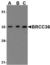
Left: Western blot analysis of BRCC36 in MCF7 cell lysate with BRCC36 antibody at (A) 0.5, (B) 1 and (C) 2 µg/ml.
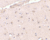
Below: Immunohistochemistry of BRCC36 in human brain tissue with BRCC36 antibody at 2.5 µg/ml.
Other Product Images
 Source : BRCC36 antibody was raised against a 18 amino acid peptide from near the amino terminus of human BRCC36.
Purification : Affinity chromatography purified via peptide column
Clonality and Clone : This is a polyclonal antibody.
Host : BRCC36 antibody was raised in rabbit. Please use anti-rabbit secondary antibodies.
Application : BRCC36 antibody can be used for detection of BRCC36 by Western blot at 0.5 – 1 µg/ml.
Tested Application(s) : E, WB, IHC
Buffer : Antibody is supplied in PBS containing 0.02% sodium azide.
Blocking Peptide : Cat.No. 4331P - BRCC36 Peptide
Long-Term Storage : BRCC36 antibody can be stored at 4ºC, stable for one year. As with all antibodies care should be taken to avoid repeated freeze thaw cycles. Antibodies should not be exposed to prolonged high temperatures.
Positive Control
1. Cat. No. 1219 - MCF7 Cell Lysate
Species Reactivity :H, M, R
GI Number : 13236583
Accession Number : NP_077308
Short Description : BRCA1/BRCA2-containing complex subunit 3
References
1. Dong Y, Hakimi MA, Chen X, et al. Regulation of BRCC, a holoenzyme complex containing BRCA1 and BRCA2, by a signalsome-like subunit and its role in DNA repair. Mol. Cell 2003; 12:1087-99.
2. Chen X, Arciero CA, Wang C, et al. BRCC36 is essential for ionizing radiation-induced BRCA1 phosphorylation and nuclear foci formation. Cancer Res. 2006; 66:5039-46.
Catalog# : 4331
Together with the breast and ovarian predisposition proteins BRCA1 and BRCA2 and RAD51 and BRCC45, BRCC36 forms a holoenzyme complex that possesses a ubiquitin E3 ligase activity. Aberrant levels of BRCC36 were detected in sporadic breast tumors and depletion of either BRCC36 or BRCC45 by siRNA resulted in increased sensitivity to ionizing radiation and defects in the G2/M checkpoint, indicating that BRCC36 acts to enhance cellular survival following DNA damage. Recent experiments have shown that BRCC36 is essential for ionizing radiation-induced BRCA1 phosphorylation and nuclear foci formation, suggesting that BRCC36 may be an important target in the treatment of radiation-resistant breast tumors. At least two isoforms of BRCC36 are known to exist.
Additional Names : BRCC36, BRCA1/BRCA2-containing complex subunit 3, BRCC3
 Description
Left: Western blot analysis of BRCC36 in MCF7 cell lysate with BRCC36 antibody at (A) 0.5, (B) 1 and (C) 2 µg/ml.
Below: Immunohistochemistry of BRCC36 in human brain tissue with BRCC36 antibody at 2.5 µg/ml.
Other Product Images
 Source : BRCC36 antibody was raised against a 18 amino acid peptide from near the amino terminus of human BRCC36.
Purification : Affinity chromatography purified via peptide column
Clonality and Clone : This is a polyclonal antibody.
Host : BRCC36 antibody was raised in rabbit. Please use anti-rabbit secondary antibodies.
Application : BRCC36 antibody can be used for detection of BRCC36 by Western blot at 0.5 – 1 µg/ml.
Tested Application(s) : E, WB, IHC
Buffer : Antibody is supplied in PBS containing 0.02% sodium azide.
Blocking Peptide : Cat.No. 4331P - BRCC36 Peptide
Long-Term Storage : BRCC36 antibody can be stored at 4ºC, stable for one year. As with all antibodies care should be taken to avoid repeated freeze thaw cycles. Antibodies should not be exposed to prolonged high temperatures.
Positive Control
1. Cat. No. 1219 - MCF7 Cell Lysate
Species Reactivity :H, M, R
GI Number : 13236583
Accession Number : NP_077308
Short Description : BRCA1/BRCA2-containing complex subunit 3
References
1. Dong Y, Hakimi MA, Chen X, et al. Regulation of BRCC, a holoenzyme complex containing BRCA1 and BRCA2, by a signalsome-like subunit and its role in DNA repair. Mol. Cell 2003; 12:1087-99.
2. Chen X, Arciero CA, Wang C, et al. BRCC36 is essential for ionizing radiation-induced BRCA1 phosphorylation and nuclear foci formation. Cancer Res. 2006; 66:5039-46.
Bonzo Antibody
Bonzo Antibody
Catalog# : 1170
Human immunodeficiency virus (HIV) and simian immunodeficiency virus (SIV) require coreceptors, in addition to CD4, to infect target cells. Some G protein-coupled receptors including CCR5, CXCR4, CCR3, and CCR2b in the chemokine receptor family have been identified as HIV coreceptors. An orphan G protein-coupled receptor was recently cloned and designated Bonzo, STRL33 and TYMSTR, and identified as HIV and SIV coreceptor. Bonzo/STRL33 is used by SIV, HIV-2 and HIV-1. The messenger RNA of Bonzo/STRL33 is expressed in lymphoid tissues and activated peripheral blood lymphocytes.
Additional Names : Bonzo (CT), STRL33
 Description
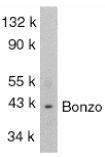
Left: Western blot analysis of Bonzo in SW1353 total cells lysate with Bonzo antibody at 1:1000 dilution.
Source : Bonzo antibody was raised against a peptide corresponding to amino acids 319 to 338 of human origin. The sequence of this peptide is identical to those
Purification : Antibody is DEAE purified
Clonality and Clone : This is a polyclonal antibody.
Host : Bonzo antibody was raised in rabbit. Please use anti-rabbit secondary antibodies.
Immunogen : Human Bonzo (C-Terminus) Peptide (Cat. No. 1170P)
Application : Bonzo antibody can be used for detection of Bonzo by Western blot at 1:1000 dilution.
Tested Application(s) : E, WB
Buffer : Antibody is supplied in PBS containing 0.02% sodium azide.
Blocking Peptide : Cat. No. 1170P - Bonzo Peptide
Long-Term Storage : Bonzo antibody can be stored at 4ºC, stable for one year. As with all antibodies care should be taken to avoid repeated freeze thaw cycles. Antibodies should not be exposed to prolonged high temperatures.
Positive Control
1. Cat. No. 1214 - SW1353 Cell Lysate
Species Reactivity :H
GI Number : 2253422
Accession Number : AAB64221
Short Description : (CT) HIV coreceptor
References
1. Deng HK, Unutmaz D, KewalRamani VN, Littman DR. Expression cloning of new receptors used by simian and human immunodeficiency viruses. Nature 1997;388:296-300
2. Liao F, Alkhatib G, Peden KW, Sharma G, Berger EA, Farber JM. STRL33, A novel chemokine receptor-like protein, functions as a fusion cofactor for both macrophage-tropic and T cell line-tropic HIV-1. J Exp Med 1997;185:2015-23
3. Alkhatib G, Liao F, Berger EA, Farber JM, Peden KW. A new SIV co-receptor, STRL33. Nature 1997;388:238
4. Loetscher M, Amara A, Oberlin E, Brass N, Legler D, Loetscher P, D'Apuzzo M, Meese E, Rousset D, Virelizier JL, Baggiolini M, Arenzana-Seisdedos F, Moser B. TYMSTR, a putative chemokine receptor selectively expressed in activated T cells, exhibits HIV-1 coreceptor function. Curr Biol 1997;7:652-60 (RD1299)
Catalog# : 1170
Human immunodeficiency virus (HIV) and simian immunodeficiency virus (SIV) require coreceptors, in addition to CD4, to infect target cells. Some G protein-coupled receptors including CCR5, CXCR4, CCR3, and CCR2b in the chemokine receptor family have been identified as HIV coreceptors. An orphan G protein-coupled receptor was recently cloned and designated Bonzo, STRL33 and TYMSTR, and identified as HIV and SIV coreceptor. Bonzo/STRL33 is used by SIV, HIV-2 and HIV-1. The messenger RNA of Bonzo/STRL33 is expressed in lymphoid tissues and activated peripheral blood lymphocytes.
Additional Names : Bonzo (CT), STRL33
 Description
Left: Western blot analysis of Bonzo in SW1353 total cells lysate with Bonzo antibody at 1:1000 dilution.
Source : Bonzo antibody was raised against a peptide corresponding to amino acids 319 to 338 of human origin. The sequence of this peptide is identical to those
Purification : Antibody is DEAE purified
Clonality and Clone : This is a polyclonal antibody.
Host : Bonzo antibody was raised in rabbit. Please use anti-rabbit secondary antibodies.
Immunogen : Human Bonzo (C-Terminus) Peptide (Cat. No. 1170P)
Application : Bonzo antibody can be used for detection of Bonzo by Western blot at 1:1000 dilution.
Tested Application(s) : E, WB
Buffer : Antibody is supplied in PBS containing 0.02% sodium azide.
Blocking Peptide : Cat. No. 1170P - Bonzo Peptide
Long-Term Storage : Bonzo antibody can be stored at 4ºC, stable for one year. As with all antibodies care should be taken to avoid repeated freeze thaw cycles. Antibodies should not be exposed to prolonged high temperatures.
Positive Control
1. Cat. No. 1214 - SW1353 Cell Lysate
Species Reactivity :H
GI Number : 2253422
Accession Number : AAB64221
Short Description : (CT) HIV coreceptor
References
1. Deng HK, Unutmaz D, KewalRamani VN, Littman DR. Expression cloning of new receptors used by simian and human immunodeficiency viruses. Nature 1997;388:296-300
2. Liao F, Alkhatib G, Peden KW, Sharma G, Berger EA, Farber JM. STRL33, A novel chemokine receptor-like protein, functions as a fusion cofactor for both macrophage-tropic and T cell line-tropic HIV-1. J Exp Med 1997;185:2015-23
3. Alkhatib G, Liao F, Berger EA, Farber JM, Peden KW. A new SIV co-receptor, STRL33. Nature 1997;388:238
4. Loetscher M, Amara A, Oberlin E, Brass N, Legler D, Loetscher P, D'Apuzzo M, Meese E, Rousset D, Virelizier JL, Baggiolini M, Arenzana-Seisdedos F, Moser B. TYMSTR, a putative chemokine receptor selectively expressed in activated T cells, exhibits HIV-1 coreceptor function. Curr Biol 1997;7:652-60 (RD1299)
Beta-actin Antibody
Beta-actin Antibody
Catalog# : 5005
Actins are highly conserved proteins that are involved in cell motility, structure and integrity, processes that are crucial for tissue development and the development of organism. The actin cytoskeleton is one of the principal drivers of cell motility and is capable of responding to complex signaling cascades. Recent evidence suggests that it may play key roles in regulating apoptosis and aging. Beta actin is one of six different actin isoforms which have been identified. Like GAPDH, -actin is constitutively expressed at high levels in almost all tissues and cell lines making it ideal for use as a loading control marker in immunoblots.
Additional Names : Beta-actin (CT), Beta-actin
 Description
Left: Western blot analysis of actin in HeLa cell lysate with beta-actin antibody at (A) 1 and (B) 2 µg/ml.
Source : Beta-actin antibody was raised against a 16 amino acid peptide from near the carboxy-terminus of human beta-actin.
Purification : Affinity chromatography purified via peptide column
Clonality and Clone : This is a polyclonal antibody.
Host : Beta-actin antibody was raised in chicken. Please use anti-chicken secondary antibodies.
Application : Beta-actin antibody can be used for the detection of beta-actin by Western blot at 1 – 2 µg/ml.
Tested Application(s) : E, WB
Buffer : Antibody is supplied in PBS containing 0.02% sodium azide.
Blocking Peptide : Cat.No. 5005P - Beta-actin Peptide
Long-Term Storage : Beta-actin antibody can be stored at 4ºC, stable for one year. As with all antibodies care should be taken to avoid repeated freeze thaw cycles. Antibodies should not be exposed to prolonged high temperatures.
Positive Control
1. Cat. No. 1201 - HeLa Cell Lysate
Species Reactivity :H, M, R
GI Number : 12803203
Accession Number : AAH02409
Short Description : (CT) A major cytoskeletal protein
References
1. Lambrechts A, Van Troys, M and Ampe C. The actin cytoskeleton in normal and pathological cell motility. Int. J. Biochem. Cell Biol. 2004; 36:1890-909.
2. Gourlay CW and Ayscough KR. The actin cytoskeleton: a key regulator of apoptosis and ageing? Nat. Rev. 2005; 6:583-9.
Wednesday, October 20, 2010
Beta-actin Antibody
Beta-actin Antibody
Catalog# : 3779
Actins are highly conserved proteins that are involved in cell motility, structure and integrity, processes that are crucial for tissue development and the development of organism (reviewed in 1). The actin cytoskeleton is one of the principal drivers of cell motility and is capable of responding to complex signaling cascades. Recent evidence suggests that it may play key roles in regulating apoptosis and aging (2). beta actin is one of six different actin isoforms which have been identified. Like GAPDH, Β-actin is constitutively expressed at high levels in almost all tissues and cell lines making it ideal for use as a loading control marker in immunoblots.
Additional Names : Beta-actin (NT), Beta actin
 Description
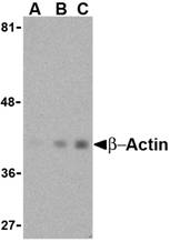
Left: Western blot analysis of beta-actin in HeLa cell lysate with beta-actin antibody at (A) 0.5, (B) 1 and (C) 2 μg/ml.
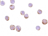
Below: Immunocytochemistry of Actin in HeLa cells with Actin antibody at 1 µg/ml.
Other Product Images
 Source : b-actin antibody was raised against a 16 amino acid peptide from near the amino-terminus of human b-actin.
Purification : Affinity chromatography purified via peptide column
Clonality and Clone : This is a polyclonal antibody.
Host : Beta-actin antibody was raised in rabbit. Please use anti-rabbit secondary antibodies.
Immunogen : Human b-actin / Beta actin (N-Terminus) Peptide (Cat. No. 3779P)
Application : b-actin antibody can be used for the detection of b-actin by Western blot at 1 – 2 µg/ml.
Tested Application(s) : E, WB, IHC
Buffer : Antibody is supplied in PBS containing 0.02% sodium azide.
Blocking Peptide : Cat. No. 3779P - B-actin Peptide
Long-Term Storage : Beta-actin antibody can be stored at 4ºC, stable for one year. As with all antibodies care should be taken to avoid repeated freeze thaw cycles. Antibodies should not be exposed to prolonged high temperatures.
Positive Control
1. Cat. No. 1201 - HeLa Cell Lysate
Species Reactivity :H, M, R
GI Number : 12803203
Accession Number : AAH02409
Short Description : (NT) a major cytoskeletal protein, N Terminus
References
1. Lambrechts A, Van Troys, M and Ampe C. The actin cytoskeleton in normal and pathological cell motility. Int. J. Biochem. Cell Biol. 2004; 36:1890-909.
2. Gourlay CW and Ayscough KR. The actin cytoskeleton: a key regulator of apoptosis and ageing. Nat. Rev. 2005; 6:583-9.
Catalog# : 3779
Actins are highly conserved proteins that are involved in cell motility, structure and integrity, processes that are crucial for tissue development and the development of organism (reviewed in 1). The actin cytoskeleton is one of the principal drivers of cell motility and is capable of responding to complex signaling cascades. Recent evidence suggests that it may play key roles in regulating apoptosis and aging (2). beta actin is one of six different actin isoforms which have been identified. Like GAPDH, Β-actin is constitutively expressed at high levels in almost all tissues and cell lines making it ideal for use as a loading control marker in immunoblots.
Additional Names : Beta-actin (NT), Beta actin
 Description
Left: Western blot analysis of beta-actin in HeLa cell lysate with beta-actin antibody at (A) 0.5, (B) 1 and (C) 2 μg/ml.
Below: Immunocytochemistry of Actin in HeLa cells with Actin antibody at 1 µg/ml.
Other Product Images
 Source : b-actin antibody was raised against a 16 amino acid peptide from near the amino-terminus of human b-actin.
Purification : Affinity chromatography purified via peptide column
Clonality and Clone : This is a polyclonal antibody.
Host : Beta-actin antibody was raised in rabbit. Please use anti-rabbit secondary antibodies.
Immunogen : Human b-actin / Beta actin (N-Terminus) Peptide (Cat. No. 3779P)
Application : b-actin antibody can be used for the detection of b-actin by Western blot at 1 – 2 µg/ml.
Tested Application(s) : E, WB, IHC
Buffer : Antibody is supplied in PBS containing 0.02% sodium azide.
Blocking Peptide : Cat. No. 3779P - B-actin Peptide
Long-Term Storage : Beta-actin antibody can be stored at 4ºC, stable for one year. As with all antibodies care should be taken to avoid repeated freeze thaw cycles. Antibodies should not be exposed to prolonged high temperatures.
Positive Control
1. Cat. No. 1201 - HeLa Cell Lysate
Species Reactivity :H, M, R
GI Number : 12803203
Accession Number : AAH02409
Short Description : (NT) a major cytoskeletal protein, N Terminus
References
1. Lambrechts A, Van Troys, M and Ampe C. The actin cytoskeleton in normal and pathological cell motility. Int. J. Biochem. Cell Biol. 2004; 36:1890-909.
2. Gourlay CW and Ayscough KR. The actin cytoskeleton: a key regulator of apoptosis and ageing. Nat. Rev. 2005; 6:583-9.
Monday, October 18, 2010
Alpha-tubulin Antibody
Alpha-tubulin Antibody
Catalog# : 5103
Alpha-tubulin belongs to the tubulin superfamily, which is composed of six distinct families. Along with beta-tubulins, alpha-tubulins are the major components of microtubules. These microtubules are involved in a wide variety of cellular activities ranging from mitosis and transport events to cell movement and the maintenance of cell shape. Alpha- and beta-tubulin dimers are assembled to 13 protofilaments that form a microtubule of 22-nm diameter. Tyrosine ligase adds a C-terminal tyrosine to monomeric alpha-tubulin. Assembled microtubules can again be detyrosinated by a cytoskeleton-associated carboxypeptidase. Another post-translational modification of detyrosinated alpha-tubulin is C-terminal polyglutamylation, which is characteristic of microtubules in neuronal cells and the mitotic spindle. Like GAPDH and beta-Actin, this antibody makes an excellent loading control in immunoblots.
Additional Names : Alpha-tubulin, Tubulin alpha-1A, TUBA1A, TUBA3, LIS3
 Description
Left: Western blot analysis of Tubulin in rat brain tissue lysate with Tubulin antibody at (A) 0.5 and (B) 1 µg/ml.
Source : Tubulin antibody was raised against a 16 amino acid peptide near the amino terminus of human Tubulin.
Purification : Affinity chromatography purified via peptide column
Clonality and Clone : This is a polyclonal antibody.
Host : Alpha-tubulin antibody was raised in chicken. Please use anti-chicken secondary antibodies.
Application : Tubulin antibody can be used for detection of Tubulin by Western blot at 1 - 2 µg/ml.
Tested Application(s) : E, WB
Buffer : Antibody is supplied in PBS containing 0.02% sodium azide.
Blocking Peptide : Cat.No. 5103P - Alpha-tubulin Peptide
Long-Term Storage : Alpha-tubulin antibody can be stored at 4ºC, stable for one year. As with all antibodies care should be taken to avoid repeated freeze thaw cycles. Antibodies should not be exposed to prolonged high temperatures.
Positive Control
1. Cat. No. 1463 - Rat Brain Tissue Lysate
Species Reactivity :H, M, R
GI Number : 37492
Accession Number : CAA25855
Short Description : Tubulin alpha-1A
References
1. McKean PG, Vaughan S, and Gull K. The extended tubulin family. J. Cell Sci. 2001; 114:2723-33.
2. Barra HA, Arce CA, and Argarana CE. Posttranslational tyrosination/detyrosination of tubulin. Mol. Neurobiol. 1988; 2:133-53.
3. Fukshima N, Furuta D, Hidaka Y, et al. Post-translational modifcations of tubulin in the nervous system. J. Neurochem. 2009; 109:683-693.
Aldh5A1 Antibody
Aldh5A1 Antibody
Catalog# : 4807
Aldh5A1 is a member of the aldehyde dehydrogenase superfamily, a group of NAD(P)(+)-dependent enzymes that catalyze the oxidation of a wide spectrum of aliphatic and aromatic aldehydes. Aldehyde dehydrogenase enzymes are thought to play a major role in the detoxification of aldehydes generated by alcohol metabolism and lipid peroxidation. Aldh5A1 is a mitochondrial NAD(+)-dependent succinic semialdehyde dehydrogenase. A deficiency of this enzyme, known as 4-hydroxybutyricaciduria, results in a disorder of the neurotransmitter 4-aminobutyric acid (GABA). Symptoms usually include static encephalopathy, associated with developmental delays, hypotonia, ataxia, speech defects, and seizures. At least two isoforms of Aldh5A1 are known to exist.
Additional Names : Aldh5A1,Aldehyde dehydrogenase family5 member A1, succinate-semialdehyde dehydrogenase, SSDH, SSADH
 Description
Left: Western blot analysis of Aldh5A1 in mouse liver lysate with Aldh5A1 antibody at (A) 1 and (B) 2 µg/ml.
Source : Aldh5A1 antibody was raised against a 22 amino acid peptide near the carboxy terminus of the human Aldh5A1.
Purification : Affinity chromatography purified via peptide column
Clonality and Clone : This is a polyclonal antibody.
Host : Aldh5A1 antibody was raised in rabbit. Please use anti-rabbit secondary antibodies.
Application : Aldh5A1 antibody can be used for detection of Aldh5A1 by Western blot at 1 – 2 µg/ml.
Tested Application(s) : E, WB
Buffer : Antibody is supplied in PBS containing 0.02% sodium azide.
Blocking Peptide : Cat.No. 4807P - Aldh5A1 Peptide
Long-Term Storage : Aldh5A1 antibody can be stored at 4ºC, stable for one year. As with all antibodies care should be taken to avoid repeated freeze thaw cycles. Antibodies should not be exposed to prolonged high temperatures.
Positive Control
1. Cat. No. 1404 - Mouse Liver Tissue Lysate
Species Reactivity :H, M, R
GI Number : 25777721
Accession Number : NP_733936
Short Description : Aldehyde dehydrogenase family5 member A1
References
1. Vasiliou V and Pappa A. Polymorphisms of human aldehyde dehydrogenases. Consequences for drug metabolism and disease. Pharmacology 2000; 61:192-8.
2. Hearl WG and Churchich JE. Interactions between4-aminobutyrate aminotransferase and succinic semialdehyde dehydrogenase, two mitochondrial enzymes. J. Biol. Chem. 1984; 259:11459-63.
3. Gibson KM, Sweetman L, Nyhan WL, et al. Succinic semialdehyde dehydrogenase deficiency: an inborn error of gamma-aminobutyric acid metabolism. Clin. Chim. Acta 1983; 133:33-42.
Aldh3A1 Antibody
Aldh3A1 Antibody
Catalog# : 4787
Aldh3A1 is a member of the aldehyde dehydrogenase superfamily, a group of NAD(P)(+)-dependent enzymes that catalyze the oxidation of a wide spectrum of aliphatic and aromatic aldehydes. Aldh3A1 is highly expressed in stomach and even more strongly in cornea, representing between 5 to 50% of the water soluble protein fraction in mammalian corneas. It is thought that Aldh3A1 acts to protect the cornea from UV-induced oxidative stress by not only detoxification of reactive aldehydes by also through the direct absorbtion of UV energy. However, corneas from Aldh3A1-null mice are indistinguishable from those from wild-type mice; mice lacking both Aldh3A1 and Aldh1A1 showed increased cataract formation following UVB exposure, suggesting that Aldh1A1 may be able to compensate for the loss of Aldh3A1. At least two isoforms of Aldh3A1 are known to exist. This antibody is predicted to have no cross-reactivity to Aldh3A2.
Additional Names : Aldh3A1,Aldehyde dehydrogenase family3 member A1, AldhIII
 Description
Left: Western blot analysis of Aldh3A1 in human stomach lysate with Aldh3A1 antibody at (A) 1 and (B) 2 µg/ml.
Source : Aldh3A1 antibody was raised against a 13 amino acid peptide near the carboxy terminus of the human Aldh3A1.
Purification : Affinity chromatography purified via peptide column
Clonality and Clone : This is a polyclonal antibody.
Host : Aldh3A1 antibody was raised in rabbit. Please use anti-rabbit secondary antibodies.
Application : Aldh3A1 antibody can be used for detection of Aldh3A1 by Western blot at 1 – 2 µg/ml.
Tested Application(s) : E, WB
Buffer : Antibody is supplied in PBS containing 0.02% sodium azide.
Blocking Peptide : Cat.No. 4787P - Aldh3A1 Peptide
Long-Term Storage : Aldh3A1 antibody can be stored at 4ºC, stable for one year. As with all antibodies care should be taken to avoid repeated freeze thaw cycles. Antibodies should not be exposed to prolonged high temperatures.
Positive Control
1. Cat. No. 1319 - Human Stomach Tissue Lysate
Species Reactivity :H, M, R
GI Number : 22907049
Accession Number : NP_000682
Short Description : Aldehyde dehydrogenase family3 member A1
References
1. Vasiliou V and Pappa A. Polymorphisms of human aldehyde dehydrogenases. Consequences for drug metabolism and disease. Pharmacology 2000; 61:192-8.
2. Hsu LC, Chang WC, Shibuya A, et al. Human stomach aldehyde dehydrogenase cDNA and genomic cloning, primary structure, and expression in Escheria coli. J. Biol. Chem. 1992; 267:3030-7.
3. Pappa A, Sophos NA and Vasiliou V. Corneal and Stomach expression of aldehyde dehydrogenases: from fish to mammals. Chem. Biol. Interact. 2001; 130:181-91.
4. Estey T, Cantore M, Weston PA, et al. Mechanisms involved in the protection of UV-induced protein inactivation by the corneal crystallin ALDH3A1. J. Biol. Chem. 2007; 282:4382-92.
Aipl1 Antibody
Aipl1 Antibody
Catalog# : 4853
Aipl1 was initially identified as a protein implicated in Leber congenital amaurosis (LCA), an autosomal recessive disorder thought to be caused by the abnormal development of photoreceptors. Aipl1 is a tetratricopeptide repeat protein that is highly homologous to ARA9, a protein involved in the HSP90-mediated nuclear translocation and transactivation of the aryl hydrocarbon receptor. Aipl1 has also been found to function as part of a chaperone heterocomplex, interacting with Hsp90 and Hsp70. Aipl1 also associates with the cell cycle regulator NUB1. It is thought that Aipl1 cooperates with Hsp70 but not Hsp90 to suppress the formation of NUB1 inclusions, and these interactions are necessary in the normal photoreceptor maturation, as mutations that lead to LCA also compromise the interactions with the Hsp chaperones. At least three isoforms of Aipl1 are known to exist.
Additional Names : Aipl1 (CT), Aryl hydrocarbon receptor interacting protein-like 1, LCA4
 Description
Left: Western blot analysis of Aipl1 in human brain tissue lysate with Aipl1 antibody at (A) 1 and (B) 2 µg/ml.
Below: Immunohistochemistry of Aipl1 in human brain tissue with Aipl1 antibody at 2.5 µg/ml.
Other Product Images

Source : Aipl1 antibody was raised against a 17 amino acid peptide near the carboxy terminus of the human Aipl1.
Purification : Affinity chromatography purified via peptide column
Clonality and Clone : This is a polyclonal antibody.
Host : Aipl1 antibody was raised in rabbit. Please use anti-rabbit secondary antibodies.
Application : Aipl1 antibody can be used for detection of Aipl1 by Western blot at 1 – 2 µg/ml.
Tested Application(s) : E, WB, IHC
Buffer : Antibody is supplied in PBS containing 0.02% sodium azide.
Blocking Peptide : Cat.No. 4853P - Aipl1 Peptide
Long-Term Storage : Aipl1 antibody can be stored at 4ºC, stable for one year. As with all antibodies care should be taken to avoid repeated freeze thaw cycles. Antibodies should not be exposed to prolonged high temperatures.
Positive Control
1. Cat. No. 1303 - Human Brain Tissue Lysate
Species Reactivity :H, M
GI Number : 74272276
Accession Number : NP_055151
Short Description : (CT) Aryl hydrocarbon receptor interacting protein-like 1
References
1. Sohocki MM, Brown SJ, Sullivan LS, et al. Mutations in a new photoreceptor-pineal gene on 17p cause Leber congenital amaurosis. Nat. Genet. 2000; 24:79-83.
2. Ma Q and Whitlock JP Jr. A novel cytoplasmic protein that interacts with the Ah receptor, contains tetratricopeptide repeat motifs, and augments the transcriptional response to 2,3,7,8-tetrachloro-dibenzo-p-dioxin. J. Biol. Chem. 1997; 272:8878-84.
3. Hidalgo-de-Quintana J, Evans RJ, Cheetham ME, et al. The Leber congenital amaurosis protein aipl1 functions as part of a chaperone complex. Invest. Ophthalmol. Vis. Sci. 2008; 49:2878-87.
4. Akey DT, Zhu X, Dyer M, et al. The inherited blindness associated protein Aipl1 interacts with the cell cycle regulator protein NUB1. Hum. Mol. Genet. 2002; 11:2723-33.
TRIM5 gamma Antibody
TRIM5 gamma Antibody
Catalog# : 3239
TRIM5 is a member of a broad family of otherwise unrelated proteins defined by the presence of a tripartite motif containing a RING domain, a B-box type 1, and a B-box type 2, followed by a coiled-coil region (1). TRIM5 has five alternately spliced isoforms, the longest of which is the ? variant which also contains a carboxy-terminal B30.2 (SPRY) domain (1). While a function has not yet been assigned to TRIM5gamma, it is known that expression of TRIM5gamma variants from humans, rhesus monkeys, and African green monkeys enable resistance to infection by various retroviruses including HIV-1 (2,3), albeit at differing efficiencies. Furthermore, the TRIM5gamma isoform appears to serve as a scaffold for the assembly of endogenous BTBD1 and BTBD2 proteins and also exhibits autoubiquitination activity in a RING finger- and UbcH5B-dependent manner (4).
Additional Names : TRIM5 gamma, TRIM5g
 Description
Left: Western blot analysis of TRIM5 gamma expression in human bladder (A) and colon (B) cell lysate with TRIM5 gamma antibody at 2 µg /ml.
Below: Immunohistochemistry of TRIM5 gamma in human bladder tissue with TRIM5 gamma antibody at 2 µg/ml.
Other Product Images
 Source : TRIM5 gamma antibody was raised against a synthetic peptide corresponding to 15 amino acids near the carboxy-terminus of human TRIM5 gamma.
Purification : Affinity chromatography purified via peptide column
Clonality and Clone : This is a polyclonal antibody.
Host : TRIM5 gamma antibody was raised in rabbit. Please use anti-rabbit secondary antibodies.
Immunogen : Human TRIM5 Gamma Peptide (Cat. No. 3239P)
Application : TRIM5 gamma antibody can be used for detection of TRIM5 gamma by Western blot at 1 to 2 µg/ml.A band at approximately 35 kDa can be detected.
Tested Application(s) : E, WB, IHC
Buffer : Antibody is supplied in PBS containing 0.02% sodium azide.
Blocking Peptide : Cat. No. 3239P - TRIM5 gamma Peptide
Long-Term Storage : TRIM5 gamma antibody can be stored at 4ºC, stable for one year. As with all antibodies care should be taken to avoid repeated freeze thaw cycles. Antibodies should not be exposed to prolonged high temperatures.
Positive Control
1. Cat. No. 1310 - Human Bladder Tissue Lysate
Species Reactivity :H
GI Number : 15011944
Accession Number : NP_149083
Short Description : Retroviral restriction factor
References
1. Reymond A, Meroni G, Fantozzi A, et al. The tripartite motif family identifies cell compartments. EMBO J. 2001; 20:2140-51.
2. Stremlau M, Owens CM, Perron MJ, et al. The cytoplasmic body component TRIM5 restricts HIV-1 infection in Old World monkeys. Nature 2004; 427:848-53.
3. Hatziioannou T, Perez-Caballero D, Yang A, et al. Retrovirus resistance factors REF1 and Lv1 are species-specific variants of TRIM5alpha. Proc. Nat’l. Acad. Sci. USA 2004; 101:10774-9
4. Xu L, Yang L, Moitra PK, et al. BTBD1 and BTBD2 colocalize to cytoplasmic bodies with the RBCC/tripartite motif protein, TRIM5. Exp. Cell Res. 2003; 288:84-93.
Thursday, October 14, 2010
TRIM5 alpha Antibody
TRIM5 alpha Antibody
Catalog# : 3251
TRIM5 is a member of a broad family of otherwise unrelated proteins defined by the presence of a tripartite motif containing a RING domain, a B-box type 1, and a B-box type 2, followed by a coiled-coil region (1). TRIM5 has six alternately spliced isoforms, the longest of which is the ? variant which also contains a carboxy-terminal B30.2 (SPRY) domain (1). Expression of TRIM5alpha variants from humans, rhesus monkeys, and African green monkeys enabled resistance to infection by various retroviruses including HIV-1 (2,3), albeit at differing efficiencies. All TRIM5alpha variants could inhibit at least two different retroviruses, but not from those viruses isolated from the same species, suggesting that TRIM5alpha acts as a natural barrier to cross-species retrovirus transmission (3)
Additional Names : TRIM5 alpha (IN2), TRIM5a
 Description
Left: Western blot analysis of TRIM5 alpha expression in human stomach (A), thymus (B), and uterus (C) cell lysate with TRIM5 alpha antibody at 2 µg /ml.
Below: Immunohistochemistry of TRIM5 alpha in human uterus tissue with TRIM5 alpha antibody at 2 µg/ml.
Other Product Images
 Source : TRIM5 alpha antibody was raised against a synthetic peptide corresponding to amino acids near the mid-region of rhesus monkey TRIM5 alpha.
Purification : Affinity chromatography purified via peptide column
Clonality and Clone : This is a polyclonal antibody.
Host : TRIM5 alpha antibody was raised in rabbit. Please use anti-rabbit secondary antibodies.
Immunogen : Human TRIM5 alpha (Intermediate Domain 2) Peptide (Cat. No. 3251P)
Application : TRIM5 alpha antibody can be used for detection of TRIM5 alpha by Western blot at 1 to 2 µg/ml.A band at approximately 55 kDa can be detected.
Tested Application(s) : E, WB, IHC
Buffer : Antibody is supplied in PBS containing 0.02% sodium azide.
Blocking Peptide : Cat. No. 3251P - TRIM5 alpha (IN2) Peptide
Long-Term Storage : TRIM5 alpha antibody can be stored at 4ºC, stable for one year. As with all antibodies care should be taken to avoid repeated freeze thaw cycles. Antibodies should not be exposed to prolonged high temperatures.
Positive Control
1. Cat. No. 1319 - Human Stomach Tissue Lysate
2. Cat. No. 1317 - Human Uterus Tissue Lysate
3. Cat. No. 1314 - Human Thymus Tissue Lysate
Species Reactivity :H
GI Number : 14719418
Accession Number : NP_149023
Short Description : (IN2) Retroviral restriction factor
References
1. Reymond A, Meroni G, Fantozzi A, et al. The tripartite motif family identifies cell compartments. EMBO J. 2001; 20:2140-51.
2. Stremlau M, Owens CM, Perron MJ, et al. The cytoplasmic body component TRIM5alpha restricts HIV-1 infection in Old World monkeys. Nature 2004; 427:848-53.
3. Hatziioannou T, Perez-Caballero D, Yang A, et al. Retrovirus resistance factors REF1 and Lv1 are species-specific variants of TRIM5alpha. Proc. Nat’l. Acad. Sci. USA 2004; 101:10774-9
TRIM5 alpha Antibody
TRIM5 alpha Antibody
Catalog# : 3249
TRIM5 is a member of a broad family of otherwise unrelated proteins defined by the presence of a tripartite motif containing a RING domain, a B-box type 1, and a B-box type 2, followed by a coiled-coil region (1). TRIM5 has six alternately spliced isoforms, the longest of which is the ? variant which also contains a carboxy-terminal B30.2 (SPRY) domain (1). Expression of TRIM5alpha variants from humans, rhesus monkeys, and African green monkeys enabled resistance to infection by various retroviruses including HIV-1 (2,3), albeit at differing efficiencies. All TRIM5alpha variants could inhibit at least two different retroviruses, but not from those viruses isolated from the same species, suggesting that TRIM5alpha acts as a natural barrier to cross-species retrovirus transmission (3).
Additional Names : TRIM5 alpha (CT), TRIM5a
 Description
Left: Western blot analysis of TRIM5 alpha expression in human stomach (A), thymus (B), and uterus (C) cell lysate with TRIM5 alpha antibody at 2 µg /ml.
Below: Immunofluorescence of TRIM5 alpha in human breast tissue with TRIM5 alpha antibody at 10 µg/ml.
Other Product Images
 Source : TRIM5 alpha antibody was raised against a synthetic peptide corresponding to amino acids near the C-terminus of rhesus monkey TRIM5 alpha.
Purification : Affinity chromatography purified via peptide column
Clonality and Clone : This is a polyclonal antibody.
Host : TRIM5 alpha antibody was raised in rabbit. Please use anti-rabbit secondary antibodies.
Immunogen : Human TRIM5 alpha (C-Terminus) Peptide (Cat. No. 3249P)
Application : TRIM5 alpha antibody can be used for detection of TRIM5 alpha by Western blot at 1 to 2 µg/ml.A band at approximately 55 kDa can be detected. Anti-TRIM5 alpha is human specific but should presumably also react with rhesus protein.
Tested Application(s) : E, WB, IF
Buffer : Antibody is supplied in PBS containing 0.02% sodium azide.
Blocking Peptide : Cat. No. 3249P - TRIM5 alpha Peptide
Long-Term Storage : TRIM5 alpha antibody can be stored at 4ºC, stable for one year. As with all antibodies care should be taken to avoid repeated freeze thaw cycles. Antibodies should not be exposed to prolonged high temperatures.
Positive Control
1. Cat. No. 1319 - Human Stomach Tissue Lysate
2. Cat. No. 1317 - Human Uterus Tissue Lysate
3. Cat. No. 1314 - Human Thymus Tissue Lysate
Species Reactivity :H
GI Number : 14719418
Accession Number : NP_149023
Short Description : (CT) Retroviral restriction factor
References
1. Reymond A, Meroni G, Fantozzi A, et al. The tripartite motif family identifies cell compartments. EMBO J. 2001; 20:2140-51.
2. Stremlau M, Owens CM, Perron MJ, et al. The cytoplasmic body component TRIM5alpha restricts HIV-1 infection in Old World monkeys. Nature 2004; 427:848-53.
3. Hatziioannou T, Perez-Caballero D, Yang A, et al. Retrovirus resistance factors REF1 and Lv1 are species-specific variants of TRIM5alpha. Proc. Nat’l. Acad. Sci. USA 2004; 101:10774-9
TRIM5 alpha Antibody
TRIM5 alpha Antibody
Catalog# : 3247
TRIM5 is a member of a broad family of otherwise unrelated proteins defined by the presence of a tripartite motif containing a RING domain, a B-box type 1, and a B-box type 2, followed by a coiled-coil region . TRIM5 has six alternately spliced isoforms, the longest of which is the alpha variant which also contains a carboxy-terminal B30.2 (SPRY) domain. Expression of TRIM5alpha variants from humans, rhesus monkeys, and African green monkeys enabled resistance to infection by various retroviruses including HIV-1, albeit at differing efficiencies. All TRIM5alpha variants could inhibit at least two different retroviruses, but not from those viruses isolated from the same species, suggesting that TRIM5alpha acts as a natural barrier to cross-species retrovirus transmission.
Additional Names : TRIM5 alpha (IN1), TRIM5a
 Description
Left: Western blot analysis of TRIM5 alpha expression in human stomach (A), thymus (B), and uterus (C) cell lysate with TRIM5 alpha antibody at 2 µg /ml. Below: Immunohistochemical staining of human stomach using TRIM5 alpha antibody at 2 µg/ml.
Below: Immunohistochemical staining of human stomach using TRIM5 alpha antibody at 2 μg/ml.
Other Product Images
 Source : TRIM5 alpha antibody was raised against a synthetic peptide corresponding to amino acids near the mid-region of human TRIM5 alpha.
Purification : Affinity chromatography purified via peptide column
Clonality and Clone : This is a polyclonal antibody.
Host : TRIM5 alpha antibody was raised in rabbit. Please use anti-rabbit secondary antibodies.
Immunogen : Human TRIM5 alpha (Intermediate Domain 1) Peptide (Cat. No. 3247P)
Application : TRIM5 alpha antibody can be used for detection of TRIM5 alpha by Western blot at 1 to 2 µg/ml.A band at approximately 55 kDa can be detected.
Tested Application(s) : E, WB, IHC
Buffer : Antibody is supplied in PBS containing 0.02% sodium azide.
Blocking Peptide : Cat. No. 3247P - TRIM5 alpha (IN1) Peptide
Long-Term Storage : TRIM5 alpha antibody can be stored at 4ºC, stable for one year. As with all antibodies care should be taken to avoid repeated freeze thaw cycles. Antibodies should not be exposed to prolonged high temperatures.
Positive Control
1. Cat. No. 1319 - Human Stomach Tissue Lysate
2. Cat. No. 1317 - Human Uterus Tissue Lysate
3. Cat. No. 1314 - Human Thymus Tissue Lysate
Species Reactivity :H
GI Number : 14719418
Accession Number : NP_149023
Short Description : (IN1) Retroviral restriction factor
References
1. Reymond A, Meroni G, Fantozzi A, et al. The tripartite motif family identifies cell compartments. EMBO J. 2001; 20:2140-51.
2. Stremlau M, Owens CM, Perron MJ, et al. The cytoplasmic body component TRIM5alpha restricts HIV-1 infection in Old World monkeys. Nature 2004; 427:848-53.
3. Hatziioannou T, Perez-Caballero D, Yang A, et al. Retrovirus resistance factors REF1 and Lv1 are species-specific variants of TRIM5alpha. Proc. Nat’l. Acad. Sci. USA 2004; 101:10774-9
Subscribe to:
Posts (Atom)