
Showing posts with label Gastric Inhibitory Peptides. Show all posts
Showing posts with label Gastric Inhibitory Peptides. Show all posts
Thursday, March 31, 2011
Modified RNA
Biosyn can provide RNA, RNA analogs and chimeras in large quantities for your diagnostic and therapeutic research. Biosyn continues the tradition of providing researchers with the highest quality RNA Oligonucleotide synthesis, in quantities ranging from multi-mg to several grams.
RNA
Ribonucleic acid or RNA is a nucleic acid made from a long chain of nucleotide units. Each nucleotide consists of a nitrogenous base, a ribose sugar, and a phosphate. RNA is very similar to DNA, but differs in a few important structural details: in the cell RNA is usually single stranded, while DNA is usually double stranded. RNA nucleotides contain ribose while DNA contains deoxyribose (a type of ribose that lacks one oxygen atom), and RNA has the nucleotide uracil rather than thymine which is present in DNA.
RNA Hybrid
RNA hybrid is a double-stranded polynucleic acids in which one strand is DNA and the other strand is the complementary RNA; formed during transcription and during multiplication of oncogenic RNA viruses.This form is also assumed by DNA-RNA hybrid helices and by regions of double-stranded RNA. It is a right-handed helix and is a more compact form than beta-DNA.
RNA Interference
RNA interference (RNAi) by double stranded RNA (dsRNAs) molecules of approximately 20-25 nucleotides termed short interfering (siRNAs) is a powerful method for preventing the expression of a particular gene. The dsRNA dominantly silences gene expression in a sequence-specific manner by causing the corresponding endogenous mRNA to be degraded. Biosyn can provide RNA, RNA analogs and chimeras in large quantities for your diagnostic and therapeutic research. Biosyn continues the tradition of providing researchers with the highest quality RNA Oligonucleotide synthesis, in quantities ranging from multi-mg to several grams.


Tuesday, November 2, 2010
GNPDA1 Antibody
GNPDA1 Antibody
Catalog# : 5049
Glucosamine-6-phosphate deaminase (GNPDA) is an allosteric enzyme that catalyzes the reversible conversion of D-glucosamine-6-phosphate into D-fructose-6-phosphate and ammonium. GNPDA1 is one of two mammalian glucosamine-6-phosphate deaminases that are thought to have arisen though gene duplication, with the GNPDA2 protein closer in structure and activity to the E. coli enzyme. GNPDA1 possesses greater affinity for ammonium than either GNPDA2 or the E. coli enzyme suggesting that the forward reaction of D-glucosamine-6-phosphate into D-fructose-6-phosphate and ammonium is catalyzed at a slower rate than GNPDA2. GNPDA1 is predicted to not cross-react with GNPDA2.
Additional Names : GNPDA1, Glucosamine-6-phosphate deaminase 1, GNPI, GNPI1
 Description
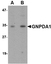
Left: Western blot analysis of GNPDA1 in mouse kidney lysate with GNPDA1 antibody at (A) 1 and (B) 2 µg/ml.
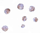
Below: Immunocytochemistry of GNPDA1 in 293 cells with GNPDA1 antibody at 5 μg/ml.
Other Product Images
 Source : GNPDA1 antibody was raised against an 18 amino acid peptide near the carboxy terminus of human GNPDA1.
Purification : Affinity chromatography purified via peptide column
Clonality and Clone : This is a polyclonal antibody.
Host : GNPDA1 antibody was raised in rabbit. Please use anti-rabbit secondary antibodies.
Application : GNPDA1 antibody can be used for detection of GNPDA1 by Western blot at 1 – 2 µg/ml.
Tested Application(s) : E, WB
Buffer : Antibody is supplied in PBS containing 0.02% sodium azide.
Blocking Peptide : Cat.No. 5049P - GNPDA1 Peptide
Long-Term Storage : GNPDA1 antibody can be stored at 4ºC, stable for one year. As with all antibodies care should be taken to avoid repeated freeze thaw cycles. Antibodies should not be exposed to prolonged high temperatures.
Positive Control
1. Cat. No. 1405 - Mouse Kidney Tissue Lysate
Species Reactivity :H, M, R
GI Number : 2935438
Accession Number : AAC05123
Short Description : Glucosamine-6-phosphate deaminase 1
References
1. Wolosker H, Kline D, Bian Y, et al. Molecularly cloned mammalian glucosamine-6-phosphate deaminase localizes to transporting epithelium and lacks oscillin activity. FASEB J. 1998; 12:91-9.
2. Zhang J, Zhang W, Zou D, et al. Cloning and functional characterization of GNPI, a novel human homolog of glucosamine-6-phosphate isomerase/oscillin. J. Cell Biochem. 2003; 88:932-40.
3. Arreola R, Valderrama B, Morante ML, et al. Two mammalian glucosamine-6-phosphate deaminases: a structural and genetic study. FEBS Lett. 2003; 551:63-70.
Catalog# : 5049
Glucosamine-6-phosphate deaminase (GNPDA) is an allosteric enzyme that catalyzes the reversible conversion of D-glucosamine-6-phosphate into D-fructose-6-phosphate and ammonium. GNPDA1 is one of two mammalian glucosamine-6-phosphate deaminases that are thought to have arisen though gene duplication, with the GNPDA2 protein closer in structure and activity to the E. coli enzyme. GNPDA1 possesses greater affinity for ammonium than either GNPDA2 or the E. coli enzyme suggesting that the forward reaction of D-glucosamine-6-phosphate into D-fructose-6-phosphate and ammonium is catalyzed at a slower rate than GNPDA2. GNPDA1 is predicted to not cross-react with GNPDA2.
Additional Names : GNPDA1, Glucosamine-6-phosphate deaminase 1, GNPI, GNPI1
 Description
Left: Western blot analysis of GNPDA1 in mouse kidney lysate with GNPDA1 antibody at (A) 1 and (B) 2 µg/ml.
Below: Immunocytochemistry of GNPDA1 in 293 cells with GNPDA1 antibody at 5 μg/ml.
Other Product Images
 Source : GNPDA1 antibody was raised against an 18 amino acid peptide near the carboxy terminus of human GNPDA1.
Purification : Affinity chromatography purified via peptide column
Clonality and Clone : This is a polyclonal antibody.
Host : GNPDA1 antibody was raised in rabbit. Please use anti-rabbit secondary antibodies.
Application : GNPDA1 antibody can be used for detection of GNPDA1 by Western blot at 1 – 2 µg/ml.
Tested Application(s) : E, WB
Buffer : Antibody is supplied in PBS containing 0.02% sodium azide.
Blocking Peptide : Cat.No. 5049P - GNPDA1 Peptide
Long-Term Storage : GNPDA1 antibody can be stored at 4ºC, stable for one year. As with all antibodies care should be taken to avoid repeated freeze thaw cycles. Antibodies should not be exposed to prolonged high temperatures.
Positive Control
1. Cat. No. 1405 - Mouse Kidney Tissue Lysate
Species Reactivity :H, M, R
GI Number : 2935438
Accession Number : AAC05123
Short Description : Glucosamine-6-phosphate deaminase 1
References
1. Wolosker H, Kline D, Bian Y, et al. Molecularly cloned mammalian glucosamine-6-phosphate deaminase localizes to transporting epithelium and lacks oscillin activity. FASEB J. 1998; 12:91-9.
2. Zhang J, Zhang W, Zou D, et al. Cloning and functional characterization of GNPI, a novel human homolog of glucosamine-6-phosphate isomerase/oscillin. J. Cell Biochem. 2003; 88:932-40.
3. Arreola R, Valderrama B, Morante ML, et al. Two mammalian glucosamine-6-phosphate deaminases: a structural and genetic study. FEBS Lett. 2003; 551:63-70.
Gle1 Antibody
Gle1 Antibody
Catalog# : 4963
The proper expression of gene products in eukaryotic cells relies on efficient transport of mRNA molecules out of the nucleus. Gle1 is an essential mRNA export factor in both human and yeast cells. It associates with the nuclear pore complex (NPC) through hCG1 and NUP155 in mammalian cells and in conjunction with inositol hexakisphosphate (IP6), stimulates Dbp5, a member of the DEAD-box helicase family, triggering mRNP remodeling and facilitating RNA export from the nucleus. Recent evidence suggests that mutations in Gle1 causing defects in mRNA export can result in human disease. At least three isoforms of Gle1 are known to exist.
Additional Names : Gle1 (CT), RNA export mediator
 Description
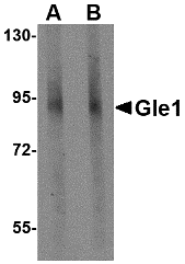
Left: Western blot analysis of Gle1 in mouse brain tissue lysate with Gle1 antibody at (A) 1 and (B) 2 µg/ml.
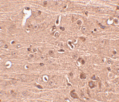
Below: Immunohistochemistry of Gle1 in mouse brain tissue with Gle1 antibody at 2.5 µg/ml.
Other Product Images
 Source : Gle1 antibody was raised against a 15 amino acid peptide near the carboxy terminus of human Gle1.
Purification : Affinity chromatography purified via peptide column
Clonality and Clone : This is a polyclonal antibody.
Host : Gle1 antibody was raised in rabbit. Please use anti-rabbit secondary antibodies.
Application : Gle1 antibody can be used for detection of Gle1 by Western blot at 1 – 2 µg/ml.
Tested Application(s) : E, WB, IHC
Buffer : Antibody is supplied in PBS containing 0.02% sodium azide.
Blocking Peptide : Cat.No. 4963P - Gle1 Peptide
Long-Term Storage : Gle1 antibody can be stored at 4ºC, stable for one year. As with all antibodies care should be taken to avoid repeated freeze thaw cycles. Antibodies should not be exposed to prolonged high temperatures.
Positive Control
1. Cat. No. 1403 - Mouse Brain Tissue Lysate
Species Reactivity :H, M, R
GI Number : 51317384
Accession Number : NP_001003722
Short Description : (CT) RNA export mediator
References
1. Murphy R and Wente SR. An RNA-export mediator with an essential nuclear export signal. Nature 1996; 383:357-60.
2. Watkins JL, Murphy R, Emtage JL, et al. The human homologue of Saccharomyces cerevisiae Gle1p is required for poly(A)+ RNA export. Proc. Natl. Acad. Sci. USA 1998; 95:6779-84.
3. Kendirgi F, Rexer DJ, Alcazar-Roman AR, et al. Interaction between the shuttling mRNA export factor Gle1 and the nucleoporin hCG1: A conserved mechanism in the export of Hsp70 mRNA. Mol. Biol. Cell 2005; 16:4304-15.
4. Rayala HJ, Kendirgi F, Barry DM, et al. The mRNA export factor human Gle1 interacts with the nuclear pore complex protein Nup155. Mol. Cell. Proteomics 2004; 3:145-55.
Catalog# : 4963
The proper expression of gene products in eukaryotic cells relies on efficient transport of mRNA molecules out of the nucleus. Gle1 is an essential mRNA export factor in both human and yeast cells. It associates with the nuclear pore complex (NPC) through hCG1 and NUP155 in mammalian cells and in conjunction with inositol hexakisphosphate (IP6), stimulates Dbp5, a member of the DEAD-box helicase family, triggering mRNP remodeling and facilitating RNA export from the nucleus. Recent evidence suggests that mutations in Gle1 causing defects in mRNA export can result in human disease. At least three isoforms of Gle1 are known to exist.
Additional Names : Gle1 (CT), RNA export mediator
 Description
Left: Western blot analysis of Gle1 in mouse brain tissue lysate with Gle1 antibody at (A) 1 and (B) 2 µg/ml.
Below: Immunohistochemistry of Gle1 in mouse brain tissue with Gle1 antibody at 2.5 µg/ml.
Other Product Images
 Source : Gle1 antibody was raised against a 15 amino acid peptide near the carboxy terminus of human Gle1.
Purification : Affinity chromatography purified via peptide column
Clonality and Clone : This is a polyclonal antibody.
Host : Gle1 antibody was raised in rabbit. Please use anti-rabbit secondary antibodies.
Application : Gle1 antibody can be used for detection of Gle1 by Western blot at 1 – 2 µg/ml.
Tested Application(s) : E, WB, IHC
Buffer : Antibody is supplied in PBS containing 0.02% sodium azide.
Blocking Peptide : Cat.No. 4963P - Gle1 Peptide
Long-Term Storage : Gle1 antibody can be stored at 4ºC, stable for one year. As with all antibodies care should be taken to avoid repeated freeze thaw cycles. Antibodies should not be exposed to prolonged high temperatures.
Positive Control
1. Cat. No. 1403 - Mouse Brain Tissue Lysate
Species Reactivity :H, M, R
GI Number : 51317384
Accession Number : NP_001003722
Short Description : (CT) RNA export mediator
References
1. Murphy R and Wente SR. An RNA-export mediator with an essential nuclear export signal. Nature 1996; 383:357-60.
2. Watkins JL, Murphy R, Emtage JL, et al. The human homologue of Saccharomyces cerevisiae Gle1p is required for poly(A)+ RNA export. Proc. Natl. Acad. Sci. USA 1998; 95:6779-84.
3. Kendirgi F, Rexer DJ, Alcazar-Roman AR, et al. Interaction between the shuttling mRNA export factor Gle1 and the nucleoporin hCG1: A conserved mechanism in the export of Hsp70 mRNA. Mol. Biol. Cell 2005; 16:4304-15.
4. Rayala HJ, Kendirgi F, Barry DM, et al. The mRNA export factor human Gle1 interacts with the nuclear pore complex protein Nup155. Mol. Cell. Proteomics 2004; 3:145-55.
Monday, October 25, 2010
CLIP170 Antibody
CLIP170 Antibody
Catalog# : 4659
CLIP170 was initially identified as a new type of intermediate filament associated protein that is highly expressed in Reed-Sternberg cells, the tumoral cells diagnostic for Hodgkin’s disease. Later experiments showed that it is located at microtubule plus ends and is required for the binding of endocytic carrier vesicles. CLIP170 has also been suggested to act with LIS1, a protein implicated in brain development, to regulate dynein/dynactin binding microtubules. Other studies suggest that CLIP170 can influence the formation of lamellipodia and cell invasion by invasive breast cancer cells by regulating the release of kinesin and IQGAP1 from a complex of those proteins, CLIP170 and Rac1. At least two isoforms of CLIP170 are known to exist.
Additional Names : CLIP170, CAP-Gly domain-containing linker protein 1, CLIP, CYLN1, Restin, RSN
 Description
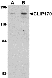
Left: Western blot analysis of CLIP170 in rat brain tissue lysate with CLIP170 antibody at (A) 0.5 and (B) 1 µg/ml.
Below: Immunofuorescence of CLIP170 in human brain tissue with CLIP170 antibody at 20 μg/ml.
Other Product Images
 Source : CLIP170 antibody was raised against a 17 amino acid peptide from near the carboxy terminus of human CLIP170.
Purification : Affinity chromatography purified via peptide column
Clonality and Clone : This is a polyclonal antibody.
Host : CLIP170 antibody was raised in rabbit. Please use anti-rabbit secondary antibodies.
Application : CLIP170 antibody can be used for detection of CLIP170 by Western blot at 0.5 – 1 µg/ml.
Tested Application(s) : E, WB
Buffer : Antibody is supplied in PBS containing 0.02% sodium azide.
Blocking Peptide : Cat.No. 4659P - CLIP170 Peptide
Long-Term Storage : CLIP170 antibody can be stored at 4ºC, stable for one year. As with all antibodies care should be taken to avoid repeated freeze thaw cycles. Antibodies should not be exposed to prolonged high temperatures.
Positive Control
1. Cat. No. 1463 - Rat Brain Tissue Lysate
Species Reactivity :H, M, R
GI Number : 4506751
Accession Number : NP_002947
Short Description : CAP-Gly domain-containing linker protein 1
References
1. Bilbe G, Delabie J, Bruggen J, et al. Restin: a novel intermediate filament-associated protein highly expressed in the Reed-Sternberg cells of Hodgkin’s disease. EMBO J. 1992; 11:2103-13.
2. Diamantopoulos GS, Perez F, Goodson HV, et al. Dynamic localization of CLIP-170 to microtubule plus ends is coupled to microtubule assembly. J. Cell Biol. 1999; 144:99-112.
3. Pierre P, Scheel J, Richard JE, et al. CLIP-170 links endocytic vesicles to microtubules. Cell 70:887-900.
4. Suzuki K and Takahashi K. Regulation of lamellipodia formation and cell invasion by CLIP-170 in invasive human breast cancer cells. Biochem. Biophys. Res. Commun. 2008; 368:199-204.
Catalog# : 4659
CLIP170 was initially identified as a new type of intermediate filament associated protein that is highly expressed in Reed-Sternberg cells, the tumoral cells diagnostic for Hodgkin’s disease. Later experiments showed that it is located at microtubule plus ends and is required for the binding of endocytic carrier vesicles. CLIP170 has also been suggested to act with LIS1, a protein implicated in brain development, to regulate dynein/dynactin binding microtubules. Other studies suggest that CLIP170 can influence the formation of lamellipodia and cell invasion by invasive breast cancer cells by regulating the release of kinesin and IQGAP1 from a complex of those proteins, CLIP170 and Rac1. At least two isoforms of CLIP170 are known to exist.
Additional Names : CLIP170, CAP-Gly domain-containing linker protein 1, CLIP, CYLN1, Restin, RSN
 Description
Left: Western blot analysis of CLIP170 in rat brain tissue lysate with CLIP170 antibody at (A) 0.5 and (B) 1 µg/ml.
Below: Immunofuorescence of CLIP170 in human brain tissue with CLIP170 antibody at 20 μg/ml.
Other Product Images
 Source : CLIP170 antibody was raised against a 17 amino acid peptide from near the carboxy terminus of human CLIP170.
Purification : Affinity chromatography purified via peptide column
Clonality and Clone : This is a polyclonal antibody.
Host : CLIP170 antibody was raised in rabbit. Please use anti-rabbit secondary antibodies.
Application : CLIP170 antibody can be used for detection of CLIP170 by Western blot at 0.5 – 1 µg/ml.
Tested Application(s) : E, WB
Buffer : Antibody is supplied in PBS containing 0.02% sodium azide.
Blocking Peptide : Cat.No. 4659P - CLIP170 Peptide
Long-Term Storage : CLIP170 antibody can be stored at 4ºC, stable for one year. As with all antibodies care should be taken to avoid repeated freeze thaw cycles. Antibodies should not be exposed to prolonged high temperatures.
Positive Control
1. Cat. No. 1463 - Rat Brain Tissue Lysate
Species Reactivity :H, M, R
GI Number : 4506751
Accession Number : NP_002947
Short Description : CAP-Gly domain-containing linker protein 1
References
1. Bilbe G, Delabie J, Bruggen J, et al. Restin: a novel intermediate filament-associated protein highly expressed in the Reed-Sternberg cells of Hodgkin’s disease. EMBO J. 1992; 11:2103-13.
2. Diamantopoulos GS, Perez F, Goodson HV, et al. Dynamic localization of CLIP-170 to microtubule plus ends is coupled to microtubule assembly. J. Cell Biol. 1999; 144:99-112.
3. Pierre P, Scheel J, Richard JE, et al. CLIP-170 links endocytic vesicles to microtubules. Cell 70:887-900.
4. Suzuki K and Takahashi K. Regulation of lamellipodia formation and cell invasion by CLIP-170 in invasive human breast cancer cells. Biochem. Biophys. Res. Commun. 2008; 368:199-204.
Friday, October 15, 2010
Custom Peptide
Bio-Synthesis, founded in 1984, is a U.S. based company whose primary emphasis is providing high quality custom products such as custom oligonucleotide, custom peptides, custom antibody, and services for life science research, diagnostics, and therapeutic applications. Our integrated manufacturing programs enable a seamless transition from the inception of an idea through product launch by providing our client with a comprehensive portfolio of services that span across genomic, proteomic, and cell biology.
As a leading supplier of genomic and proteomic research products, BSI is able to offer its customers integrated solutions for custom DNA synthesis, custom RNA synthesis, custom polyclonal antibody production, custom peptide synthesis, molecular diagnostic detection kits, DNA Real-Time PCR products, DNA microarray services and a wide range of custom DNA-based diagnostic kits and consumables.
The proteomic division is experienced in the design of peptides, both for antibody production and bioactive peptide synthesis, e.g cosmetic peptides. While the custom peptide synthesis division has the ability to produce thousands of modified or non-modified peptides per day for research applications, BSI also provides diagnostic/ therapeutic GMP custom peptide production. Synthesis of custom peptides results in quantities ranging from milligrams to multi-kilograms. The staff in the custom antibody division provide expert support in the selection of peptide antigens for target antibody production and offer convenient custom-bundled packages with a wide range of host animals. In addition, peptide, antibody and protein arrays are also amongst its expertise.
BSI is a U.S. based company whose primary emphasis is on the synthesis of high quality custom products for a variety of applications and a full-service contract manufacturing organization (CMO) delivering research, development and production services for cGMP clinical batch manufacturing.
As a leading supplier of genomic and proteomic research products, BSI is able to offer its customers integrated solutions for custom DNA synthesis, custom RNA synthesis, custom polyclonal antibody production, custom peptide synthesis, molecular diagnostic detection kits, DNA Real-Time PCR products, DNA microarray services and a wide range of custom DNA-based diagnostic kits and consumables.
The proteomic division is experienced in the design of peptides, both for antibody production and bioactive peptide synthesis, e.g cosmetic peptides. While the custom peptide synthesis division has the ability to produce thousands of modified or non-modified peptides per day for research applications, BSI also provides diagnostic/ therapeutic GMP custom peptide production. Synthesis of custom peptides results in quantities ranging from milligrams to multi-kilograms. The staff in the custom antibody division provide expert support in the selection of peptide antigens for target antibody production and offer convenient custom-bundled packages with a wide range of host animals. In addition, peptide, antibody and protein arrays are also amongst its expertise.
BSI is a U.S. based company whose primary emphasis is on the synthesis of high quality custom products for a variety of applications and a full-service contract manufacturing organization (CMO) delivering research, development and production services for cGMP clinical batch manufacturing.
Thursday, October 14, 2010
MED28 Antibody
MED28 Antibody
Catalog# : 4731
The mediator complex is a multi-protein transcriptional co-activator that is expressed ubiquitously in eukaryotes from yeast to mammals and is required for induction of RNA polymerase II (pol II) transcription by DNA binding transcription factor. One of the proteins in this complex is MED28, also known as Magicin. MED28 is expressed in many cell lines and tissues. It has been shown that a down-regulation of MED28 expression in NIH3T3 cells results in a significant induction of several genes associated with smooth muscle cell differentiation and conversely its over-expression represses expression of SMC genes. MED28 can also form a ternary complex with Grb2 and Merlin, the neurofibromatosis 2 tumor suppressor protein, indicating that MED28 may play a role in Merlin’s tumor suppressive activity. MED28 has also been recently identified as an HIV dependency factor (HDF), suggesting that MED28 may be an important drug target in HIV treatment. At least two isoforms of MED28 are known to exist.
Additional Names : MED28, Mediator of RNA polymerase II transcription subunit 28, magicin, EG1
 Description
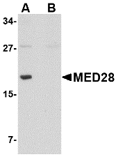
Left: Western blot analysis of MED28 in human brain tissue lysate with MED28 antibody at 1 µg/ml in (A) the absence and (B) the presence of blocking peptide.
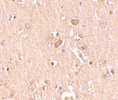
Below: Immunohistochemistry of MED28 in human brain tissue with MED28 antibody at 2.5 μg/ml.
Other Product Images
 Source : MED28 antibody was raised against a 17 amino acid peptide from near the center of human MED28.
Purification : Affinity chromatography purified via peptide column
Clonality and Clone : This is a polyclonal antibody.
Host : MED28 antibody was raised in rabbit. Please use anti-rabbit secondary antibodies.
Application : MED28 antibody can be used for detection of MED28 by Western blot at 0.5 – 1 µg/ml.
Tested Application(s) : E, WB
Buffer : Antibody is supplied in PBS containing 0.02% sodium azide.
Blocking Peptide : Cat.No. 4731P - MED28 Peptide
Long-Term Storage : MED28 antibody can be stored at 4ºC, stable for one year. As with all antibodies care should be taken to avoid repeated freeze thaw cycles. Antibodies should not be exposed to prolonged high temperatures.
Positive Control
1. Cat. No. 1303 - Human Brain Tissue Lysate
Species Reactivity :H, M
GI Number : 52851391
Accession Number : NP_079481
Short Description : Mediator of RNA polymerase II transcription subunit 28
References
1. Sato S, Tomomori-Sato C, Parmely TJ, et al. A set of consensus mammalian mediator subunits identified by multidimensional protein identification technology. Mol. Cell 2004; 14:685-91.
2. Beyer KS, Beauchamp RL, Lee MF, et al. Mediator subunit MED28 (Magicin) is a repressor of smooth muscle cell differentiation. J. Biol. Chem. 2007; 282:32152-7.
3. Wiederhold T, Lee MF, James M, et al. Magicin, a novel cytoskeletal protein associates with the NF2 tumor suppressor merlin and Grb2. Oncogene 2004; 23:8815-25.
4. Brass AL, Dykxhoorn DM, Benita Y, et al. Identification of host proteins required for HIV infection through a functional genomic screen. Science 2008; 319:921-6.
Catalog# : 4731
The mediator complex is a multi-protein transcriptional co-activator that is expressed ubiquitously in eukaryotes from yeast to mammals and is required for induction of RNA polymerase II (pol II) transcription by DNA binding transcription factor. One of the proteins in this complex is MED28, also known as Magicin. MED28 is expressed in many cell lines and tissues. It has been shown that a down-regulation of MED28 expression in NIH3T3 cells results in a significant induction of several genes associated with smooth muscle cell differentiation and conversely its over-expression represses expression of SMC genes. MED28 can also form a ternary complex with Grb2 and Merlin, the neurofibromatosis 2 tumor suppressor protein, indicating that MED28 may play a role in Merlin’s tumor suppressive activity. MED28 has also been recently identified as an HIV dependency factor (HDF), suggesting that MED28 may be an important drug target in HIV treatment. At least two isoforms of MED28 are known to exist.
Additional Names : MED28, Mediator of RNA polymerase II transcription subunit 28, magicin, EG1
 Description
Left: Western blot analysis of MED28 in human brain tissue lysate with MED28 antibody at 1 µg/ml in (A) the absence and (B) the presence of blocking peptide.
Below: Immunohistochemistry of MED28 in human brain tissue with MED28 antibody at 2.5 μg/ml.
Other Product Images
 Source : MED28 antibody was raised against a 17 amino acid peptide from near the center of human MED28.
Purification : Affinity chromatography purified via peptide column
Clonality and Clone : This is a polyclonal antibody.
Host : MED28 antibody was raised in rabbit. Please use anti-rabbit secondary antibodies.
Application : MED28 antibody can be used for detection of MED28 by Western blot at 0.5 – 1 µg/ml.
Tested Application(s) : E, WB
Buffer : Antibody is supplied in PBS containing 0.02% sodium azide.
Blocking Peptide : Cat.No. 4731P - MED28 Peptide
Long-Term Storage : MED28 antibody can be stored at 4ºC, stable for one year. As with all antibodies care should be taken to avoid repeated freeze thaw cycles. Antibodies should not be exposed to prolonged high temperatures.
Positive Control
1. Cat. No. 1303 - Human Brain Tissue Lysate
Species Reactivity :H, M
GI Number : 52851391
Accession Number : NP_079481
Short Description : Mediator of RNA polymerase II transcription subunit 28
References
1. Sato S, Tomomori-Sato C, Parmely TJ, et al. A set of consensus mammalian mediator subunits identified by multidimensional protein identification technology. Mol. Cell 2004; 14:685-91.
2. Beyer KS, Beauchamp RL, Lee MF, et al. Mediator subunit MED28 (Magicin) is a repressor of smooth muscle cell differentiation. J. Biol. Chem. 2007; 282:32152-7.
3. Wiederhold T, Lee MF, James M, et al. Magicin, a novel cytoskeletal protein associates with the NF2 tumor suppressor merlin and Grb2. Oncogene 2004; 23:8815-25.
4. Brass AL, Dykxhoorn DM, Benita Y, et al. Identification of host proteins required for HIV infection through a functional genomic screen. Science 2008; 319:921-6.
Thursday, October 7, 2010
Bst2 Antibody
Bst2 Antibody
Catalog# : 4661
Bst2 was first identified as a novel terminal B-cell-restricted antigen termed HM1.24 that is expressed on certain bone marrow cells as well as other tissues. The surface expression of Bst2 on fibroblast cell lines facilitated the stromal cell-dependent growth of DW34, a pre-B-cell line, suggesting that this protein is involved in pre-B-cell growth. Later experiments also showed it to be upregulated in several myeloma cells. More recently, Bst2 was identified an inhibitor of retroviral release from human cells whose activity is antagonized by the HIV-1 accessory protein Vpu. While Bst2 caused retention of virions on cell surfaces or endocytosis into Bst2-positive compartments, its depletion abolished the viral requirement for Vpu for virus release. This activity may represent a potential therapeutic strategy for the treatment of HIV/AIDS.
Additional Names : Bst2 (NT), Bone marrow stromal cell antigen 2, CD317, HM1.24, tetherin
 Description
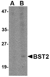
Left: Western blot analysis of Bst2 in Daudi cell lysate with Bst2 antibody at (A) 1 and (B) 2 µg/ml.
Source : Bst2 antibody was raised against a 18 amino acid peptide near the amino terminus of the mouse Bst2.
Purification : Affinity chromatography purified via peptide column
Clonality and Clone : This is a polyclonal antibody.
Host : Bst2 antibody was raised in rabbit. Please use anti-rabbit secondary antibodies.
Application : Bst2 antibody can be used for detection of Bst2 by Western blot at 1 – 2 µg/ml.
Tested Application(s) : E, WB
Buffer : Antibody is supplied in PBS containing 0.02% sodium azide.
Blocking Peptide : Cat.No. 4661P - Bst2 Peptide
Long-Term Storage : Bst2 antibody can be stored at 4ºC, stable for one year. As with all antibodies care should be taken to avoid repeated freeze thaw cycles. Antibodies should not be exposed to prolonged high temperatures.
Positive Control
1. Cat. No. 1224 - Daudi Cell Lysate
Species Reactivity :H, M
GI Number : 56971124
Accession Number : AAH87949
Short Description : (NT) Bone marrow stromal cell antigen 2
References
1. Goto T, Kennel SJ, Abe M, et al. A novel membrane antigen selectively expressed on terminally differentiated human B cells. Blood 1994; 84:1922-30.
2. Ishikawa J, Kaisho T, Tomizawa H, et al. Molecular cloning and chromosomal mapping of a bone marrow stromal cell surface gene, BST2, that may be involved in pre-B-cell growth. Genomics 1995; 26:527-34.
3. Ohtomo T, Sugamata Y, Ozaki Y, et al. Molecular cloning and characterization of a surface antigen preferentially overexpressed on multiple myeloma cells. Biochem. Biophys. Res. Commun. 1999; 258:583-91.
4. Neil SJ, Zang T, and Bieniasz PD. Tetherin inhibits retrovirus release and is antagonized by HIV-1 Vpu. Nature 2008; 451:425-30.
Catalog# : 4661
Bst2 was first identified as a novel terminal B-cell-restricted antigen termed HM1.24 that is expressed on certain bone marrow cells as well as other tissues. The surface expression of Bst2 on fibroblast cell lines facilitated the stromal cell-dependent growth of DW34, a pre-B-cell line, suggesting that this protein is involved in pre-B-cell growth. Later experiments also showed it to be upregulated in several myeloma cells. More recently, Bst2 was identified an inhibitor of retroviral release from human cells whose activity is antagonized by the HIV-1 accessory protein Vpu. While Bst2 caused retention of virions on cell surfaces or endocytosis into Bst2-positive compartments, its depletion abolished the viral requirement for Vpu for virus release. This activity may represent a potential therapeutic strategy for the treatment of HIV/AIDS.
Additional Names : Bst2 (NT), Bone marrow stromal cell antigen 2, CD317, HM1.24, tetherin
 Description
Left: Western blot analysis of Bst2 in Daudi cell lysate with Bst2 antibody at (A) 1 and (B) 2 µg/ml.
Source : Bst2 antibody was raised against a 18 amino acid peptide near the amino terminus of the mouse Bst2.
Purification : Affinity chromatography purified via peptide column
Clonality and Clone : This is a polyclonal antibody.
Host : Bst2 antibody was raised in rabbit. Please use anti-rabbit secondary antibodies.
Application : Bst2 antibody can be used for detection of Bst2 by Western blot at 1 – 2 µg/ml.
Tested Application(s) : E, WB
Buffer : Antibody is supplied in PBS containing 0.02% sodium azide.
Blocking Peptide : Cat.No. 4661P - Bst2 Peptide
Long-Term Storage : Bst2 antibody can be stored at 4ºC, stable for one year. As with all antibodies care should be taken to avoid repeated freeze thaw cycles. Antibodies should not be exposed to prolonged high temperatures.
Positive Control
1. Cat. No. 1224 - Daudi Cell Lysate
Species Reactivity :H, M
GI Number : 56971124
Accession Number : AAH87949
Short Description : (NT) Bone marrow stromal cell antigen 2
References
1. Goto T, Kennel SJ, Abe M, et al. A novel membrane antigen selectively expressed on terminally differentiated human B cells. Blood 1994; 84:1922-30.
2. Ishikawa J, Kaisho T, Tomizawa H, et al. Molecular cloning and chromosomal mapping of a bone marrow stromal cell surface gene, BST2, that may be involved in pre-B-cell growth. Genomics 1995; 26:527-34.
3. Ohtomo T, Sugamata Y, Ozaki Y, et al. Molecular cloning and characterization of a surface antigen preferentially overexpressed on multiple myeloma cells. Biochem. Biophys. Res. Commun. 1999; 258:583-91.
4. Neil SJ, Zang T, and Bieniasz PD. Tetherin inhibits retrovirus release and is antagonized by HIV-1 Vpu. Nature 2008; 451:425-30.
Wednesday, October 6, 2010
GFR alpha 1 Antibody
GFR alpha 1 Antibody
Catalog# : 1133
Glial cell line-derived neurotrophic factor (GDNF) is a potent survival factor for central and peripheral neurons and is essential for the development of kidneys and the enteric nerves system. Physiological responses to GDNF require the presence of a novel glycosylphosphadidylinositol linked protein GDNFRa, which is a cell surface receptor for GDNF. The cDNAs encoding GDNFRalpha from human, rat, chicken and mouse have been cloned recently. GDNFRalpha was also termed Ret ligand 1 (RETL1) or TGF-beta-related neurotrophic factor receptor 1 (TrnR1) and nominated as GFRalpha-1 recently. GFRalpha-1 binds GDNF specifically and mediates activation of the Ret protein tyrosine kinase (PTK). Thus, GDNF, GFRalpha and the Ret PTK form a complex to transduce GDNF signal and to mediate GDNF function.
Additional Names : GFR alpha 1 (IN), GFR alpha 1, GFRa-1, GDNFRa, GDNFRalpha
 Description
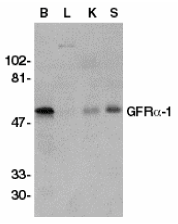
Left: Western blot analysis of GFR alpha 1 in crude membrane fractions of human brain (B), liver (L), kidney (K), and spleen (S), respectively, with GFR alpha 1 antibody at 1:500 dilution.
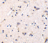
Below: Immunohistochemistry of GFR alpha 1 in human brain tissue with GFR alpha 1 antibody at 1 µg/ml.
Other Product Images
 Source : GFR alpha 1 antibody was raised against a peptide corresponding to amino acids 369 to 382 of human GFR alpha 1.
Purification : Antibody is DEAE purified
Clonality and Clone : This is a polyclonal antibody.
Host : GFR alpha 1 antibody was raised in rabbit. Please use anti-rabbit secondary antibodies.
Immunogen : Human GFR alpha 1 (Intermediate Domain) Peptide (Cat. No. 1133P)
Application : GFR alpha 1 antibody can be used for detection of GFR alpha 1 by Western blot. The amino acid sequence of immunogenic peptide is identical to those of mouse and rat and the antibody recognizes GFR alpha 1 from human, mouse and rat origins.
Tested Application(s) : E, WB, IHC
Buffer : Antibody is supplied in PBS containing 0.02% sodium azide.
Blocking Peptide : Cat. No. 1133P - GFR alpha 1 Peptide
Long-Term Storage : GFR alpha 1 antibody can be stored at 4ºC, stable for one year. As with all antibodies care should be taken to avoid repeated freeze thaw cycles. Antibodies should not be exposed to prolonged high temperatures.
Positive Control
1. Cat. No. 1303 - Human Brain Tissue Lysate
2. Cat. No. 1304 - Human Liver Tissue Lysate
3. Cat. No. 1305 - Human Kidney Tissue Lysate
4. Cat. No. 1306 - Human Spleen Tissue Lysate
Species Reactivity :H, M, R
GI Number : 20141405
Accession Number : P56159
Short Description : (IN) GDNF receptor
References
1. Jing S; Wen D; Yu Y; Holst PL ; Luo Y; Fang M; Tamir R; Antonio L; Hu Z; Cupples R; Louis JC; Hu S; Altrock BW; Fox GM. GDNF-induced activation of the Ret protein tyrosien kinase is mediated by GDNFR-a, a novel receptor for GDNF. Cell, 1996;85:1113-24.
2. Treanor JJS; GoodmanL; Sauvage FD; Stone DM; Poulsen KT; Beck CD; Gray C; Armanini MP; Pollock RA; Herti F; Phillips HS; Goddard A; Moore MW; Buj-Bello A;Davies AM; Asai N; Takahashi M; Vandlen R; Henderson CE; Rosenthal A. Characterization of a multicomponent receptor for GDNF. Nature, 1996;382:80-83.
3. Sanicola M, Hession C, Worley D, Carmillo P, Ehrenfels C, Walus L, Robinson S, Jaworski G, Wei H, Tizard R, Whitty A, Pepinsky RB, Cate RL. Glial cell line-derived neurotrophic factor-dependent RET activation can be mediated by two different cell-surface accessory proteins. Proc Natl Acad Sci U S. 1997;94:6238-6243
4. Buj-Bello A, Adu J, Pinon LG, Horton A, Thompson J, Rosenthal A, Chinchetru M, Buchman VL, Davies AM. Neurturin responsiveness requires a GPI-linked receptor and the Ret receptor tyrosine kinase. Nature 1997;387:721-724
Catalog# : 1133
Glial cell line-derived neurotrophic factor (GDNF) is a potent survival factor for central and peripheral neurons and is essential for the development of kidneys and the enteric nerves system. Physiological responses to GDNF require the presence of a novel glycosylphosphadidylinositol linked protein GDNFRa, which is a cell surface receptor for GDNF. The cDNAs encoding GDNFRalpha from human, rat, chicken and mouse have been cloned recently. GDNFRalpha was also termed Ret ligand 1 (RETL1) or TGF-beta-related neurotrophic factor receptor 1 (TrnR1) and nominated as GFRalpha-1 recently. GFRalpha-1 binds GDNF specifically and mediates activation of the Ret protein tyrosine kinase (PTK). Thus, GDNF, GFRalpha and the Ret PTK form a complex to transduce GDNF signal and to mediate GDNF function.
Additional Names : GFR alpha 1 (IN), GFR alpha 1, GFRa-1, GDNFRa, GDNFRalpha
 Description
Left: Western blot analysis of GFR alpha 1 in crude membrane fractions of human brain (B), liver (L), kidney (K), and spleen (S), respectively, with GFR alpha 1 antibody at 1:500 dilution.
Below: Immunohistochemistry of GFR alpha 1 in human brain tissue with GFR alpha 1 antibody at 1 µg/ml.
Other Product Images
 Source : GFR alpha 1 antibody was raised against a peptide corresponding to amino acids 369 to 382 of human GFR alpha 1.
Purification : Antibody is DEAE purified
Clonality and Clone : This is a polyclonal antibody.
Host : GFR alpha 1 antibody was raised in rabbit. Please use anti-rabbit secondary antibodies.
Immunogen : Human GFR alpha 1 (Intermediate Domain) Peptide (Cat. No. 1133P)
Application : GFR alpha 1 antibody can be used for detection of GFR alpha 1 by Western blot. The amino acid sequence of immunogenic peptide is identical to those of mouse and rat and the antibody recognizes GFR alpha 1 from human, mouse and rat origins.
Tested Application(s) : E, WB, IHC
Buffer : Antibody is supplied in PBS containing 0.02% sodium azide.
Blocking Peptide : Cat. No. 1133P - GFR alpha 1 Peptide
Long-Term Storage : GFR alpha 1 antibody can be stored at 4ºC, stable for one year. As with all antibodies care should be taken to avoid repeated freeze thaw cycles. Antibodies should not be exposed to prolonged high temperatures.
Positive Control
1. Cat. No. 1303 - Human Brain Tissue Lysate
2. Cat. No. 1304 - Human Liver Tissue Lysate
3. Cat. No. 1305 - Human Kidney Tissue Lysate
4. Cat. No. 1306 - Human Spleen Tissue Lysate
Species Reactivity :H, M, R
GI Number : 20141405
Accession Number : P56159
Short Description : (IN) GDNF receptor
References
1. Jing S; Wen D; Yu Y; Holst PL ; Luo Y; Fang M; Tamir R; Antonio L; Hu Z; Cupples R; Louis JC; Hu S; Altrock BW; Fox GM. GDNF-induced activation of the Ret protein tyrosien kinase is mediated by GDNFR-a, a novel receptor for GDNF. Cell, 1996;85:1113-24.
2. Treanor JJS; GoodmanL; Sauvage FD; Stone DM; Poulsen KT; Beck CD; Gray C; Armanini MP; Pollock RA; Herti F; Phillips HS; Goddard A; Moore MW; Buj-Bello A;Davies AM; Asai N; Takahashi M; Vandlen R; Henderson CE; Rosenthal A. Characterization of a multicomponent receptor for GDNF. Nature, 1996;382:80-83.
3. Sanicola M, Hession C, Worley D, Carmillo P, Ehrenfels C, Walus L, Robinson S, Jaworski G, Wei H, Tizard R, Whitty A, Pepinsky RB, Cate RL. Glial cell line-derived neurotrophic factor-dependent RET activation can be mediated by two different cell-surface accessory proteins. Proc Natl Acad Sci U S. 1997;94:6238-6243
4. Buj-Bello A, Adu J, Pinon LG, Horton A, Thompson J, Rosenthal A, Chinchetru M, Buchman VL, Davies AM. Neurturin responsiveness requires a GPI-linked receptor and the Ret receptor tyrosine kinase. Nature 1997;387:721-724
Tuesday, September 28, 2010
CXCR4 Antibody
CXCR4 Antibody
Catalog# : 1009
Human immunodeficiency virus (HIV) and related viruses require coreceptors, in addition to CD4, to infect target cells. Some G protein-coupled receptors including CCR5, CXCR4, CCR3, CCR2b and CCR8 in the chemokine receptor family, and four new human molecules GPR15, STRL33, GPR1 and V28 were recently identified as HIV coreceptors. Among them, CXCR4 (fusin, LESTR or HUMSTR) is a principal coreceptor for T-cell tropic strains of HIV-1 fusion and entry of human white blood cells. CXCR4 is also required for the infection by dual-tropic strains of HIV-1 and mediates CD-4 independent infection by HIV-2. The a-chemokine SDF-1 is the ligand for CXCR4 and prevents infection by T-tropic HIV-1. CXCR4 associates with the surface CD4-gp120 complex before HIV enters target cells. CXCR4 messenger RNA levels correlated with HIV-1 permissiveness in diverse human cell types. Antibodies to CXCR4 block HIV-1 and HIV-2 fusion and infection of human target cells. The amino-terminal domain and the second extracellular loop of CXCR4 serve as HIV binding sites.
Additional Names : CXCR4 (NT), CXCR4
 Description
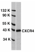
Left: Western blot analysis of CXCR4 in HeLa whole cell lysate with CXCR4 antibody at 0.5 µg/ml.
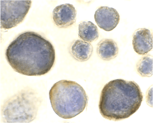
Below: Immunocytochemistry of CXCR4 in HeLa cells with CXCR4 antibody at 2 µg/ml.
Other Product Images

Source : CXCR4 antibody was raised against a peptide corresponding to amino acids near the amino terminus of human CXCR4.
Purification : Antibody is DEAE purified
Clonality and Clone : This is a polyclonal antibody.
Host : CXCR4 antibody was raised in rabbit. Please use anti-rabbit secondary antibodies.
Immunogen : Human CXCR4 (N-Terminus) Peptide (Cat. No. 1009P)
Application : CXCR-4 antibody can be used for Western blot at 1 – 2 µg/ml dilution and for immunoprecipitation and immunocytochemistry at 10 µg/ml.
Tested Application(s) : E, WB, ICC, IP
Buffer : Antibody is supplied in PBS containing 0.02% sodium azide.
Blocking Peptide : Cat. No. 1009P - CXCR4 Peptide
Long-Term Storage : CXCR4 antibody can be stored at 4ºC, stable for one year. As with all antibodies care should be taken to avoid repeated freeze thaw cycles. Antibodies should not be exposed to prolonged high temperatures.
Positive Control
1. Cat. No. 1201 - HeLa Cell Lysate
Species Reactivity :H, M
GI Number : 4503175
Accession Number : NP_003458
Short Description : (NT) HIV & chemokine receptor
References
1. Dimitrov DS. Cell 1997;91:721-730
2. Feng Y et al. Science 1996;272:872-7
3. Berson JF et al. J Virol 1996;70:6288-95
4. Doranz BJ et al. Cell 1996;85:1149-1158
Catalog# : 1009
Human immunodeficiency virus (HIV) and related viruses require coreceptors, in addition to CD4, to infect target cells. Some G protein-coupled receptors including CCR5, CXCR4, CCR3, CCR2b and CCR8 in the chemokine receptor family, and four new human molecules GPR15, STRL33, GPR1 and V28 were recently identified as HIV coreceptors. Among them, CXCR4 (fusin, LESTR or HUMSTR) is a principal coreceptor for T-cell tropic strains of HIV-1 fusion and entry of human white blood cells. CXCR4 is also required for the infection by dual-tropic strains of HIV-1 and mediates CD-4 independent infection by HIV-2. The a-chemokine SDF-1 is the ligand for CXCR4 and prevents infection by T-tropic HIV-1. CXCR4 associates with the surface CD4-gp120 complex before HIV enters target cells. CXCR4 messenger RNA levels correlated with HIV-1 permissiveness in diverse human cell types. Antibodies to CXCR4 block HIV-1 and HIV-2 fusion and infection of human target cells. The amino-terminal domain and the second extracellular loop of CXCR4 serve as HIV binding sites.
Additional Names : CXCR4 (NT), CXCR4
 Description
Left: Western blot analysis of CXCR4 in HeLa whole cell lysate with CXCR4 antibody at 0.5 µg/ml.
Below: Immunocytochemistry of CXCR4 in HeLa cells with CXCR4 antibody at 2 µg/ml.
Other Product Images
Source : CXCR4 antibody was raised against a peptide corresponding to amino acids near the amino terminus of human CXCR4.
Purification : Antibody is DEAE purified
Clonality and Clone : This is a polyclonal antibody.
Host : CXCR4 antibody was raised in rabbit. Please use anti-rabbit secondary antibodies.
Immunogen : Human CXCR4 (N-Terminus) Peptide (Cat. No. 1009P)
Application : CXCR-4 antibody can be used for Western blot at 1 – 2 µg/ml dilution and for immunoprecipitation and immunocytochemistry at 10 µg/ml.
Tested Application(s) : E, WB, ICC, IP
Buffer : Antibody is supplied in PBS containing 0.02% sodium azide.
Blocking Peptide : Cat. No. 1009P - CXCR4 Peptide
Long-Term Storage : CXCR4 antibody can be stored at 4ºC, stable for one year. As with all antibodies care should be taken to avoid repeated freeze thaw cycles. Antibodies should not be exposed to prolonged high temperatures.
Positive Control
1. Cat. No. 1201 - HeLa Cell Lysate
Species Reactivity :H, M
GI Number : 4503175
Accession Number : NP_003458
Short Description : (NT) HIV & chemokine receptor
References
1. Dimitrov DS. Cell 1997;91:721-730
2. Feng Y et al. Science 1996;272:872-7
3. Berson JF et al. J Virol 1996;70:6288-95
4. Doranz BJ et al. Cell 1996;85:1149-1158
Monday, September 27, 2010
CCR5 Antibody
CCR5 Antibody
Catalog# : 1112
Human immunodeficiency virus (HIV) and related virus require coreceptors, in addition to CD4, to infect target cells. Some G protein-coupled receptors including CCR5, CXCR4, CCR3, CCR2b and CCR8 in the chemokine receptor family, and four new human molecules GPR15, STRL33, GPR1 and V28 were recently identified as HIV coreceptors. Among them, CCR5 (CC-CKR-5) is a principal coreceptor for macrophage- and dual-tropic HIV-1 strains fusion and entry of human white blood cells. CCR5 is required for the infection by HIV-1, HIV-2, and SIV. The b-chemokines RANTES, MIP-1alpha and MIP-1beta are the ligands for CCR5 and prevent infection by M-tropic HIV-1. CXC5 associates with the surface CD4-gp120 of HIV complex and leads to membrane fusion and virus entry of target cells. The amino-terminal domain and the extracellular loops of CCR5 serve as HIV biding sites. CCR5 messenger RNA is expressed in lymphoid organs and monocytes.
Additional Names : CCR5 (NT), CCR5
 Description
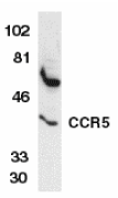
Left: Western blot analysis of CCR5 in THP-1 whole cell lysate with CCR5 antibody at 1:1000 dilution.
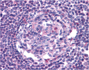
Below: Immunohistochemistry of CCR5 in human lymph node tissue with CCR5 antibody at 20 µg/ml.
Other Product Images
 Source : CCR5 antibody was raised against a peptide corresponding to amino acids 6 to 20 of human CCR5 .
Purification : Antibody is DEAE purified
Clonality and Clone : This is a polyclonal antibody.
Host : CCR5 antibody was raised in rabbit. Please use anti-rabbit secondary antibodies.
Immunogen : Human CCR5 (N-Terminus) Peptide (Cat. No. 1112P)
Application : CCR5 antibody can be used for Western blot at 1:1000 to 1:2000 dilution. An approximate 40 kDa band can be detected. CCR5 antibody can also detect CCR5 by immunohistochemistry at 20 µg/ml.
Tested Application(s) : E, WB, IHC
Buffer : Antibody is supplied in PBS containing 0.02% sodium azide.
Blocking Peptide : Cat. No. 1112P - CCR5 Peptide
Long-Term Storage : CCR5 antibody can be stored at 4ºC, stable for one year. As with all antibodies care should be taken to avoid repeated freeze thaw cycles. Antibodies should not be exposed to prolonged high temperatures.
Positive Control
1. Cat. No. 1208 - THP-1 Cell Lysate
2. Cat. No. 1369 - Human Lymph Node Tissue Lysate
Species Reactivity :H
GI Number : 2104520
Accession Number : AAB57793
Short Description : (NT) HIV & chemokine receptor
References
1. Dimitrov, DS. Cell 1997;91:721-30
2. Littman, DR. Cell 1998;93:677-80
3. Deng, H, et al. Nature 1996;381:661-6
4. Dragic, T, et al. Nature 1996;381:667-73
Catalog# : 1112
Human immunodeficiency virus (HIV) and related virus require coreceptors, in addition to CD4, to infect target cells. Some G protein-coupled receptors including CCR5, CXCR4, CCR3, CCR2b and CCR8 in the chemokine receptor family, and four new human molecules GPR15, STRL33, GPR1 and V28 were recently identified as HIV coreceptors. Among them, CCR5 (CC-CKR-5) is a principal coreceptor for macrophage- and dual-tropic HIV-1 strains fusion and entry of human white blood cells. CCR5 is required for the infection by HIV-1, HIV-2, and SIV. The b-chemokines RANTES, MIP-1alpha and MIP-1beta are the ligands for CCR5 and prevent infection by M-tropic HIV-1. CXC5 associates with the surface CD4-gp120 of HIV complex and leads to membrane fusion and virus entry of target cells. The amino-terminal domain and the extracellular loops of CCR5 serve as HIV biding sites. CCR5 messenger RNA is expressed in lymphoid organs and monocytes.
Additional Names : CCR5 (NT), CCR5
 Description
Left: Western blot analysis of CCR5 in THP-1 whole cell lysate with CCR5 antibody at 1:1000 dilution.
Below: Immunohistochemistry of CCR5 in human lymph node tissue with CCR5 antibody at 20 µg/ml.
Other Product Images
 Source : CCR5 antibody was raised against a peptide corresponding to amino acids 6 to 20 of human CCR5 .
Purification : Antibody is DEAE purified
Clonality and Clone : This is a polyclonal antibody.
Host : CCR5 antibody was raised in rabbit. Please use anti-rabbit secondary antibodies.
Immunogen : Human CCR5 (N-Terminus) Peptide (Cat. No. 1112P)
Application : CCR5 antibody can be used for Western blot at 1:1000 to 1:2000 dilution. An approximate 40 kDa band can be detected. CCR5 antibody can also detect CCR5 by immunohistochemistry at 20 µg/ml.
Tested Application(s) : E, WB, IHC
Buffer : Antibody is supplied in PBS containing 0.02% sodium azide.
Blocking Peptide : Cat. No. 1112P - CCR5 Peptide
Long-Term Storage : CCR5 antibody can be stored at 4ºC, stable for one year. As with all antibodies care should be taken to avoid repeated freeze thaw cycles. Antibodies should not be exposed to prolonged high temperatures.
Positive Control
1. Cat. No. 1208 - THP-1 Cell Lysate
2. Cat. No. 1369 - Human Lymph Node Tissue Lysate
Species Reactivity :H
GI Number : 2104520
Accession Number : AAB57793
Short Description : (NT) HIV & chemokine receptor
References
1. Dimitrov, DS. Cell 1997;91:721-30
2. Littman, DR. Cell 1998;93:677-80
3. Deng, H, et al. Nature 1996;381:661-6
4. Dragic, T, et al. Nature 1996;381:667-73
CCR3 Antibody
CCR3 Antibody
Catalog# : 1105
Human immunodeficiency virus (HIV) and related virus require coreceptors to infect target cells. Some G protein-coupled receptors including CCR5, CXCR4, CCR3, CCR2b, CCR8, GPR15, STRL33, and CX3CR1 in the chemokine receptor family were recently identified as HIV coreceptors. CCR5, CXCR4 and CCR3 are the principal receptors for HIV fusion and entry of target cells. CCR3 facilitates infection by a subset of virus. CCR3 and CCR5 promote efficient infection of microglia, the major target cells in the CNS (3,4). High levels of CCR3 and CXCR4 expression were found on the neurons from both the central and peripheral nervous systems. The CCR3 ligand, eotaxin, and an anti-CCR3 antibody inhibited HIV infection of microglia. These results indicate CCR3 plays an important role in HIV infection of CNS.
Additional Names : CCR3 (CT), CC chemokine receptor 3, eosinophil eotaxin receptor
 Description
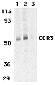
Left: Western blot analysis of CCR3 in human spleen tissue lysates with CCR3 antibody at 1 (lane 1) and 2 µg/ml (lane 2), and 2 µg/ml in the presence of blocking peptide (lane 3).
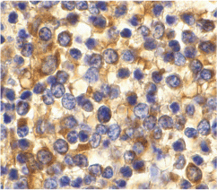
Below: Immunohistochemistry of CCR3 in human spleen tissue with CCR3 antibody at 10 µg/ml.
Other Product Images
 Source : CCR3 antibody was raised against a 14 amino acid peptide from near the carboxy terminus of human CCR3.
Purification : Immunoaffinity purified IgG
Clonality and Clone : This is a polyclonal antibody.
Host : CCR3 antibody was raised in rabbit. Please use anti-rabbit secondary antibodies.
Immunogen : Human CCR3 (C-Terminus) Peptide (Cat. No. 1105P)
Application : CCR3 antibody can be used for the detection of CCR3 by Western blot at 1 – 2 µg/ml.
Tested Application(s) : E, WB, IHC
Buffer : Antibody is supplied in PBS containing 0.02% sodium azide.
Blocking Peptide : Cat. No. 1105P - CCR3 Peptide
Long-Term Storage : CCR3 antibody can be stored at 4ºC, stable for one year. As with all antibodies care should be taken to avoid repeated freeze thaw cycles. Antibodies should not be exposed to prolonged high temperatures.
Positive Control
1. Cat. No. 1306 - Human Spleen Tissue Lysate
Species Reactivity :H, M, R
GI Number : 30581170
Accession Number : NP_847899
Short Description : (CT) HIV and chemokine receptor
References
1. Feng Y, Broder CC, Kennedy PE, et al. HIV-1 entry cofactor: functional cDNA cloning of a seven-transmembrane, G protein-coupled receptor. Science 1996; 272:872-7.
2. Deng H, Liu R, Ellmeier W, et al. Identification of a major co-receptor for primary isolates of HIV-1. Nature 1996; 381:661-6.
3. Choe H, Farzan M, Sun Y, et al. The beta-chemokine receptors CCR3 and CCR5 facilitate infection by primary HIV-1 isolates. Cell 1996; 85:1135-48.
4. He J, Chen Y, Farzan M, et al. CCR3 and CCR5 are co-receptors for HIV-1 infection of microglia. Nature 1997; 385:645-9.
Catalog# : 1105
Human immunodeficiency virus (HIV) and related virus require coreceptors to infect target cells. Some G protein-coupled receptors including CCR5, CXCR4, CCR3, CCR2b, CCR8, GPR15, STRL33, and CX3CR1 in the chemokine receptor family were recently identified as HIV coreceptors. CCR5, CXCR4 and CCR3 are the principal receptors for HIV fusion and entry of target cells. CCR3 facilitates infection by a subset of virus. CCR3 and CCR5 promote efficient infection of microglia, the major target cells in the CNS (3,4). High levels of CCR3 and CXCR4 expression were found on the neurons from both the central and peripheral nervous systems. The CCR3 ligand, eotaxin, and an anti-CCR3 antibody inhibited HIV infection of microglia. These results indicate CCR3 plays an important role in HIV infection of CNS.
Additional Names : CCR3 (CT), CC chemokine receptor 3, eosinophil eotaxin receptor
 Description
Left: Western blot analysis of CCR3 in human spleen tissue lysates with CCR3 antibody at 1 (lane 1) and 2 µg/ml (lane 2), and 2 µg/ml in the presence of blocking peptide (lane 3).
Below: Immunohistochemistry of CCR3 in human spleen tissue with CCR3 antibody at 10 µg/ml.
Other Product Images
 Source : CCR3 antibody was raised against a 14 amino acid peptide from near the carboxy terminus of human CCR3.
Purification : Immunoaffinity purified IgG
Clonality and Clone : This is a polyclonal antibody.
Host : CCR3 antibody was raised in rabbit. Please use anti-rabbit secondary antibodies.
Immunogen : Human CCR3 (C-Terminus) Peptide (Cat. No. 1105P)
Application : CCR3 antibody can be used for the detection of CCR3 by Western blot at 1 – 2 µg/ml.
Tested Application(s) : E, WB, IHC
Buffer : Antibody is supplied in PBS containing 0.02% sodium azide.
Blocking Peptide : Cat. No. 1105P - CCR3 Peptide
Long-Term Storage : CCR3 antibody can be stored at 4ºC, stable for one year. As with all antibodies care should be taken to avoid repeated freeze thaw cycles. Antibodies should not be exposed to prolonged high temperatures.
Positive Control
1. Cat. No. 1306 - Human Spleen Tissue Lysate
Species Reactivity :H, M, R
GI Number : 30581170
Accession Number : NP_847899
Short Description : (CT) HIV and chemokine receptor
References
1. Feng Y, Broder CC, Kennedy PE, et al. HIV-1 entry cofactor: functional cDNA cloning of a seven-transmembrane, G protein-coupled receptor. Science 1996; 272:872-7.
2. Deng H, Liu R, Ellmeier W, et al. Identification of a major co-receptor for primary isolates of HIV-1. Nature 1996; 381:661-6.
3. Choe H, Farzan M, Sun Y, et al. The beta-chemokine receptors CCR3 and CCR5 facilitate infection by primary HIV-1 isolates. Cell 1996; 85:1135-48.
4. He J, Chen Y, Farzan M, et al. CCR3 and CCR5 are co-receptors for HIV-1 infection of microglia. Nature 1997; 385:645-9.
Thursday, September 23, 2010
OCC-1 Antibody
OCC-1 Antibody
Catalog# : 4997
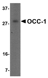
OCC-1 (Overexpressed in colon carcinoma-1) was initially identified as a novel human cDNA overexpressed in a colon carcinoma cell line. Elevated levels of OCC-1 mRNA were observed in three of eight colon carcinomas, suggesting that OCC-1 may be a hallmark of only a subset of colon carcinomas. It is highly expressed in skeletal muscle, kidney, placenta, and pancreas tissues, with low to no expression in heart, brain, lung and liver tissues. At least two different OCC-1 mRNA species are known to exist. In vitro transcription and translation of the OCC-1 cDNA yields polypeptides less than 9 kDa. Despite this, OCC-1 from cell lines is detected at ~28 kDa in SDS-PAGE.
Additional Names : OCC-1 (NT), Overexpressed in colon carcinoma-1
 Description
Left: Western blot analysis of OCC-1 in human spleen tissue lysate with OCC-1 antibody at 2 µg/ml.
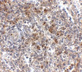
Below: Immunohistochemistry of OCC-1 in human spleen tissue with OCC-1 antibody at 2.5 μg/ml.
Other Product Images
 Source : OCC-1 antibody was raised against a 16 amino acid peptide near the amino terminus of human OCC-1.
Purification : Affinity chromatography purified via peptide column
Clonality and Clone : This is a polyclonal antibody.
Host : OCC-1 antibody was raised in rabbit. Please use anti-rabbit secondary antibodies.
Application : OCC-1 antibody can be used for detection of OCC-1 by Western blot at 2 – 4 µg/ml.
Tested Application(s) : E, WB
Buffer : Antibody is supplied in PBS containing 0.02% sodium azide.
Blocking Peptide : Cat.No. 4997P - OCC-1 Peptide
Long-Term Storage : OCC-1 antibody can be stored at 4ºC, stable for one year. As with all antibodies care should be taken to avoid repeated freeze thaw cycles. Antibodies should not be exposed to prolonged high temperatures.
Positive Control
1. Cat. No. 1210 - 293 Cell Lysate
Species Reactivity :H
GI Number : 112180576
Accession Number : AAH61920
Short Description : (NT) Overexpressed in colon carcinoma-1
References
1. Pibouin L, Villaudy J, Ferbus D, et al. Cloning of the mRNA of overexpression in colon-carcinoma-1: a sequence overexpressed in a subset of colon carcinomas. Cancer Genet. Cytogenet. 2002; 133:55-60.
Catalog# : 4997
OCC-1 (Overexpressed in colon carcinoma-1) was initially identified as a novel human cDNA overexpressed in a colon carcinoma cell line. Elevated levels of OCC-1 mRNA were observed in three of eight colon carcinomas, suggesting that OCC-1 may be a hallmark of only a subset of colon carcinomas. It is highly expressed in skeletal muscle, kidney, placenta, and pancreas tissues, with low to no expression in heart, brain, lung and liver tissues. At least two different OCC-1 mRNA species are known to exist. In vitro transcription and translation of the OCC-1 cDNA yields polypeptides less than 9 kDa. Despite this, OCC-1 from cell lines is detected at ~28 kDa in SDS-PAGE.
Additional Names : OCC-1 (NT), Overexpressed in colon carcinoma-1
 Description
Left: Western blot analysis of OCC-1 in human spleen tissue lysate with OCC-1 antibody at 2 µg/ml.
Below: Immunohistochemistry of OCC-1 in human spleen tissue with OCC-1 antibody at 2.5 μg/ml.
Other Product Images
 Source : OCC-1 antibody was raised against a 16 amino acid peptide near the amino terminus of human OCC-1.
Purification : Affinity chromatography purified via peptide column
Clonality and Clone : This is a polyclonal antibody.
Host : OCC-1 antibody was raised in rabbit. Please use anti-rabbit secondary antibodies.
Application : OCC-1 antibody can be used for detection of OCC-1 by Western blot at 2 – 4 µg/ml.
Tested Application(s) : E, WB
Buffer : Antibody is supplied in PBS containing 0.02% sodium azide.
Blocking Peptide : Cat.No. 4997P - OCC-1 Peptide
Long-Term Storage : OCC-1 antibody can be stored at 4ºC, stable for one year. As with all antibodies care should be taken to avoid repeated freeze thaw cycles. Antibodies should not be exposed to prolonged high temperatures.
Positive Control
1. Cat. No. 1210 - 293 Cell Lysate
Species Reactivity :H
GI Number : 112180576
Accession Number : AAH61920
Short Description : (NT) Overexpressed in colon carcinoma-1
References
1. Pibouin L, Villaudy J, Ferbus D, et al. Cloning of the mRNA of overexpression in colon-carcinoma-1: a sequence overexpressed in a subset of colon carcinomas. Cancer Genet. Cytogenet. 2002; 133:55-60.
Wednesday, September 22, 2010
JMJD1B Antibody
JMJD1B Antibody
Catalog# : 5369
The jumonji domain containing 1B protein (JMJD1B) was originally discovered as a candidate for the myeloid leukemia tumor suppressor gene. Besides CD34+ cells and acute myeloid leukemia (AML) cell lines, JMJD1B mRNA is highly expressed in liver, heart, kidney, skeletal muscle, and placenta tissues. The JMJD1B gene is localized to a region of chromosome 5q31, which is frequently deleted in myeloid leukemias and myelodysplasias and expression of JMJD1B in a del(5q) cell line results in suppression of clonogenic growth suggesting that JMJD1B may function as a tumor suppressor. In contrast, JMJD1B gene copy number and mRNA expression level was increased in several non-small cell lung cancers indicating that the role of JMJD1B in cancer formation and progression is more complex than originally postulated. At least three isoforms of JMJD1B are known to exist. This antibody will not cross-react with JMJD1A or JMJD1C.
Additional Names : JMJD1B,5qNCA, Lysine-specific demethylase 3B, KDM3B
 Description
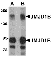
Left: Western blot analysis of JMJD1B in rat liver tissue lysate with JMJD1B antibody at (A) 1 and (B) 2 µg/ml.
Source : JMJD1B antibody was raised against a 20 amino acid peptide from near the center of human JMJD1B.
Purification : Affinity chromatography purified via peptide column
Clonality and Clone : Polyclonal
Host : JMJD1B antibody was raised in rabbit. Please use anti-rabbit secondary antibodies.
Application : JMJD1B antibody can be used for detection of JMJD1B by Western blot at 1 - 2 µg/ml.
Tested Application(s) : E, WB
Buffer : Antibody is supplied in PBS containing 0.02% sodium azide.
Blocking Peptide : Cat.No. 5369P - JMJD1B Peptide
Long-Term Storage : JMJD1B antibody can be stored at 4ºC, stable for one year. As with all antibodies care should be taken to avoid repeated freeze thaw cycles. Antibodies should not be exposed to prolonged high temperatures.
Positive Control
1. Cat. No. 1464 - Rat Liver Tissue Lysate
Species Reactivity :H, M, R
GI Number : 119582545
Accession Number : EAW62141
Short Description : Lysine-specific demethylase 3B
References
1. Lai F, Godley LA, Fernald AA, et al. cDNA cloning and genomic structure of three genes localized to human chromosome band 5q31 encoding potential nuclear proteins. Genomics 2000; 70:123-30.
2. Hu Z, Gomes I, Horrigan SK, et al. A novel nuclear protein, 5qNCA (LOC51780) is a candidate for the myeloid leukemia tumor suppressor gene on chromosome band q31. Oncogene 2001; 20:6946-54.
3. Baik S-H, Jee B-K, Choi J-S, et al. DNA profiling by array comparative genomic hybridization (CGH) of peripheral blood mononuclear cells (PBMC) and tumor tissue cell in non-small cell lung cancer (NSCLC). Mol. Biol. Rep. 2009; 36:1767-78.
Catalog# : 5369
The jumonji domain containing 1B protein (JMJD1B) was originally discovered as a candidate for the myeloid leukemia tumor suppressor gene. Besides CD34+ cells and acute myeloid leukemia (AML) cell lines, JMJD1B mRNA is highly expressed in liver, heart, kidney, skeletal muscle, and placenta tissues. The JMJD1B gene is localized to a region of chromosome 5q31, which is frequently deleted in myeloid leukemias and myelodysplasias and expression of JMJD1B in a del(5q) cell line results in suppression of clonogenic growth suggesting that JMJD1B may function as a tumor suppressor. In contrast, JMJD1B gene copy number and mRNA expression level was increased in several non-small cell lung cancers indicating that the role of JMJD1B in cancer formation and progression is more complex than originally postulated. At least three isoforms of JMJD1B are known to exist. This antibody will not cross-react with JMJD1A or JMJD1C.
Additional Names : JMJD1B,5qNCA, Lysine-specific demethylase 3B, KDM3B
 Description
Left: Western blot analysis of JMJD1B in rat liver tissue lysate with JMJD1B antibody at (A) 1 and (B) 2 µg/ml.
Source : JMJD1B antibody was raised against a 20 amino acid peptide from near the center of human JMJD1B.
Purification : Affinity chromatography purified via peptide column
Clonality and Clone : Polyclonal
Host : JMJD1B antibody was raised in rabbit. Please use anti-rabbit secondary antibodies.
Application : JMJD1B antibody can be used for detection of JMJD1B by Western blot at 1 - 2 µg/ml.
Tested Application(s) : E, WB
Buffer : Antibody is supplied in PBS containing 0.02% sodium azide.
Blocking Peptide : Cat.No. 5369P - JMJD1B Peptide
Long-Term Storage : JMJD1B antibody can be stored at 4ºC, stable for one year. As with all antibodies care should be taken to avoid repeated freeze thaw cycles. Antibodies should not be exposed to prolonged high temperatures.
Positive Control
1. Cat. No. 1464 - Rat Liver Tissue Lysate
Species Reactivity :H, M, R
GI Number : 119582545
Accession Number : EAW62141
Short Description : Lysine-specific demethylase 3B
References
1. Lai F, Godley LA, Fernald AA, et al. cDNA cloning and genomic structure of three genes localized to human chromosome band 5q31 encoding potential nuclear proteins. Genomics 2000; 70:123-30.
2. Hu Z, Gomes I, Horrigan SK, et al. A novel nuclear protein, 5qNCA (LOC51780) is a candidate for the myeloid leukemia tumor suppressor gene on chromosome band q31. Oncogene 2001; 20:6946-54.
3. Baik S-H, Jee B-K, Choi J-S, et al. DNA profiling by array comparative genomic hybridization (CGH) of peripheral blood mononuclear cells (PBMC) and tumor tissue cell in non-small cell lung cancer (NSCLC). Mol. Biol. Rep. 2009; 36:1767-78.
Monday, September 20, 2010
FAM120A Antibody
FAM120A Antibody
Catalog# : 5307
FAM120A (C9orf10) is a member of the constitutive coactivator of PPAR gamma family and the gene was mapped to chromosome 9q22.31. FAM120A was recently detected within the Pur-alpha-containing mRNA-protein complex in the brain. As a novel RNA-binding protein, FAM120A is a critical component of the oxidative stress-induced survival signaling. It may participate in mRNA transport in the cytoplasm. FAM120A activates src family kinases and acts as a scaffolding protein enabling src family kinases to phosphorylate and activate PI3-kinase. FAM120A protects cells from apoptosis through activation of SRKs in response to oxidative stress. Blocking of the survival signaling mediated by FAM120A, which sensitizes the cancer cells to stress-induced apoptosis, may be a novel therapeutic approach for gastric scirrhous carcinoma cells. This antibody will not cross-reacti with FAM120B / PGCC1.
Additional Names : FAM120A,Constitutive coactivator of PPAR-gamma-like protein 1, DNAPTP1, DNAPTP5, Oxidative stress-associated Src activator, OSSA
 Description
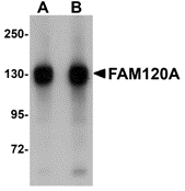
Left: Western blot analysis of FAM120A in SK-N-SH cell lysate with FAM120A antibody at (A) 0.5 and (B) 1 µg/ml.
Source : FAM120A antibody was raised against an 18 amino acid peptide from near the center of human FAM120A.
Purification : Affinity chromatography purified via peptide column
Clonality and Clone : Polyclonal
Host : FAM120A antibody was raised in rabbit. Please use anti-rabbit secondary antibodies.
Application : FAM120A antibody can be used for detection of FAM120A by Western blot at 0.5 - 1 µg/ml.
Tested Application(s) : E, WB
Buffer : Antibody is supplied in PBS containing 0.02% sodium azide.
Blocking Peptide : Cat.No. 5307P - FAM120A Peptide
Long-Term Storage : FAM120A antibody can be stored at 4ºC, stable for one year. As with all antibodies care should be taken to avoid repeated freeze thaw cycles. Antibodies should not be exposed to prolonged high temperatures.
Positive Control
1. Cat. No. 1220 - SK-N-SH Cell Lysate
Species Reactivity :H, M, R
GI Number : 39652628
Accession Number : NP_055427
Short Description : Constitutive coactivator of PPAR-gamma-like protein 1
References
1. Holden S and Raymond FL. The human gene CXorf17 encodes a member of a novel family of putative transmembrane proteins: cDNA cloning and characterization of CXorf17 and its mouse ortholog orf34. Gene 2003; 318:149-61.
2. Kobayashi Y, Suzuki K, Kobayashi H, et al. C9orf10 protein, a novel protein component of Puralpha-containing mRNA-protein particles (Puralpha-mRNPs): characterization of developmental and regional expressions in the mouse brain. J. Histochem. Cytochem. 2008; 56:723-31.
3. Tanaka M, Sasaki K, Kamata R, et al. A novel RNA-binding protein, Ossa/C9orf10, regulates activity of Src kinases to protect cells from oxidative stress-induced apoptosis. Mol. Cell. Biol. 2009; 29:402-13.
Catalog# : 5307
FAM120A (C9orf10) is a member of the constitutive coactivator of PPAR gamma family and the gene was mapped to chromosome 9q22.31. FAM120A was recently detected within the Pur-alpha-containing mRNA-protein complex in the brain. As a novel RNA-binding protein, FAM120A is a critical component of the oxidative stress-induced survival signaling. It may participate in mRNA transport in the cytoplasm. FAM120A activates src family kinases and acts as a scaffolding protein enabling src family kinases to phosphorylate and activate PI3-kinase. FAM120A protects cells from apoptosis through activation of SRKs in response to oxidative stress. Blocking of the survival signaling mediated by FAM120A, which sensitizes the cancer cells to stress-induced apoptosis, may be a novel therapeutic approach for gastric scirrhous carcinoma cells. This antibody will not cross-reacti with FAM120B / PGCC1.
Additional Names : FAM120A,Constitutive coactivator of PPAR-gamma-like protein 1, DNAPTP1, DNAPTP5, Oxidative stress-associated Src activator, OSSA
 Description
Left: Western blot analysis of FAM120A in SK-N-SH cell lysate with FAM120A antibody at (A) 0.5 and (B) 1 µg/ml.
Source : FAM120A antibody was raised against an 18 amino acid peptide from near the center of human FAM120A.
Purification : Affinity chromatography purified via peptide column
Clonality and Clone : Polyclonal
Host : FAM120A antibody was raised in rabbit. Please use anti-rabbit secondary antibodies.
Application : FAM120A antibody can be used for detection of FAM120A by Western blot at 0.5 - 1 µg/ml.
Tested Application(s) : E, WB
Buffer : Antibody is supplied in PBS containing 0.02% sodium azide.
Blocking Peptide : Cat.No. 5307P - FAM120A Peptide
Long-Term Storage : FAM120A antibody can be stored at 4ºC, stable for one year. As with all antibodies care should be taken to avoid repeated freeze thaw cycles. Antibodies should not be exposed to prolonged high temperatures.
Positive Control
1. Cat. No. 1220 - SK-N-SH Cell Lysate
Species Reactivity :H, M, R
GI Number : 39652628
Accession Number : NP_055427
Short Description : Constitutive coactivator of PPAR-gamma-like protein 1
References
1. Holden S and Raymond FL. The human gene CXorf17 encodes a member of a novel family of putative transmembrane proteins: cDNA cloning and characterization of CXorf17 and its mouse ortholog orf34. Gene 2003; 318:149-61.
2. Kobayashi Y, Suzuki K, Kobayashi H, et al. C9orf10 protein, a novel protein component of Puralpha-containing mRNA-protein particles (Puralpha-mRNPs): characterization of developmental and regional expressions in the mouse brain. J. Histochem. Cytochem. 2008; 56:723-31.
3. Tanaka M, Sasaki K, Kamata R, et al. A novel RNA-binding protein, Ossa/C9orf10, regulates activity of Src kinases to protect cells from oxidative stress-induced apoptosis. Mol. Cell. Biol. 2009; 29:402-13.
CUEDC2 Antibody
CUEDC2 Antibody
Catalog# : 4839
The CUE (coupling of ubiquitin conjugation to endoplasmic reticulum degradation) domain is an evolutionarily conserved, ~40 amino acid monoubiquitin-binding domain that mediates intramolecular monoubiquitylation. CUE domains are present in eukaryotic proteins that are involved in ubiquitination and protein trafficking pathways and may be required for ubiquitination of the proteins in which they are found. CUEDC2 (CUE domain-containing protein 2) was found through a yeast two-hybrid screening as a protein that interacts with the progesterone receptor (PR) and promotes progesterone-induced PR degradation by the ubiquitin-proteasome pathway. CUEDC2 also decreases the sumoylation of PR. CUEDC2 has also been found to interact with IKK-a and IKK-b and decrease the activation of NF-kB by decreasing the activation of IKK. CUEDC2 antibody will not cross-react with CUEDC1.
Additional Names : CUEDC2, CUE Domain-Containing Protein 2
 Description
Left: Western blot analysis of CUEDC2 in HeLa cell lysate with CUEDC2 antibody at (A) 1 and (B) 2 µg/ml.
Below: Immunocytochemistry of CUEDC2 in HeLa cells with CUEDC2 antibody at 5 µg/ml.
Other Product Images
 Source : CUEDC2 antibody was raised against a 19 amino acid peptide from near the carboxy terminus of human CUEDC2.
Purification : Affinity chromatography purified via peptide column
Clonality and Clone : This is a polyclonal antibody.
Host : CUEDC2 antibody was raised in rabbit. Please use anti-rabbit secondary antibodies.
Application : CUEDC2 antibody can be used for detection of CUEDC2 by Western blot at 1 – 2 µg/ml.
Tested Application(s) : E, WB, ICC
Buffer : Antibody is supplied in PBS containing 0.02% sodium azide.
Blocking Peptide : Cat.No. 4839P - CUEDC2 Peptide
Long-Term Storage : CUEDC2 antibody can be stored at 4ºC, stable for one year. As with all antibodies care should be taken to avoid repeated freeze thaw cycles. Antibodies should not be exposed to prolonged high temperatures.
Positive Control
1. Cat. No. 1201 - HeLa Cell Lysate
Species Reactivity :H, M, R
GI Number : 148596996
Accession Number : NP_076945
Short Description : CUE Domain-Containing Protein 2
References
1. Shih SC, Prag G, Francis SA, et al. A ubiquitin-binding motif required for intramolecular monoubiquitylation, the CUE domain. EMBO J. 2003; 22:1273-81.
2. Ponting CP. Proteins of the endoplasmic-reticulum-associated degradation pathway: domain detection and function prediction. Biochem. J. 2000; 351:527-35.
3. Prag G, Misra S, Jones EA, et al. Mechanism of ubiquitin recognition by the CUE domain of Vps9p. Cell 2003; 113:609-20.
4. Kang RS, Daniels CM, Francis SA, et al. Solution structure of a CUE-ubiquitin complex reveals a conserved mode of ubiquitin binding. Cell 2003; 113:621-30.
Catalog# : 4839
The CUE (coupling of ubiquitin conjugation to endoplasmic reticulum degradation) domain is an evolutionarily conserved, ~40 amino acid monoubiquitin-binding domain that mediates intramolecular monoubiquitylation. CUE domains are present in eukaryotic proteins that are involved in ubiquitination and protein trafficking pathways and may be required for ubiquitination of the proteins in which they are found. CUEDC2 (CUE domain-containing protein 2) was found through a yeast two-hybrid screening as a protein that interacts with the progesterone receptor (PR) and promotes progesterone-induced PR degradation by the ubiquitin-proteasome pathway. CUEDC2 also decreases the sumoylation of PR. CUEDC2 has also been found to interact with IKK-a and IKK-b and decrease the activation of NF-kB by decreasing the activation of IKK. CUEDC2 antibody will not cross-react with CUEDC1.
Additional Names : CUEDC2, CUE Domain-Containing Protein 2
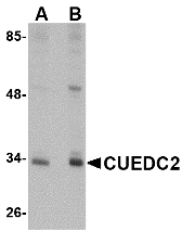 Description
Left: Western blot analysis of CUEDC2 in HeLa cell lysate with CUEDC2 antibody at (A) 1 and (B) 2 µg/ml.
Below: Immunocytochemistry of CUEDC2 in HeLa cells with CUEDC2 antibody at 5 µg/ml.
Other Product Images
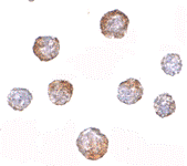 Source : CUEDC2 antibody was raised against a 19 amino acid peptide from near the carboxy terminus of human CUEDC2.
Purification : Affinity chromatography purified via peptide column
Clonality and Clone : This is a polyclonal antibody.
Host : CUEDC2 antibody was raised in rabbit. Please use anti-rabbit secondary antibodies.
Application : CUEDC2 antibody can be used for detection of CUEDC2 by Western blot at 1 – 2 µg/ml.
Tested Application(s) : E, WB, ICC
Buffer : Antibody is supplied in PBS containing 0.02% sodium azide.
Blocking Peptide : Cat.No. 4839P - CUEDC2 Peptide
Long-Term Storage : CUEDC2 antibody can be stored at 4ºC, stable for one year. As with all antibodies care should be taken to avoid repeated freeze thaw cycles. Antibodies should not be exposed to prolonged high temperatures.
Positive Control
1. Cat. No. 1201 - HeLa Cell Lysate
Species Reactivity :H, M, R
GI Number : 148596996
Accession Number : NP_076945
Short Description : CUE Domain-Containing Protein 2
References
1. Shih SC, Prag G, Francis SA, et al. A ubiquitin-binding motif required for intramolecular monoubiquitylation, the CUE domain. EMBO J. 2003; 22:1273-81.
2. Ponting CP. Proteins of the endoplasmic-reticulum-associated degradation pathway: domain detection and function prediction. Biochem. J. 2000; 351:527-35.
3. Prag G, Misra S, Jones EA, et al. Mechanism of ubiquitin recognition by the CUE domain of Vps9p. Cell 2003; 113:609-20.
4. Kang RS, Daniels CM, Francis SA, et al. Solution structure of a CUE-ubiquitin complex reveals a conserved mode of ubiquitin binding. Cell 2003; 113:621-30.
Friday, July 9, 2010
Bioconjugates of Nucleic Acids
The availability of synthetic nucleic acids/oligonucleotides and peptides, areas where Bio Synthesis has extensive experience, has promoted the development of conjugates of these molecules cross-linked to compounds such as lipids, carbohydrates and small molecules (drugs) to yield products with distinct properties. Bio Synthesis has developed several approaches for the production of different bioconjugates. From the point of view of therapeutic applications, because of their potential uses in genetic therapy and blocking gene expression, the bioconjugates of nucleic acids are the most important ones. In general these bioconjugates acquire new properties, such as resistance to degradation and passage of the hydrophilic and anionic oligonucleotide across the hydrophobic negatively charged cell membrane or transduction. Yet, a suitable bioconjugate should maintain practically all of the oligonucleotide's biological properties.
 Conjugation Services:
 Conjugation Services:
 - Oligonucleotide Peptide or protein conjugation
- Fluorescent labeling of peptide, proteins, drugs, DNA probes, etc.
- Protein-Protein conjugation
- Antibody conjugation
- Drug-Protein, peptide oliognucleotide conjugation
- Coupling of small molecules to carrier proteins
- Coupling of small molecules or proteins to solid support
- Chemical synthesis and modification of small molecules, proteins and drugs
Subscribe to:
Posts (Atom)