Bio-Synthesis provides comprehensive custom antibody packages and immunological services, including epitope prediction, custom antibody synthesis
,carrier protein conjugation, immunization and purification, ELISA analysis, antibody labeling, and antibody microarrays. BSI offers peptide antigen design, using a proprietary Antigen Identifier TM design tool. Anti-peptide polyclonal antibody production is performed with the best titer guarantee in the industry, by implementing a proprietary SP-MaxTM adjuvant, a new family of adjuvants with improved properties, but minimal toxicity. BSI works with host animals under USDA license and holds an NIH Animal Welfare Assurance from the Office of Laboratory Animal Welfare for immunization. All procedures performed during immunization are described in our Standard Operation Procedures (SOPs) to ensure a high-quality product.
All custom polyclonal antibody projects are treated with strict confidentiality. Used animals, developed antigens remain the exclusive property of the customer.
From antigen selection to antibody purfication, BSI has optimized each step of the antibody production process to provided the best antibodies available.
We offer 8 standard custom antibody protocols (combinations of hosts and protocol timeframe) and 6 standard antibody packages (combinations of provisions, antigen and deliverables). For information about a particular protocol, see our Polyclonal Antibody Services below or request a quote today.
- Single anti-peptide polyclonal antibody
- Peptide phospho-specific polyclonal antibody
- Multiple-peptide co-immunization
- Multiple antigen peptide (MAP) polyclonal antibody
- User-supplied antigen
- Create-your-own antibody packages
Why choose Bio-Synthesis for your custom polyclonal antibody services?
Advanced antigen design- Proprietary Antigen Identifier design tool with team of expert scientist to assist client for best peptide antigen selection
- Supreme adjuvant SP-MAX technology
- A new family of adjuvants with improved properties, but minimal toxicity which ensures high specificity and titer of the produced antibodies.
- ELISA Guarantee
- Our polyclonal antibodies have an ELISA titer of 1:20,000 or better for any host if peptide antigens were designed, synthesized, and conjugated by Bio-Synthesis.
Comprehensive antibody packages
- Single anti-peptide polyclonal antibody
- Peptide phospho-specific polyclonal antibody
- Multiple-peptide co-immunization
- Multiple antigen peptide (MAP) polyclonal antibody
- User-supplied antigen
- Create-your-own antibody packages
Acinus Antibody
Catalog# : 2217
Chromatin condensation and nuclear fragmentation (CCNF) is the hallmark of apoptosis. CCNF is triggered by the activation of members of caspase family, caspase activated DNase (CAD/DFF40), and several novel proteins including AIF and CIDE . A new inducer of chromatin condensation was recently identified and designated Acinus (for apoptotic chromatin condensation inducer in the nucleus). Acinus is cleaved by caspase-3 and an additional unknown protease generating a small active peptide p17, which causes chromatin condensation in vitro when it is added to purified nuclei. Acinus also induces apoptotic chromatin condensation in cells. Acinus is ubiquitously expressed. Three different spliced forms of Acinus have been identified in human and mouse and designated AcinusL, AcinusS and AcinusS’ (2).
Additional Names : Acinus (CT), Acinus
 Description
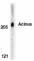
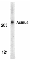
Left: Western blot analysis of Acinus in K562 whole cell lysate with Acinus antibody at 0.5 µg/ml. Below: Immunocytochemistry of Acinus in K562 cells with Acinus antibody at 0.5 µg/ml.
Source : Acinus antibody was raised against a peptide corresponding to amino acids near the carbosy terminus of human AcinusL, which are identical to those of mouse Acinus.
Purification : Affinity chromatography purified via peptide column
Clonality and Clone : This is a polyclonal antibody.
Host : Acinus antibody was raised in rabbit.
Immunogen : Murine Acinus (C-Terminus) Peptide (Cat. No. 2217P)
Application : Acinus antibody antibody can be for
Tested Application(s) : E, WB, ICC
Buffer : Antibody is supplied in PBS containing 0.02% sodium azide.
Blocking Peptide : Cat. No. 2217P - Acinus Peptide
Long-Term Storage : Acinus antibody can be stored at 4ºC, stable for one year. As with all antibodies care should be taken to avoid repeated freeze thaw cycles. Antibodies should not be exposed to prolonged high temperatures.
Positive Control
- Cat. No. 1204 - K562 Cell Lysate
Species Reactivity : H
GI Number : 5931959
Accession Number : AAD56724
Short Description : (CT) Inducer of chromatin condensation
References
- Zamzami N, Kroemer G. Condensed matter in cell death. Nature 1999 ;401:127-8.
- Sahara S, Aoto M, Eguchi Y, Imamoto N, Yoneda Y, Tsujimoto Y. Acinus is a caspase-3-activated protein required for apoptotic chromatin condensation. Nature 1999 401:168-73.
For more detail regarding Peptide please visit here : Custom Peptide Synthesis
Acinus Antibody
Catalog# : 2215
Chromatin condensation and nuclear fragmentation (CCNF) is the hallmark of apoptosis. CCNF is triggered by the activation of members of caspase family, caspase activated DNase (CAD/DFF40), and several novel proteins including AIF and CIDE . A new inducer of chromatin condensation was recently identified and designated Acinus (for apoptotic chromatin condensation inducer in the nucleus). Acinus is cleaved by caspase-3 and an additional unknown protease generating a small active peptide p17, which causes chromatin condensation in vitro when it is added to purified nuclei. Acinus also induces apoptotic chromatin condensation in cells. Acinus is ubiquitously expressed. Three different spliced forms of Acinus have been identified in human and mouse and designated AcinusL, AcinusS and AcinusS’ (2).
Additional Names : Acinus (CP), Acinus
 Description
Left: Western blot analysis of Acinus in K562 whole cell lysate with Acinus antibody (CP) at 1 µg/ml. Below: Immunocytochemistry staining of K562 cells using Acinus antibody at 10 µg/ml.
Source : Acinus antibody was raised against a peptide corresponding to amino acids near the C-terminus of the cleaved active peptide p17, which are identical to those of mouse Acinus.
Purification : Affinity chromatography purified via peptide column
Clonality and Clone : This is a polyclonal antibody.
Host : Acinus antibody was raised in rabbit.
Immunogen : Human Acinus (CP) Peptide (Cat. No. 2215P)
Application : Acinus antibody can be used for
Tested Application(s) : E, WB, ICC
Buffer : Antibody is supplied in PBS containing 0.02% sodium azide.
Blocking Peptide : Cat. No. 2215P - Acinus Peptide
Long-Term Storage : Acinus antibody can be stored at 4ºC, stable for one year. As with all antibodies care should be taken to avoid repeated freeze thaw cycles. Antibodies should not be exposed to prolonged high temperatures.
Positive Control
- Cat. No. 1204 - K562 Cell Lysate
Species Reactivity :H
GI Number : 5931959
Accession Number : AAD56724
Short Description : (CP) Inducer of chromatin condensation
References
- Zamzami N, Kroemer G. Condensed matter in cell death. Nature 1999 ;401:127-8.
- Sahara S, Aoto M, Eguchi Y, Imamoto N, Yoneda Y, Tsujimoto Y. Acinus is a caspase-3-activated protein required for apoptotic chromatin condensation. Nature 1999 401:168-73.
For more detail regarding Peptide please visit here : Custom Peptide Synthesis
Acinus Antibody
Catalog# : 2219
Chromatin condensation and nuclear fragmentation (CCNF) is the hallmark of apoptosis. CCNF is triggered by the activation of members of caspase family, caspase activated DNase (CAD/DFF40), and several novel proteins including AIF and CIDE . A new inducer of chromatin condensation was recently identified and designated Acinus (for apoptotic chromatin condensation inducer in the nucleus). Acinus is cleaved by caspase-3 and an additional unknown protease generating a small active peptide p17, which causes chromatin condensation in vitro when it is added to purified nuclei. Acinus also induces apoptotic chromatin condensation in cells. Acinus is ubiquitously expressed. Three different spliced forms of Acinus have been identified in human and mouse and designated AcinusL, AcinusS and AcinusS’ (2).
Additional Names : Acinus (IN), Acinus
 Description
Left: Western blot analysis of Acinus in K562 whole cell lysate with Acinus antibody (IN) at 1 µg/ml. Below: Immunocytochemistry of Acinus in K562 cells with Acinus antibody at 5 µg/ml.
Source : Acinus antibody was raised against a peptide (dtsenrpendvpepp) corresponding to amino acids 775 to 789 of human AcinusL, 48 to 62 of human AcinusS’, or 17 to 31 of human AcinusS, which differ from those of mouse Acinus by one amino acid.
Purification : Affinity chromatography purified via peptide column
Clonality and Clone : This is a polyclonal antibody.
Host : Acinus antibody was raised in rabbit.
Immunogen : Murine Acinus (Intermediate Domain) Peptide (Cat. No. 2219P)
Application : Acinus antibody can be used for
Tested Application(s) : E, WB, ICC
Buffer : Antibody is supplied in PBS containing 0.02% sodium azide.
Blocking Peptide : Cat. No. 2219P - Acinus Peptide
Long-Term Storage : Acinus antibody can be stored at 4ºC, stable for one year. As with all antibodies care should be taken to avoid repeated freeze thaw cycles. Antibodies should not be exposed to prolonged high temperatures.
Positive Control
- Cat. No. 1204 - K562 Cell Lysate
Species Reactivity : H
GI Number : 5931959
Accession Number : AAD56724
Short Description : (IN) Inducer of chromatin condensation
References
- Zamzami N, Kroemer G. Condensed matter in cell death. Nature 1999 ;401:127-8.
- Sahara S, Aoto M, Eguchi Y, Imamoto N, Yoneda Y, Tsujimoto Y. Acinus is a caspase-3-activated protein required for apoptotic chromatin condensation. Nature 1999 401:168-73. (WD0800
AATF Antibody
Catalog# : 3039
AATF (apoptosis antagonizing transcription factor) was initially discovered as an interaction partner of ZIP kinase (ZIPK) (1), a member of death-associated protein (DAP) kinase family of pro-apoptotic serine/threonine kinases (2). AATF is a phosphoprotein containing an acidic region and a putative leucine zipper domain and nuclear localization signal, features which are typical of transcription factors. AATF inhibits the ZIPK-mediated pro-apoptotic pathway and may activate other anti-apoptotic pathways (3). Recently, it has also been shown to protect neural cells against oxidative damage induced by amyloid b-peptide and to inhibit aberrant production of the b-peptide by interacting with Par-4 (prostate apoptosis response-4), another pro-apoptotic leucine zipper protein that is associated with neuronal degeneration in Alzheimer’s disease (AD) (4,5), suggesting that AATF may have potential therapeutic applications in both familial and sporadic forms of AD.
Additional Names : AATF, Apoptosis antagonizing transcription factor, Che-1
 Description
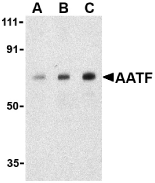
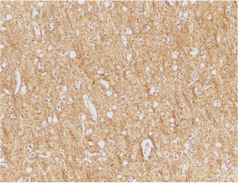
Left: Western blot analysis of AATF in human small intestine cell lysate with AATF antibody at (A) 0.5, (B) 1 and (C) 2 µg/ml. Below: Immunohistochemistry of AATF in human small intestine tissue with AATF antibody at 10 µg/ml.
Source : AATF antibody was raised against a 12 amino acid peptide from near the carboxy terminus of human AATF.
Purification : Immunoaffinity purified IgG
Clonality and Clone : This is a polyclonal antibody.
Host : AATF antibody was raised in rabbit.
Immunogen : Human AATF Peptide (Cat. No. 3039P)
Application : AATF antibody can be used for
Tested Application(s) : E, WB, IHC
Buffer : Antibody is supplied in PBS containing 0.02% sodium azide.
Blocking Peptide : Cat. No. 3039P - AATF Peptide
Long-Term Storage : AATF antibody can be stored at 4ºC, stable for one year. As with all antibodies care should be taken to avoid repeated freeze thaw cycles. Antibodies should not be exposed to prolonged high temperatures.
Positive Control
- Cat. No. 1308 - Human Small Intestine Tissue Lysate
Species Reactivity : H, M
GI Number : 7657013
Accession Number : NP_036270
Short Description : An anti-apoptotic transcription factor
References
- AATF antibody can be stored at 4ºC, stable for one year. Page G, Lodige I, Kogel D et al. AATF, a novel transcription factor that interacts with Dlk/ZIP kinase and interferes with apoptosis. FEBS Lett. 1999; 462:187-191.
- Kawai T, Matsumoto M, Takeda K, et al. ZIP kinase, a novel serine/threonine kinase which mediates apoptosis. Mol. Cell Biol. 1998; 18:1642-51.
- Lindfors K, Halttunen T, Huotari P, et al. Identification of novel transcription factor-like gene from human intestinal cells. Biochem. Biophys. Res. Commun. 2000; 276:660-6.
- Xie J and Guo Q. AATF protects neural cells against oxidative damage induced by amyloid b-peptide. Neurobiol. of Dis. 2004; 16:150-7.
For more detail regarding Peptide please visit here : Custom Peptide Synthesis
ATR Antibody
The Anthrax toxin receptor (ATR) was initially discovered as the tumor endothelial marker 8 (TEM8) (1). This protein, which exists in three isoforms (36, 40, and 60 kDa), is highly expressed in tumor vessels as well as in the vasculature of developing embryos, suggesting that it may normally play a role in angiogenesis. However, it also acts as the receptor for anthrax toxin (2). Following the binding of this protein by the protective antigen (PA) of anthrax, PA is cleaved and heptamerizes to form the binding site for both edema factor (EF) and lethal factor (LF) (3). This complex is then endocytosed by the cell; acidification in endosomes allows the release of EF and LF into the cytoplasm where they interfere with MAPK signaling and induce apoptosis (4).
Additional Names : ATR (IN), TEM8
 Description
Left: Western blot analysis of ATR in HepG2 cell lysates with ATR antibody (IN) at (A) 0.5, (B) 1, and (C) 2 µg/ml. Below: Immunohistochemical staining of human brain tissue using ATR antibody at 2 µg/ml.
Source : ATR antibody was raised against a peptide corresponding to 13 amino acids near the center of human ATR.
Purification : Ion exchange chromatography purified
Clonality and Clone : This is a polyclonal antibody.
Host : ATR antibody was raised in rabbit.
Immunogen : Human ATR / TEM8 (Intermediate Domain) Peptide (Cat. No. 3117P)
Application : ATR antibody can be used for d
Tested Application(s) : E, WB, IHC
Buffer : Antibody is supplied in PBS containing 0.02% sodium azide.
Blocking Peptide : Cat. No. 3117P - ATR Peptide
Long-Term Storage : ATR antibody can be stored at 4ºC, stable for one year. As with all antibodies care should be taken to avoid repeated freeze thaw cycles. Antibodies should not be exposed to prolonged high temperatures.
Positive Control
- Cat. No. 1211 - HepG2 Cell Lysate
- Cat. No. 1303 - Human Brain Tissue Lysate
Species Reactivity : H, M, R
GI Number : 16933551
Accession Number : NP_444262
Short Description : (IN) Anthrax receptor
For more detail regarding Peptide please visit here : Custom Peptide Synthesis
Anthrax PA Antibody
Catalog# : 3411
Anthrax infection is initiated by the inhalation, ingestion, or cutaneous contact with Bacillus anthracis endospores. B. anthracis produces three polypeptides that comprise the anthrax toxin: protective antigen (PA), lethal factor (LF), and edema factor (EF) (1,2 for review). PA binds to two related proteins on the cell surface; these are termed tumor epithelial marker 8 (TEM8)/anthrax toxin receptor (ATR) (3) and capillary morphogenesis protein 2 (CMG2) (4), although it is still unclear which is physiologically relevant. Following PA binding to its receptor, PA is cleaved into two fragments by a furin-like protease. The bound fragment binds both LF and EF; the resulting complex is then endocytosed which allows the translocation of LF and EF into the cytoplasm (5). These toxins are usually sufficient to cause rapid cell death, and often the death of the organism.
Additional Names : Anthrax PA (IN), Anthrax PA
Source : Anthrax PA antibody was raised against a synthetic peptide corresponding to 16 amino acids in the middle of the Anthrax protective antigen protein.
Purification : Affinity chromatography purified via peptide column
Clonality and Clone : This is a polyclonal antibody.
Host : Anthrax PA antibody was raised in rabbit.
Immunogen : Human Anthrax PA (Intermediate Domain) Peptide (Cat. No. 3411P)
Application : Anthrax PA antibody can be use
Tested Application(s) : E
Buffer : Antibody is supplied in PBS containing 0.02% sodium azide.
Blocking Peptide : Cat. No. 3411P - Anthrax PA Peptide
Long-Term Storage : Anthrax PA antibody can be stored at 4ºC, stable for one year. As with all antibodies care should be taken to avoid repeated freeze thaw cycles. Antibodies should not be exposed to prolonged high temperatures.
Species Reactivity :Bac
GI Number : 9280533
Accession Number : AAF86457
Short Description : (IN) Anthrax protective antigen
References
- Schwartz MN. Recognition and management of anthrax – an update. New Engl. J. Med. 2001; 345:1621-6.
- Moayeri M and Leppla SH. The roles of anthrax toxin in pathogenesis. Curr. Opin. Microbiol. 2004; 7:19-24.
- Bradley KA, Mogridge J, Mourez M, et al. Identification of the cellular receptor for anthrax toxin. Nature 2001; 414:225-9.
- Scobie HM, Rainey GJ, Bradley KA, et al. Human capillary morphogenesis protein 2 functions as an anthrax toxin receptor. Proc. Natl. Acad. Sci. USA 2003; 100:5170-4.
For more detail regarding Peptide please visit here : Custom Peptide Synthesis