Thursday, October 21, 2010

Beta-actin Antibody

Beta-actin Antibody

Catalog# : 5005

Actins are highly conserved proteins that are involved in cell motility, structure and integrity, processes that are crucial for tissue development and the development of organism. The actin cytoskeleton is one of the principal drivers of cell motility and is capable of responding to complex signaling cascades. Recent evidence suggests that it may play key roles in regulating apoptosis and aging. Beta actin is one of six different actin isoforms which have been identified. Like GAPDH, -actin is constitutively expressed at high levels in almost all tissues and cell lines making it ideal for use as a loading control marker in immunoblots.
Additional Names : Beta-actin (CT), Beta-actin
Description
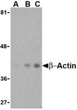
Left: Western blot analysis of actin in HeLa cell lysate with beta-actin antibody at (A) 1 and (B) 2 µg/ml.


Source : Beta-actin antibody was raised against a 16 amino acid peptide from near the carboxy-terminus of human beta-actin.
Purification : Affinity chromatography purified via peptide column
Clonality and Clone : This is a polyclonal antibody.
Host : Beta-actin antibody was raised in chicken. Please use anti-chicken secondary antibodies.
Application : Beta-actin antibody can be used for the detection of beta-actin by Western blot at 1 – 2 µg/ml.
Tested Application(s) : E, WB
Buffer : Antibody is supplied in PBS containing 0.02% sodium azide.
Blocking Peptide : Cat.No. 5005P - Beta-actin Peptide
Long-Term Storage : Beta-actin antibody can be stored at 4ºC, stable for one year. As with all antibodies care should be taken to avoid repeated freeze thaw cycles. Antibodies should not be exposed to prolonged high temperatures.
Positive Control
1. Cat. No. 1201 - HeLa Cell Lysate
Species Reactivity :H, M, R
GI Number : 12803203
Accession Number : AAH02409
Short Description : (CT) A major cytoskeletal protein
References
1. Lambrechts A, Van Troys, M and Ampe C. The actin cytoskeleton in normal and pathological cell motility. Int. J. Biochem. Cell Biol. 2004; 36:1890-909.
2. Gourlay CW and Ayscough KR. The actin cytoskeleton: a key regulator of apoptosis and ageing? Nat. Rev. 2005; 6:583-9.

Beta-actin Antibody

Beta-actin Antibody

Catalog# : 5009

Actins are highly conserved proteins that are involved in cell motility, structure and integrity, processes that are crucial for tissue development and the development of organism. The actin cytoskeleton is one of the principal drivers of cell motility and is capable of responding to complex signaling cascades. Recent evidence suggests that it may play key roles in regulating apoptosis and aging. Beta actin is one of six different actin isoforms which have been identified. Like GAPDH, b-actin is constitutively expressed at high levels in almost all tissues and cell lines making it ideal for use as a loading control marker in immunoblots.
Additional Names : beta-Actin (NT), Beta actin
Description
Left: Western blot analysis of actin in HeLa cell lysate with b-actin antibody at (A) 1 and (B) 2 µg/ml.

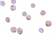
Below: Immunocytochemistry of Actin in HeLa cells with Actin antibody at 2 µg/ml.

Other Product Images

Source : Beta-actin antibody was raised against a 16 amino acid peptide from near the amino terminus of human beta-actin.
Purification : Affinity chromatography purified via peptide column
Clonality and Clone : This is a polyclonal antibody.
Host : Beta-actin antibody was raised in chicken. Please use anti-chicken secondary antibodies.
Application : beta-actin antibody can be used for the detection of beta-actin by Western blot at 1 – 2 µg/ml.
Tested Application(s) : E, WB, ICC
Buffer : Antibody is supplied in PBS containing 0.02% sodium azide.
Blocking Peptide : Cat.No. 5009P - Beta-actin Peptide
Long-Term Storage : Beta-actin antibody can be stored at 4ºC, stable for one year. As with all antibodies care should be taken to avoid repeated freeze thaw cycles. Antibodies should not be exposed to prolonged high temperatures.
Positive Control
1. Cat. No. 1201 - HeLa Cell Lysate
Species Reactivity :H, M, R
GI Number : 12803203
Accession Number : AAH02409
Short Description : (NT) a major cytoskeletal protein
References

1. Lambrechts A, Van Troys, M and Ampe C. The actin cytoskeleton in normal and pathological cell motility. Int. J. Biochem. Cell Biol. 2004; 36:1890-909.
2. Gourlay CW and Ayscough KR. The actin cytoskeleton: a key regulator of apoptosis and ageing? Nat. Rev. 2005; 6:583-9.

Beta-actin Antibody

Beta-actin Antibody

Catalog# : 3779
Actins are highly conserved proteins that are involved in cell motility, structure and integrity, processes that are crucial for tissue development and the development of organism (reviewed in 1). The actin cytoskeleton is one of the principal drivers of cell motility and is capable of responding to complex signaling cascades. Recent evidence suggests that it may play key roles in regulating apoptosis and aging (2). beta actin is one of six different actin isoforms which have been identified. Like GAPDH, Β-actin is constitutively expressed at high levels in almost all tissues and cell lines making it ideal for use as a loading control marker in immunoblots.
Additional Names : Beta-actin (NT), Beta actin
Description
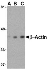
Left: Western blot analysis of beta-actin in HeLa cell lysate with beta-actin antibody at (A) 0.5, (B) 1 and (C) 2 μg/ml.

Below: Immunocytochemistry of Actin in HeLa cells with Actin antibody at 1 µg/ml.

Other Product Images

Source : b-actin antibody was raised against a 16 amino acid peptide from near the amino-terminus of human b-actin.
Purification : Affinity chromatography purified via peptide column
Clonality and Clone : This is a polyclonal antibody.
Host : Beta-actin antibody was raised in rabbit. Please use anti-rabbit secondary antibodies.
Immunogen : Human b-actin / Beta actin (N-Terminus) Peptide (Cat. No. 3779P)
Application : b-actin antibody can be used for the detection of b-actin by Western blot at 1 – 2 µg/ml.
Tested Application(s) : E, WB, IHC
Buffer : Antibody is supplied in PBS containing 0.02% sodium azide.
Blocking Peptide : Cat. No. 3779P - B-actin Peptide
Long-Term Storage : Beta-actin antibody can be stored at 4ºC, stable for one year. As with all antibodies care should be taken to avoid repeated freeze thaw cycles. Antibodies should not be exposed to prolonged high temperatures.
Positive Control
1. Cat. No. 1201 - HeLa Cell Lysate
Species Reactivity :H, M, R
GI Number : 12803203
Accession Number : AAH02409
Short Description : (NT) a major cytoskeletal protein, N Terminus
References
1. Lambrechts A, Van Troys, M and Ampe C. The actin cytoskeleton in normal and pathological cell motility. Int. J. Biochem. Cell Biol. 2004; 36:1890-909.
2. Gourlay CW and Ayscough KR. The actin cytoskeleton: a key regulator of apoptosis and ageing. Nat. Rev. 2005; 6:583-9.

Polypeptides

Polypeptides
Amino acids can be joined together to form a peptide or polypeptide. They are called peptides because when the carboxyl group of one amino acid joins to the amino group of another, a peptide bond is formed. Chemically this is an amide bond but when it occurs in proteins it is given the name peptide bond.

Polypeptides
Polypeptides are chains of amino acids. Proteins are made up of one or more polypeptide molecules. The distinction is that peptides are short and polypeptides/proteins are long. There are several different conventions to determine these, all of which have caveats and nuances. In a polypeptide, there are many peptide bonds. These bonds are rigid and planar, due to electron sharing between the carboxyl carbon and the amide nitrogen which contribute to the bond and give it a partial double-bond character.

Primary Structure
A protein has a completely defined order of amino acids, called its sequence. The primary structure is specified by the sequence of the piece of DNA containing a gene for that protein, and the sequence is unique to the individual protein. The primary structure of a protein (amino acid sequence) contains all the information needed to make the protein into a complicated 3 dimensional shape (similar to those you saw in the first tutorial). This shape is essential to the function of the protein.

Wednesday, October 20, 2010

Beta-actin Antibody

Beta-actin Antibody

Catalog# : 3779
Actins are highly conserved proteins that are involved in cell motility, structure and integrity, processes that are crucial for tissue development and the development of organism (reviewed in 1). The actin cytoskeleton is one of the principal drivers of cell motility and is capable of responding to complex signaling cascades. Recent evidence suggests that it may play key roles in regulating apoptosis and aging (2). beta actin is one of six different actin isoforms which have been identified. Like GAPDH, Β-actin is constitutively expressed at high levels in almost all tissues and cell lines making it ideal for use as a loading control marker in immunoblots.
Additional Names : Beta-actin (NT), Beta actin
Description
Left: Western blot analysis of beta-actin in HeLa cell lysate with beta-actin antibody at (A) 0.5, (B) 1 and (C) 2 μg/ml.

Below: Immunocytochemistry of Actin in HeLa cells with Actin antibody at 1 µg/ml.

Other Product Images

Source : b-actin antibody was raised against a 16 amino acid peptide from near the amino-terminus of human b-actin.
Purification : Affinity chromatography purified via peptide column
Clonality and Clone : This is a polyclonal antibody.
Host : Beta-actin antibody was raised in rabbit. Please use anti-rabbit secondary antibodies.
Immunogen : Human b-actin / Beta actin (N-Terminus) Peptide (Cat. No. 3779P)
Application : b-actin antibody can be used for the detection of b-actin by Western blot at 1 – 2 µg/ml.
Tested Application(s) : E, WB, IHC
Buffer : Antibody is supplied in PBS containing 0.02% sodium azide.
Blocking Peptide : Cat. No. 3779P - B-actin Peptide
Long-Term Storage : Beta-actin antibody can be stored at 4ºC, stable for one year. As with all antibodies care should be taken to avoid repeated freeze thaw cycles. Antibodies should not be exposed to prolonged high temperatures.
Positive Control
1. Cat. No. 1201 - HeLa Cell Lysate
Species Reactivity :H, M, R
GI Number : 12803203
Accession Number : AAH02409
Short Description : (NT) a major cytoskeletal protein, N Terminus
References
1. Lambrechts A, Van Troys, M and Ampe C. The actin cytoskeleton in normal and pathological cell motility. Int. J. Biochem. Cell Biol. 2004; 36:1890-909.
2. Gourlay CW and Ayscough KR. The actin cytoskeleton: a key regulator of apoptosis and ageing. Nat. Rev. 2005; 6:583-9.

Beta-actin Antibody

Beta-actin Antibody

Catalog# : 3777

Actins are highly conserved proteins that are involved in cell motility, structure and integrity, processes that are crucial for tissue development and the development of organism (reviewed in 1). The actin cytoskeleton is one of the principal drivers of cell motility and is capable of responding to complex signaling cascades. Recent evidence suggests that it may play key roles in regulating apoptosis and aging (2). beta actin is one of six different actin isoforms which have been identified. Like GAPDH, Β-actin is constitutively expressed at high levels in almost all tissues and cell lines making it ideal for use as a loading control marker in immunoblots.
Additional Names : Beta-actin (CT), Beta actin
Description
Left: Western blot analysis of beta-actin in HeLa cell lysate with beta-actin antibody at (A) 0.5, (B) 1 and (C) 2 µg/ml.

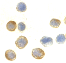
Below: Immunocytochemistry of beta-actin in HeLa cells with beta-actin antibody at 10µg/ml.

Other Product Images
Source : b-actin antibody was raised against a 16 amino acid peptide from near the carboxy-terminus of human b-actin.
Purification : Affinity chromatography purified via peptide column
Clonality and Clone : This is a polyclonal antibody.
Host : Beta-actin antibody was raised in rabbit. Please use anti-rabbit secondary antibodies.
Immunogen : Human b-actin / Beta actin (C-Terminus) Peptide (Cat. No. 3777P)
Application : b-actin antibody can be used for the detection of b-actin by Western blot at 1 – 2 µg/ml.
Tested Application(s) : E, WB, ICC
Buffer : Antibody is supplied in PBS containing 0.02% sodium azide.
Blocking Peptide : Cat. No. 3777P - B-actin Peptide
Long-Term Storage : Beta-actin antibody can be stored at 4ºC, stable for one year. As with all antibodies care should be taken to avoid repeated freeze thaw cycles. Antibodies should not be exposed to prolonged high temperatures.
Positive Control
1. Cat. No. 1201 - HeLa Cell Lysate
Species Reactivity :H, M, R
GI Number : 12803203
Accession Number : AAH02409
Short Description : (CT) a major cytoskeletal protein, C Terminus
References
1. Lambrechts A, Van Troys, M and Ampe C. The actin cytoskeleton in normal and pathological cell motility. Int. J. Biochem. Cell Biol. 2004; 36:1890-909.
2. Gourlay CW and Ayscough KR. The actin cytoskeleton: a key regulator of apoptosis and ageing. Nat. Rev. 2005; 6:583-9.

Tuesday, October 19, 2010

ATM Antibody

ATM Antibody

Catalog# : 3945

DNA double strand breaks represent a major threat to an organism’s genome. Eukaryotic cells have developed mechanisms that sense the presence this damage and initiate suitable responses that can include DNA repair, cell cycle delay, and programmed cell death. The ATM (mutated in Ataxia-Telangiectasia) protein kinase is activated following the formation of DNA double strand breaks, phosphorylating p53 and another kinase CHK2. This initiates a signaling cascade leading to the phosphorylation and inhibition of Cdc25, ultimately preventing cell cycle progression. In some cell types, such as the hemapoietic system, this leads to apoptosis instead of cell cycle arrest. Multiple isoforms of ATM are known to exist.
Additional Names : ATM, mutated in Ataxia-Telangiectasia
Description
Left: Western blot analysis of ATM in Daudi whole cell lysate with ATM antibody at (A) 1 and (B) 2 µg/ml.

Below: Immunohistochemistry of ATM in human lymph node tissue with ATM antibody at 2.5 µg/ml.

Other Product Images

Source : ATM antibody was raised against a peptide corresponding to 14 amino acids near the carboxy terminus of human ATM.
Purification : Affinity chromatography purified via peptide column
Clonality and Clone : This is a polyclonal antibody.
Host : ATM antibody was raised in rabbit. Please use anti-rabbit secondary antibodies.
Application : ATM antibody can be used for detection of ATM by Western blot at 1 – 2 µg/ml.
Tested Application(s) : E, WB, IHC
Buffer : Antibody is supplied in PBS containing 0.02% sodium azide.
Blocking Peptide : Cat.No. 3945P - ATM Peptide
Long-Term Storage : ATM antibody can be stored at 4ºC, stable for one year. As with all antibodies care should be taken to avoid repeated freeze thaw cycles. Antibodies should not be exposed to prolonged high temperatures.
Positive Control
1. Cat. No. 1224 - Daudi Cell Lysate
2. Cat. No. 1369 - Human Lymph node Tissue Lysate
Species Reactivity :H
GI Number : 2304971
Accession Number : AAB65827
Short Description : DNA double-stranded break signaling molecule
References

1. Cahill D, Connor B, and Carney JP. Mechanisms of eukaryotic DNA double strand break repair. Front. Biosci. 2006 ; 11 :1958-76.
2. 2. Dasika GK, Lis SC, Zhao S, et al. DNA damage-induced cell cycle checkpoints and DNA strand break repair in development and tumorigenesis. Oncogene 1999; 18:7883-99.

ASAH1 Antibody

ASAH1 Antibody

Catalog# : 4741
Sphingolipids are hydrolyzed by ceramidases to yield sphingosine and fatty acids. These ceramidases are classified according to the pH range that supports their optimal activity. ASAH1 is an acid ceramidase and key regulator of ceramide metabolism. Mutations in this gene results in Farber Lipogranulomatosis, a fatal human genetic disorder that results in the painful swelling of the joints and tendons and pulminary insufficiency, while a complete knockout of its expression is lethal in mice. Recent studies have shown elevated levels of ASAH1 in Alzheimer’s disease (AD) patients correlating with a reduction in sphingomyelin and elevation of ceramide. Pretreatment of cultured neurons with recombinant AHAH1 prevented the cells from undergoing A-beta (Ab)-induced apoptosis. Multiple isoforms of this protein are known to exist.
Additional Names : ASAH1, N-acylsphingosine amidohydrolase (non-lysosomal ceramidase) 1, acid ceramidase

Description
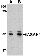
Left: Western blot analysis of ASAH1 in mouse heart tissue lysate with ASAH1 antibody at (A) 1 and (B) 2 µg/ml.

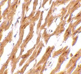
Below: Immunohistochemistry of ASAH1 in human heart tissue with ASAH1 antibody at 2.5 μg/ml.

Other Product Images

Source : ASAH1 antibody was raised against a 16 amino acid peptide near the carboxy terminus of the human ASAH1.
Purification : Affinity chromatography purified via peptide column
Clonality and Clone : This is a polyclonal antibody.
Host : ASAH1 antibody was raised in rabbit. Please use anti-rabbit secondary antibodies.
Application : ASAH1 antibody can be used for detection of ASAH1 by Western blot at 1 µg/ml.
Tested Application(s) : E, WB
Buffer : Antibody is supplied in PBS containing 0.02% sodium azide.
Blocking Peptide : Cat.No. 4741P - ASAH1 Peptide
Long-Term Storage : ASAH1 antibody can be stored at 4ºC, stable for one year. As with all antibodies care should be taken to avoid repeated freeze thaw cycles. Antibodies should not be exposed to prolonged high temperatures.
Positive Control
1. Cat. No. 1401 - Mouse Heart Tissue Lysate
Species Reactivity :H, M, R
GI Number : 119584199
Accession Number : EAW63795
Short Description : an acid ceramidase
References
1. Nilsson A and Duan RD. Alkaline sphingomyelinases and ceramidases of the gastrointestinal tract. Chem. Phys. Lipids 1999; 102:97-105.
2. Koch J, Gartner S, Li CM, et al. Molecular cloning and characterization of a full-length complementary DNA encoding human acid ceramidase. Identification of the first molecular lesion causing Farber’s disease. J. Biol. Chem. 1996; 271:33110-5.
3. Li CM, Park JH, Simonaro CM, et al. Insertional mutagenesis of the mouse acid ceramidase gene leads to early embryonic lethality in homozygotes and progressive lipid storage disease in heterozygotes. Genomics 2002; 79:218-24.
4. He X, Huang Y, Li B. Deregulation of sphingolipid metabolism in Alzheimer’s disease. Neurobiol. Aging 2008; epub.

ApoA1 Antibody

ApoA1 Antibody

Catalog# : 5097

Apolipoprotein A1 (ApoA1) is the major protein component of high density lipoprotein (HDL) in plasma. ApoA1 is synthesized in the liver and small intestine and promotes cholesterol efflux from tissues to the liver for excretion. It is a cofactor for lecithin cholesterolacyltransferase (LCAT), the enzyme responsible for the formation of most plasma cholesteryl esters. Defects in ApoA1 are associated with HDL deficiency, Tangier disease, and systemic non-neuropathic amyloidosis.
Additional Names : ApoA1, Apolipoprotein A1, ApoAI

Description
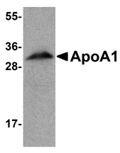
Left: Western blot analysis of ApoA1 in human liver tissue lysate with ApoA1 antibody at 1 µg/ml.

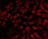
Below: Immunofluorescence of ApoA1 in human liver tissue with ApoA1 antibody at 20 µg/ml.

Other Product Images


Source : ApoA1 antibody was raised against a 17 amino acid peptide from near the amino terminus of human ApoA1.
Purification : Affinity chromatography purified via peptide column
Clonality and Clone : This is a polyclonal antibody.
Host : ApoA1 antibody was raised in chicken. Please use anti-chicken secondary antibodies.
Application : ApoA1 antibody can be used for detection of ApoA1 by Western blot at 1 - 2 µg/ml.
Tested Application(s) : E, WB, IF
Buffer : Antibody is supplied in PBS containing 0.02% sodium azide.
Blocking Peptide : Cat.No. 5097P - ApoA1 Peptide
Long-Term Storage : ApoA1 antibody can be stored at 4ºC, stable for one year. As with all antibodies care should be taken to avoid repeated freeze thaw cycles. Antibodies should not be exposed to prolonged high temperatures.
Positive Control
1. Cat. No. 1304 - Human Liver Tissue Lysate
Species Reactivity :H, M, R
GI Number : 113992
Accession Number : P02647
Short Description : Apolipoprotein A1
References
1. Sorci-Thomas MG, Prack MM, Dashti N, et al. Differential effects of dietary fat on the tissue-specific expression of the apolipoprotein A-I gene: relationship to plasma concentration of high density lipoproteins. J. Lipid Res. 1989; 30:1397-403.
2. Lai C-Q, Parnell LD, and Ordovas JM. The APOA1/C3/A4/A5 gene cluster, lipid metabolism and cardiovascular disease risk. Curr. Opin. Lipid. 2005; 16:153-66.

Polypeptide Synthesis


Polypeptide synthesis at high temperature directed by single strand DNA as a messenger was investigated using cell-free extracts of an extremely thermophilic bacterium; Thermos thermophilus strain HB27, and a hyperthermophilic, acidophilic archaeon, Sulfolobus tokodaii strain 7. Aminoglycoside antibiotics enhanced the reaction; neomycin stimulated it most effectively when the extract of the thermophilic bacterium was used, and paromomycin was the best among the antibiotics tested for the extract of the hyperthermophilic archaeon.

Polypeptide Synthesis

Polypeptide synthesis at high temperature directed by single strand DNA as a messenger was investigated using cell-free extracts of an extremely thermophilic bacterium; Thermos thermophilus strain HB27, and a hyperthermophilic, acidophilic archaeon, Sulfolobus tokodaii strain 7. Aminoglycoside antibiotics enhanced the reaction; neomycin stimulated it most effectively when the extract of the thermophilic bacterium was used, and paromomycin was the best among the antibiotics tested for the extract of the hyperthermophilic archaeon.

Polypeptide Synthesis Using An Expressed Peptide

An expressed peptide proved to be useful as a building block for the synthesis of a polypeptide via the thioester method. A partially protected peptide segment, for use as a C-terminal building block, could be prepared from a recombinant protein; its N-terminal amino acid residue was transaminated to an alpha-oxoacyl group, the side-chain amino groups were then protected with t-butoxycarbonyl (Boc) groups, and. finally, the alpha-oxoacyl group was removed. On the other hand, an O-phosphoserine-containing peptide thioester was synthesized via a solid-phase method using Boc chemistry. These building blocks were then condensed in the presence of silver ions and an active ester component.