Showing posts with label Cosmetic Peptides. Show all posts

Thursday, December 16, 2010

op18 Antibody

op18 Antibody

Catalog# :4237

Op18 belongs to the stathmin family of genes and encodes a ubiquitous cytosolic phosphoprotein that may function as an intracellular relay integrating several signaling pathways such as those involved in cell proliferation and differentiation. Op18 has also been shown to be involved in the regulation of the microtubule filament system by destabilizing microtubules, thereby preventing assembly and promoting the disassembly of microtubules. More recently, op18 has been implicated as a potential target of the ASK1-p38 MAP kinase cascade, suggesting that the ASK1-p38 cascade may regulate microtubule dynamics through op18. Op18 is highly expressed in a wide variety of human malignancies, including leukemia, prostate cancer, ovarian carcinoma, and breast carcinoma, suggesting that op18 may be an ideal target for anti-cancer therapeutics.

Additional Names : op18 (NT), oncoprotein 18, STMN1, Stathmin 1, Lag, LAP18
Description
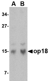
Left: Western blot analysis of op18 in human brain tissue lysate with op18 antibody at (A) 0.5 and (B) 1 µg/ml.

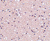
Below: Immunohistochemistry of op18 in human brain tissue with op18 antibody at 2.5 µg/ml.

Other Product Images


Source : Op18 antibody was raised against a 17 amino acid peptide from near the amino terminus of human op18.
Purification : Affinity chromatography purified via peptide column
Clonality and Clone : This is a polyclonal antibody.
Host : op18 antibody was raised in rabbit. Please use anti-rabbit secondary antibodies.
Application : Op18 antibody can be used for detection of op18 by Western blot at 0.5 – 1 µg/ml.
Tested Application(s) : E, WB, IHC
Buffer : Antibody is supplied in PBS containing 0.02% sodium azide.
Blocking Peptide :Cat.No. 4237P - op18 Peptide
Long-Term Storage : op18 antibody can be stored at 4ºC, stable for one year. As with all antibodies care should be taken to avoid repeated freeze thaw cycles. Antibodies should not be exposed to prolonged high temperatures.
Positive Control :
1. Cat. No. 1303 - Human Brain Tissue Lysate
Species Reactivity : H, M, R
GI Number : 51895905
Accession Number : AAH82228
Short Description : (NT) a member of the Stahmin family of proteins
References
1. Curmi PA, Gavet O, Charbaut E, et al. Stathmin and its phosphoprotein family: general properties, biochemical and functional interaction with tubulin. Cell Structure and Function 1999; 24:345-57.
2. Belmont LD and Mitchison TJ. Identification of a protein that interacts with tubulin dimers and increases catastrophe rate of microtubules. Cell 1996; 84:623-31.
3. Mizumura K, Takeda K, Hashimoto S, et al. Identification of op18/stathmin as a potential target of ASK1-p38 MAP kinase cascade. J. Cell Physiol. 2006; 206:363-70.
4. Mistry SJ and Atweh GF. Role of stathmin in the regulation of the mitotic spindle. Mount Sinai J. Med. 2002; 69:299-304

Monday, November 8, 2010

GSTP1 Antibody

GSTP1 Antibody

Catalog# : 4413

Glutathione S-transferases (GSTs) are a family of enzymes that play an important role in detoxification by catalyzing the conjugation of many hydrophobic and electrophilic compounds with reduced glutathione. Based on their biochemical, immunologic, and structural properties, the soluble GSTs are categorized into 4 main classes: alpha, mu, pi, and theta. The glutathione S-transferase pi gene (GSTP1) is a polymorphic gene encoding active, functionally different GSTP1 variant proteins that are thought to function in xenobiotic metabolism (i.e., the metabolism of environmental mutagens and carcinogens) and may play a role in susceptibility to cancer. More recent experiments have suggested that differential expression of GSTP1 also contributes to the sensitivity of xenobiotics in the substantia nigra and may influence the pathogenesis of reactive oxygen species-induced neurological disorders such as Parkinson’s disease. CpG island hypermethylation of the GSTP1 promoter leading to the silencing of the GSTP1 gene has also been linked to cancer.
Additional Names : GSTP1, Glutathione S-transferase pi

[Image]Description
Left: Western blot analysis of GSTP1 in Jurkat cell lysate with GSTP1 antibody at (A) 0.5 and (B) 1 µg/ml.

Below: Immunohistochemical staining of human lung tissue using GSTP1 antibody at 2.5 µg/ml.

Other Product Images
[Image]Source : GSTP1 antibody was raised against a 14 amino acid peptide from near the center of human GSTP1.
Purification : Affinity chromatography purified via peptide column
Clonality and Clone : This is a polyclonal antibody.
Host : GSTP1 antibody was raised in rabbit. Please use anti-rabbit secondary antibodies.
Application : GSTP1 antibody can be used for the detection of ATG10 by Western blot at 0.5 – 1 µg/ml.
Tested Application(s) : E, WB, IHC
Buffer : Antibody is supplied in PBS containing 0.02% sodium azide.
Blocking Peptide : Cat.No. 4413P - GSTP1 Peptide
Long-Term Storage : GSTP1 antibody can be stored at 4ºC, stable for one year. As with all antibodies care should be taken to avoid repeated freeze thaw cycles. Antibodies should not be exposed to prolonged high temperatures.
Positive Control
1. Cat. No. 1205 - Jurkat Cell Lysate
Species Reactivity :H, M, R
GI Number : 4504183
Accession Number : NP_000843
Short Description : Glutathione S-transferase pi
References

1. Pearson WR. Phylogenies of glutathione transferase families. Methods Enzymol. 2005; 401:186-204.
2. Clapper ML. Genetic polymorphism and cancer risk. Curr. Oncol. Rep. 2000; 2:251-6.
3. Smeyne M, Boyd J, Shepherd KR, etc. GSTpi expression mediates dopaminergic neuron sensitivity in experimental parkinsonism. Proc. Natl. Acad. Sci. USA 2007; 104:1977-82.
4. Ellinger J, Bastian PJ, Jurgan T, et al. CpG island hypermethylation at multiple gene sites in diagnosis and prognosis of prostate cancer. Urology 2008; 71:161-7.

Thursday, November 4, 2010

DNA Primer Synthesis


A primer is a strand of nucleic acid that serves as a starting point for DNA replication. They are required because the enzymes that catalyze replication, DNA polymerases, can only add new nucleotides to an existing strand of DNA. The polymerase starts replication at the 3'-end of the primer, and copies the opposite strand. Bio Synthesis has provided custom DNA primer synthesis since 1984. We offer wide variety of oligo modifications and labelings from small discovery research scale to high throughput production. As always, quality is guaranteed!.

DNA Primer

In most cases of natural DNA replication, the primer for DNA synthesis and replication is a short strand of RNA (which can be made de novo). This RNA is produced by primase, and is later removed and replaced with DNA by a repair polymerase. Many of the laboratory techniques of biochemistry and molecular biology that involve DNA polymerase, such as DNA sequencing and the polymerase chain reaction (PCR), require primers. These primers are usually short, chemically synthesized oligonucleotides, with a length of about twenty bases. They are hybridized to a target DNA, which is then copied by the polymerase.

DNA Sequencing

The term DNA sequencing encompasses biochemical methods for determining the order of the nucleotide bases, adenine, guanine, cytosine, and thymine, in a DNA oligonucleotide. The sequence of DNA constitutes the heritable genetic information in nuclei, plasmids, mitochondria, and chloroplasts that forms the basis for the developmental programs of all living organisms.

Read More:
DNA Primer Synthesis

Wednesday, November 3, 2010

GPVI Antibody

GPVI Antibody

Catalog# : 4769

Glycoprotein VI (GP6) is a 58kD platelet membrane glycoprotein that plays a crucial role in the collagen-induced activation and aggregation of platelets. It is uniquely expressed by cells of the megakaryocytic/platelet lineage, and is a member of the immunoglobulin gene superfamily, closely related to Fc receptor gamma chain (FcRγ) and natural killer receptors. Glycoprotein VI plays a key role in platelet procoagulant activity and subsequent thrombin and fibrin formation. This procoagulant function may contribute to arterial and venous thrombus formation. The signaling pathway involves the FcRγ, the Src kinases (likely Fyn/Lyn), the adapter protein LAT and leads to the activation of phospholipase C gamma2. GPVI deficiency can result in bleeding disorders. Further study should reveal the extent of GPVI involvement in thrombotic disease and allow the development of alternative anti-thrombotic compounds.
Additional Names : GPVI (CT), Glycoprotein VI, GPIV, Platelet glycoprotein VI precursor
Description
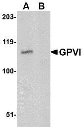
Left: Western blot analysis of GPVI in A20 lysate with GPVI antibody at 1µg/m in either the absence or (B) the presence of blocking peptide.

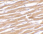
Below: Immunohistochemistry of GPVI in human heart tissue with GPVI antibody at 10 μg/ml.

Other Product Images

Source : GPVI antibody was raised against a 15 amino acid peptide near the carboxy terminus of the human GPVI.
Purification : Affinity chromatography purified via peptide column
Clonality and Clone : This is a polyclonal antibody.
Host : GPVI antibody was raised in rabbit. Please use anti-rabbit secondary antibodies.
Application : GPVI antibody can be used for detection of GPVI by Western blot at 1 µg/ml.
Tested Application(s) : E, WB
Buffer : Antibody is supplied in PBS containing 0.02% sodium azide.
Blocking Peptide : Cat.No. 4769P - GPVI Peptide
Long-Term Storage : GPVI antibody can be stored at 4ºC, stable for one year. As with all antibodies care should be taken to avoid repeated freeze thaw cycles. Antibodies should not be exposed to prolonged high temperatures.
Positive Control
1. Cat. No. 1288 - A20 Cell Lysate
Species Reactivity :H, M, R
GI Number : 143770755
Accession Number : NP_057447
Short Description : (CT) Platelet glycoprotein VI precursor
References
1. Jarvis GE, Atkinson BT, Snell DC, et al. Distinct roles of GPVI and integrin alpha(2) beta(1) in platelet shape change and aggregation induced by different collagens. Br. J. Pharmacol. 2002; 137:107-17.
2. Inoue O, Suzuki-Inoue K, Dean WL, et al. Integrin alpha2beta1 mediates outside-in regulation of platelet spreading on collagen through activation of Src kinases and PLCgamma2. J. Cell Biol. 2003;160:769-80.
3. Clemetson JM, Polgar J, Magnenat E, et al. The platelet collagen receptor glycoprotein VI is a member of the immunoglobulin superfamily closely related to FcαR and the natural killer receptors. J Biol. Chem. 1999; 274:29019-24.
4. Jandrot-Perrus M, Busfield S, Lagrue AH, et al. Cloning, characterization, and functional studies of human and mouse glycoprotein VI: a platelet-specific collagen receptor from the immunoglobulin superfamily. Blood 2000; 96:1798-807.

Friday, October 22, 2010

Large Scale Custom Peptide Synthesis


Bio-Synthesis has been producing synthetic peptides for over 25 years. Our expertise in custom synthetic polypeptide manufacturing allows us to produce the high-quality, large-scale, and GMP peptides with the highest success rate with long standing records. We have been delivered more than 100,000 peptides to customers worldwide, including very hydrophobic polypeptide, peptide with multiple disulfide bonds, multi-phosph0rylated peptides and extremely long peptides. Our large scale non-GMP ever delivered 5 kilograms of peptides on a single order and has the capacity of 10,000 peptides per month. Our capacity of GMP peptide is 10 kilograms.

Peptide Applications

Peptide applications play an important role in biochemistry, molecular biology, immunology and medicine. In the human body, most if not all biological/physiological processes are regulated by various forms of molecular recognition. Most of these processes involve initiation or inhibition trough protein-protein interaction. As we know peptides and proteins due to the vast number of conformational possibilities are ideal to carry out such complex control functions.

Use of Synthetic Peptides

Synthetic peptides have been widely used for many purposes like: structure-function studies of polypeptides, as peptide hormones and hormone analogues, in the preparation of cross-reacting antibodies, in the design of novel enzyme and in drug discovery.

Biologically Active Peptides

The last 40 years have seen an enormous growth in the methodologies available to obtain peptide and protein molecules. Through recombinant methods, most labs can now assemble genes, subcloned into expression vectors and obtain a wide range of endogenous proteins; likewise the pioneering work of Bruce Merrifield, makes it possible to obtain large scale with multikilo amounts of a number of biologically active peptides.

Thursday, October 21, 2010

Bora Antibody

Bora Antibody

Catalog# : 5117

Bora (Protein aurora borealis) is a key activator of Aurora Related Protein Kinase A (ARK-1), which is a centrosome-associated serine/threonine kinase that regulates centrosome maturation, bipolar spindle assembly and chromosome segregation during mitosis. Bora is localized to the nucleus until mitosis is initiated, then translocates to the cytoplasm in a Cdc2 dependent manner. Activation of Cdc2 initiates the release of Bora into the cytoplasm where it can bind and activate ARK-1. PLK1 (polo-like kinase-1) interacts with Bora to control the accessibility of its activation loop for phosphorylation and activation by ARK-1. Bora and ARK-1 cooperatively activate PLK1 and control mitotic entry. Bora mutants result in multipolar spindles in mitosis identical to those observed when ARK-1 function is blocked. Thus, the ARK1-Bora-PLK1 regulatory circuit in mammalian cells elucidates a key mechanism in cell cycle regulation. At least three isoforms of Bora are known to exist.
Additional Names : Bora (NT), Protein aurora borealis, C13orf34, Chromosome 13 open reading frame 34

Description
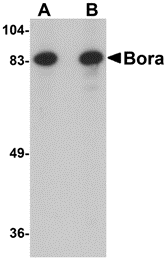
Left: Western blot analysis of Bora in Jurkat cell lysate with Bora antibody at (A) 1 and (B) 2 µg/ml.

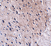
Below: Immunohistochemistry of Bora in mouse brain tissue with Bora antibody at 2.5 µg/ml.

Other Product Images

Source : Bora antibody was raised against a 15 amino acid peptide from near the amino terminus of human Bora.
Purification : Affinity chromatography purified via peptide column
Clonality and Clone : This is a polyclonal antibody.
Host : Bora antibody was raised in rabbit. Please use anti-rabbit secondary antibodies.
Application : Bora antibody can be used for detection of Bora by Western blot at 1-2 µg/ml.
Tested Application(s) : E, WB, IHC
Buffer : Antibody is supplied in PBS containing 0.02% sodium azide.
Blocking Peptide : Cat.No. 5117P - Bora Peptide
Long-Term Storage : Bora antibody can be stored at 4ºC, stable for one year. As with all antibodies care should be taken to avoid repeated freeze thaw cycles. Antibodies should not be exposed to prolonged high temperatures.
Positive Control
1. Cat. No. 1205 - Jurkat Cell Lysate
Species Reactivity :H, M, R
GI Number : 74737659
Accession Number : Q6PGQ7
Short Description : (NT) Protein aurora borealis
References
1. Berdnik D and Knoblich JA. Drosophila Aurora A is required for centrosome maturation and Actin-dependent asymmetric protein localization during mitosis. Curr. Biol. 2002; 12:640-647.
2. Wiese C and O’Brien LL. What's so Bor(a)ing about Aurora A activation? Dev. Cell 2006; 11:133-134.
3. Hutterer A, Berdnik D, Wirtz-Peitz F, et al. Mitotic activation of the kinase Aurora A requires its binding partner Bora. Dev. Cell 2006; 11:147-157.
4. Fu J, Bian M, Jiang Q, et al.. Roles of Aurora kinases in mitosis and tumorigenesis. Mol. Cancer Res. 2007; 5:1-10.

Beta-actin Antibody

Beta-actin Antibody

Catalog# : 3779
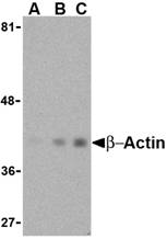
Actins are highly conserved proteins that are involved in cell motility, structure and integrity, processes that are crucial for tissue development and the development of organism (reviewed in 1). The actin cytoskeleton is one of the principal drivers of cell motility and is capable of responding to complex signaling cascades. Recent evidence suggests that it may play key roles in regulating apoptosis and aging (2). beta actin is one of six different actin isoforms which have been identified. Like GAPDH, Β-actin is constitutively expressed at high levels in almost all tissues and cell lines making it ideal for use as a loading control marker in immunoblots.
Additional Names : Beta-actin (NT), Beta actin
Description
Left: Western blot analysis of beta-actin in HeLa cell lysate with beta-actin antibody at (A) 0.5, (B) 1 and (C) 2 μg/ml.

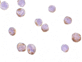
Below: Immunocytochemistry of Actin in HeLa cells with Actin antibody at 1 µg/ml.

Other Product Images

Source : b-actin antibody was raised against a 16 amino acid peptide from near the amino-terminus of human b-actin.
Purification : Affinity chromatography purified via peptide column
Clonality and Clone : This is a polyclonal antibody.
Host : Beta-actin antibody was raised in rabbit. Please use anti-rabbit secondary antibodies.
Immunogen : Human b-actin / Beta actin (N-Terminus) Peptide (Cat. No. 3779P)
Application : b-actin antibody can be used for the detection of b-actin by Western blot at 1 – 2 µg/ml.
Tested Application(s) : E, WB, IHC
Buffer : Antibody is supplied in PBS containing 0.02% sodium azide.
Blocking Peptide : Cat. No. 3779P - B-actin Peptide
Long-Term Storage : Beta-actin antibody can be stored at 4ºC, stable for one year. As with all antibodies care should be taken to avoid repeated freeze thaw cycles. Antibodies should not be exposed to prolonged high temperatures.
Positive Control
1. Cat. No. 1201 - HeLa Cell Lysate
Species Reactivity :H, M, R
GI Number : 12803203
Accession Number : AAH02409
Short Description : (NT) a major cytoskeletal protein, N Terminus
References
1. Lambrechts A, Van Troys, M and Ampe C. The actin cytoskeleton in normal and pathological cell motility. Int. J. Biochem. Cell Biol. 2004; 36:1890-909.
2. Gourlay CW and Ayscough KR. The actin cytoskeleton: a key regulator of apoptosis and ageing. Nat. Rev. 2005; 6:583-9.

Polypeptides

Polypeptides
Amino acids can be joined together to form a peptide or polypeptide. They are called peptides because when the carboxyl group of one amino acid joins to the amino group of another, a peptide bond is formed. Chemically this is an amide bond but when it occurs in proteins it is given the name peptide bond.

Polypeptides
Polypeptides are chains of amino acids. Proteins are made up of one or more polypeptide molecules. The distinction is that peptides are short and polypeptides/proteins are long. There are several different conventions to determine these, all of which have caveats and nuances. In a polypeptide, there are many peptide bonds. These bonds are rigid and planar, due to electron sharing between the carboxyl carbon and the amide nitrogen which contribute to the bond and give it a partial double-bond character.

Primary Structure
A protein has a completely defined order of amino acids, called its sequence. The primary structure is specified by the sequence of the piece of DNA containing a gene for that protein, and the sequence is unique to the individual protein. The primary structure of a protein (amino acid sequence) contains all the information needed to make the protein into a complicated 3 dimensional shape (similar to those you saw in the first tutorial). This shape is essential to the function of the protein.

Tuesday, October 19, 2010

Polypeptide Synthesis


Polypeptide synthesis at high temperature directed by single strand DNA as a messenger was investigated using cell-free extracts of an extremely thermophilic bacterium; Thermos thermophilus strain HB27, and a hyperthermophilic, acidophilic archaeon, Sulfolobus tokodaii strain 7. Aminoglycoside antibiotics enhanced the reaction; neomycin stimulated it most effectively when the extract of the thermophilic bacterium was used, and paromomycin was the best among the antibiotics tested for the extract of the hyperthermophilic archaeon.

Polypeptide Synthesis

Polypeptide synthesis at high temperature directed by single strand DNA as a messenger was investigated using cell-free extracts of an extremely thermophilic bacterium; Thermos thermophilus strain HB27, and a hyperthermophilic, acidophilic archaeon, Sulfolobus tokodaii strain 7. Aminoglycoside antibiotics enhanced the reaction; neomycin stimulated it most effectively when the extract of the thermophilic bacterium was used, and paromomycin was the best among the antibiotics tested for the extract of the hyperthermophilic archaeon.

Polypeptide Synthesis Using An Expressed Peptide

An expressed peptide proved to be useful as a building block for the synthesis of a polypeptide via the thioester method. A partially protected peptide segment, for use as a C-terminal building block, could be prepared from a recombinant protein; its N-terminal amino acid residue was transaminated to an alpha-oxoacyl group, the side-chain amino groups were then protected with t-butoxycarbonyl (Boc) groups, and. finally, the alpha-oxoacyl group was removed. On the other hand, an O-phosphoserine-containing peptide thioester was synthesized via a solid-phase method using Boc chemistry. These building blocks were then condensed in the presence of silver ions and an active ester component.

Monday, October 18, 2010

AP2M1 Antibody

AP2M1 Antibody

Catalog# : 5161

The heterotetrameric coat assembly protein complex, also known as the adaptor-related protein complex 2 (AP-2), belongs to the adaptor complexes medium subunits family. The mu 1 subunit of the AP-2 complex (AP2M1) is required for the activity of a vacuolar ATPase, which is responsible for proton pumping occurring in the acidification of endosomes and lysosomes. AP2M1 has also been shown to associate with the HIV-1 protein Nef, suggesting that Nef may use AP-2 complex to enhance the rate of endocytosis of both CD4 and class I MHC. AP2M1 may also play an important role in regulating the intracellular trafficking and function of cytotoxic T-lymphocyte associated (CTLA)-4 protein. At least two isoforms of AP2M1 are known to exist.
Additional Names : AP2M1, Adaptor-related protein complex 2 mu 1, AP-2 mu-1, mu2, AP50, CLAPM1
Description
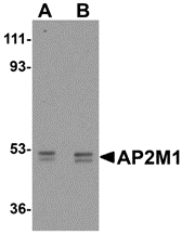
Left: Western blot analysis of AP2M1 in human kidney tissue lysate with AP2M1 antibody at (A) 1 and (B) 2 µg/ml.

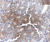
Below: Immunohistochemistry of AP2M1 in mouse kidney tissue with AP2M1 antibody at 2.5 μg/ml.

Other Product Images
Source : AP2M1 antibody was raised against a 16 amino acid peptide near the carboxy terminus of human AP2M1.
Purification : Affinity chromatography purified via peptide column
Clonality and Clone : This is a polyclonal antibody.
Host : AP2M1 antibody was raised in chicken. Please use anti-chicken secondary antibodies.
Application : AP2M1 antibody can be used for detection of AP2M1 by Western blot at 1 – 2 µg/ml.
Tested Application(s) : E, WB
Buffer : Antibody is supplied in PBS containing 0.02% sodium azide.
Blocking Peptide : Cat.No. 5161P - AP2M1 Peptide
Long-Term Storage : AP2M1 antibody can be stored at 4ºC, stable for one year. As with all antibodies care should be taken to avoid repeated freeze thaw cycles. Antibodies should not be exposed to prolonged high temperatures.
Positive Control
1. Cat. No. 1305 - Human Kidney Tissue Lysate
Species Reactivity :H, M, R
GI Number : 14917109
Accession Number : NP_004059
Short Description : Adaptor-related protein complex 2 mu 1
References
1. Robinson MS. Adaptable adaptors for coated vesicles. Trends Cell Biol. 2004; 14:167-74.
2. Aguilar RC, Ohno H, Roche KW, et al. Functional domain mapping of the clathrin-associated adaptor medium chains mu1 and mu J. Biol. Chem. 1997; 272:27160-6.
3. Liu Q, Feng Y, and Forgac M. Activity and in vitro reassembly of the coated vesicle (H+)-ATPase requires the 50-kDa subunit of the clathrin assembly complex AP-2. J. Biol. Chem. 1994; 269:31592-7.
4. Craig HM, Reddy TR, Riggs NL, et al. Interactions of HIV-1 Nef with the m subunits of adaptor protein complexes 1, 2, and 3: role of the dileucine-based sorting motif. Virology 271:9-17.

Conjugates in Signal Transduction


Use of conjugates in elucidating the roles on the different components that participate in signal transduction is a powerful tool in the development of new therapeutic agents to up-regulate or down-regulate certain biological responses. The design and production of these compounds frequently requires a deep knowledge of the chemistry used in their synthesis. Bio Synthesis, with 25 years of experience in the areas of peptide and nucleic acid synthesis is well positioned to assist any researcher in the design and production of these agents. More Info

Hyaluronic Acid (HA) Peptide Conjugates
Hyaluronic acid (HA) peptide conjugates for formyl peptide receptor like 1 (FPRL1) receptor. FPRL1 is a G protein-receptor that in phagocytes binds chemotactic peptides, some of them formylated, and mediate important biological functions, e.g. regulation of immune responses against pathogens by engulfing and destroying bacteria. These peptidic ligands can be either agonistic or antagonistic and therefore have a significant therapeutic potential. A potential application of antagonistic peptides would be the down-regulation of immunity to reduce inflammatory disease, e.g. arthritis and asthma.

Glycopeptide
The study of glycopeptide ligands to cell surface receptors has been advanced by the use of neoglycopolymers of which neoglycopeptides are an example. For instance, while some monomeric oligosaccharide act as inhibitors of L-selectin shedding, attachment of multiple copies of that oligosaccharide to a polymeric carrier, usually a polypeptide, resulted in the selectin’s shedding but independent of activation. An elegant example of how conjugates can help to dissociate the functions of the different moieties of a ligand.

Lipid-modified Peptides
Signal transducing proteins frequently have covalently attached lipids required for their biological functions. Lipidation of peptides derived from some G protein-coupled receptors, e.g. ras proteins, has provided information about the effectiveness of different lipid residues. For instance, it has been shown that geranylgeranylated or palmitoylated peptides have a higher membrane affinity than their analogous myristoylated or farnesylated peptides. Also increasing the degree of lipidation of a peptide showed that addition of a second lipid residue resulted in its stable insertion in the membrane. Thus, like in the glycosylation case the addition of lipids to model peptides allow the elucidation of the roles for the different protein’s moieties.

Oligonucleotide-peptide Conjugates
The objective of signal transduction is to stimulate a cell response that is usually the expression of a protein by transcription of DNA. In some viruses replication the formation of nucleopeptides is an obligatory step, also some processing of DNA by certain enzymes require the formation of intermediary nucleopeptides. Usually oligonucleotides are linked via the hydroxyl group of tyrosine, serine or threonine, either directly or via a spacer, depending on the requirements of the system. Synthesis of these conjugates is usually carried out by cross-linking an oligonucleotide to a peptide where both components had been prepared separately.

Mass Spectrometry of Biomolecules


Mass spectrometers used for the analysis of macromolecules from biological sources, such as proteins, peptides, DNA or RNA oligomers, have experienced tremendious improvements in recent years. Electro spray ionization mass spectrometry (ESI-MS) and matrix assisted laser desorption time-of-flight mass spectrometry (MALDI-TOF MS) have become the methods of choice. Both techniques are powerful analytical tools by themself, but are most powerful if used in combination with techniques such as gel electrophoresis, electroblotting, LC, CE or protein sequencing. This is the major technology used for proteomic approaches.

Mass Spectrometer

All mass spectrometers designed to analyze proteins consist of two essential components, the ion source and the mass analyzer. In the ion source, a seemingly unlikely phase transition is effected: proteins introduced as solids or in solution are converted into intact, naked ionized molecules in the gas phase. Subsequently, in the mass analyzer, the mass-to-charge (m/z) ratios of the naked protein molecule ions are determined.

Protein Analysis

The determination of the carbohydrate portion of glycoproteins provides an analytical challenge to researchers, especially when only small amounts of sample are available. The reason for this is the very large number of isomers that are possible when these structures are built up from their constituent monosaccharides. Phosphoproteins play a central role in many intracellular processes, including signal transduction and regulation of cell division. The site and extent of phosphorylation of key proteins are believed to play an important regulatory role in many intracellular signaling pathways.

Thursday, October 14, 2010

TRIM5 delta Antibody

TRIM5 delta Antibody

Catalog# : 3241

TRIM5 is a member of a broad family of otherwise unrelated proteins defined by the presence of a tripartite motif containing a RING domain, a B-box type 1, and a B-box type 2, followed by a coiled-coil region (1). TRIM5 has five alternately spliced isoforms, the longest of which is the ? variant which also contains a carboxy-terminal B30.2 (SPRY) domain (1). Expression of TRIM5alpha variants from humans, rhesus monkeys, and African green monkeys enabled resistance to infection by various retroviruses including HIV-1 (2,3), albeit at differing efficiencies. The TRIM5delta isoform has been shown to colocalize with the topoisomerase I-interacting proteins BTBD1 and BTBD2 in punctate or elongated cytoplasmic bodies in several mouse and human cells where it appears to serve as a scaffold for the assembly of endogenous BTBD proteins. TRIM5delta also exhibits autoubiquitination activity in a RING finger- and UbcH5B-dependent manner (4).
Additional Names : TRIM5 delta, TRIM5d
Description
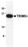
Left: Western blot analysis of TRIM5 delta expression in mouse brain cell lysate with TRIM5 delta antibody at 2 µg/ml.

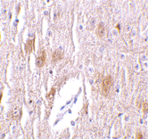
Below: Immunohistochemistry of TRIM5 delta in mouse brain tissue with TRIM5 delta antibody at 1 µg/ml.

Other Product Images

Source : TRIM5 delta antibody was raised against a synthetic peptide corresponding to 13 amino acids near the carboxy-terminus of human TRIM5 delta.
Purification : Affinity chromatography purified via peptide column
Clonality and Clone : This is a polyclonal antibody.
Host : TRIM5 delta antibody was raised in rabbit. Please use anti-rabbit secondary antibodies.
Immunogen : Human TRIM5 delta Peptide (Cat. No. 3241P)
Application : TRIM5 delta antibody can be used for detection of TRIM5 delta by Western blot at 1 to 2 µg/ml.A band at approximately 25 kDa can be detected.
Tested Application(s) : E, WB, IHC
Buffer : Antibody is supplied in PBS containing 0.02% sodium azide.
Blocking Peptide : Cat. No. 3241P - TRIM5d Peptide
Long-Term Storage : TRIM5 delta antibody can be stored at 4ºC, stable for one year. As with all antibodies care should be taken to avoid repeated freeze thaw cycles. Antibodies should not be exposed to prolonged high temperatures.
Positive Control
1. Cat. No. 1403 - Mouse Brain Tissue Lysate
Species Reactivity :H
GI Number : 15011946
Accession Number : NP_149084
Short Description : retroviral restriction factor
References
1. Reymond A, Meroni G, Fantozzi A, et al. The tripartite motif family identifies cell compartments. EMBO J. 2001; 20:2140-51.
2. Stremlau M, Owens CM, Perron MJ, et al. The cytoplasmic body component TRIM5 restricts HIV-1 infection in Old World monkeys. Nature 2004; 427:848-53.
3. Hatziioannou T, Perez-Caballero D, Yang A, et al. Retrovirus resistance factors REF1 and Lv1 are species-specific variants of TRIM5alpha. Proc. Nat’l. Acad. Sci. USA 2004; 101:10774-9
4. Xu L, Yang L, Moitra PK, et al. BTBD1 and BTBD2 colocalize to cytoplasmic bodies with the RBCC/tripartite motif protein, TRIM5. Exp. Cell Res. 2003; 288:84-93.

Peptidomimetics


Many research groups, both in academia and in pharmaceutical companies search constantly for non-peptide compounds that have better bioavailability and stability, perhaps even with greater receptor selectivity. The known structure-activity interactions and conformational foldings of peptide structures aid a great deal in the design of novel peptidomimetics.

Peptidomimetics

Peptidomimetics are compounds specially derived from peptides and protein by structural modifications using unnatural amino acids and conformational restraints. Peptides are becoming extremely popular with researchers because they affect such a wide range of cellular activity. Once a peptide crosses the cell membrane, it often loses stability. Peptidomimetics, however, may be the answer. There are several different ways that organic chemists can alter and modify the peptides. The modifications involve changes to the peptide that do not occur naturally, such as incorporating unnatural amino acids, replacing the peptide bond, and altering backbones.

Peptide Design

There are a number of factors that help in the rational design of PM’s such us: binding site optimal fit, conformational stabilization, (given by rigid elements and the positioning of specific functional groups), polar or hydrophobic regions (inside strategic reactive pockets) that favor the basic atomic interactions provided by hydrogen , electrostatic and hydrophobic bonding.

Natural Peptide

There are many instances where the native information within a natural peptide ligand can be conferred/duplicated or mimetized into a non-peptide molecule, preferably of low molecular weight, hence the basis for the field of peptidomimetics(PM’s). The desire to convey the three dimensional information present in a peptide into small nonpeptide molecules is what encompasses the field of peptidomimetics.

Peptide Mimetics

The goal in PM’s is to obtain molecules that mimic the specific molecular interactions of natural proteins and their ligands. The protein to protein interaction of biologivally active peptides at the receptor level can be obtained by small molecules, in an agonistic fashion or can be blocked, in an antagonistic fashion.

TRBP1 Antibody

TRBP1 Antibody

Catalog# : 4011

The human trans-activation response (TAR) RNA binding protein 1 (TRBP1) was initially identified as a protein that binds to the HIV-1 TAR RNA and activates the long terminal repeat (LTR) expression in the absence and presence of the viral trans-activator Tat. This binding is enhanced by the presence of co-factors such as elongation factor 1alpha (EF-1alpha), polypyrimidine tract-binding protein (PTB), and the chaperonin-like protein stimulator of TAR RNA-binding proteins (SRB). TRBP1 may act to disengage RNA polymerase II from TAR during transcriptional elongation if the RNA polymerase stalls at the TAR during transcriptional elongation.
Additional Names : TRBP1 (IN), TAR RNA binding protein 1, TARBP1, TRP-185, TRP-1
Description
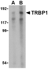
Left: Western blot analysis of TRBP1 in 3T3 cell lysate with TRBP1 antibody at (A) 1 and (B) 2 µg/ml.


Source : TRBP1 antibody was raised against a 17 amino acid peptide from near the center of human TRBP1.
Purification : Affinity chromatography purified via peptide column
Clonality and Clone : This is a polyclonal antibody.
Host : TRBP1 antibody was raised in rabbit. Please use anti-rabbit secondary antibodies.
Application : TRBP1 antibody can be used for detection of TRBP1 by Western blot at 1 – 2 µg/ml.
Tested Application(s) : E, WB
Buffer : Antibody is supplied in PBS containing 0.02% sodium azide.
Blocking Peptide : Cat.No. 4011P - TRBP1 Peptide
Long-Term Storage : TRBP1 antibody can be stored at 4ºC, stable for one year. As with all antibodies care should be taken to avoid repeated freeze thaw cycles. Antibodies should not be exposed to prolonged high temperatures.
Positive Control
1. Cat. No. 1212 - 3T3 Cell Lysate
Species Reactivity :H, M
GI Number : 110825988
Accession Number : NP_005637
Short Description : (IN) TAR RNA binding protein 1
References
1. Gatignol A, Buckler-White A, Berkhout B, et al. Characterization of a human TAR REN-binding protein that activates the HIV-1 LTR. Science 1991; 251:1597-1600.
2. Wu-Baer F, Lane WS, and Gaynor RB. Identification of a group of cellular cofactors that stimulate the binding of RNA polymerase II and TRP-185 to human immunodeficiency virus 1 TAR RNA. J. Biol. Chem. 1996; 271:4201-8.
3. Wu-Baer F, Lane WS, and Gaynor RB. The cellular factor TRP-185 regulates RNA polymerase II binding to HIV-1 TAR RNA. EMBO J. 1995; 14:5995-6009. (07-01D)

Monday, October 11, 2010

DC-SIGN Antibody

DC-SIGN Antibody

Catalog# : 2347

Dendritic cells (DCs) that control immune responses were recently found to capture and transport HIV from the mucosal area to remote lymph nodes (1), where DCs hand over HIV to CD4+ T lymphocytes. DCs also amplify the amount of virus and extend the duration of viral infectivity. Multiple strains of HIV-1, HIV-2 and SIV bind to DCs via DC-SIGN (2). ICAM-3 is the natural ligand for DC-SIGN (3). A DC-SIGN homologue (termed DC-SIGNR, L-SIGN, and DC-SIGN2) was identified recently (4-8). DC-SIGN forms a novel gene family with DC-SIGNR and many alternatively spliced isoforms of DC-SIGN and DC-SIGNR (8). The expression of DC-SIGN was found in mucosal tissues including placenta, small intestine, and rectum.
Additional Names : DC-SIGN (CT), DC-SIGN
Description
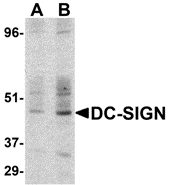
Left: Western blot detection of DC-SIGN in human small intestine at (A) 1 and (B) 2 µg /ml.

Below: Immunohistochemistry of DC-SIGN in human lymph node tissue with DC-SIGN antibody at 10 µg/ml.

Other Product Images
Source : DC-SIGN antibody was raised against a synthetic peptide corresponding to amino acids near the center of human DC-DIGN .
Purification : Affinity chromatography purified via peptide column
Clonality and Clone : This is a polyclonal antibody.
Host : DC-SIGN antibody was raised in rabbit. Please use anti-rabbit secondary antibodies.
Immunogen : Human DC-SIGN (C-Terminus) Peptide (Cat. No. 2347P)
Application : DC-SIGN antibody can be used for detection of DC-SIGN by Western blot at 1 to 2 µg/ml.A band at approximately 44 kDa can be detected.
Tested Application(s) : E, WB, IHC
Buffer : Antibody is supplied in PBS containing 0.02% sodium azide.
Blocking Peptide : Cat. No. 2347P - DC-SIGN Peptide
Long-Term Storage : DC-SIGN antibody can be stored at 4ºC, stable for one year. As with all antibodies care should be taken to avoid repeated freeze thaw cycles. Antibodies should not be exposed to prolonged high temperatures.
Positive Control
1. Cat. No. 1308 - Human Small Intesting Tissue Lysate
Species Reactivity :H
GI Number : 46396012
Accession Number : Q9NNX6
Short Description : (CT) A novel HIV biding protein
References
1. Geijtenbeek TB, Kwon DS, Torensma R, et al. DC-SIGN, a dendritic cell-specific HIV-1-binding protein that enhances trans-infection of T cells. Cell. 2000;100:587-97.
2. Pohlmann S, Baribaud F, Lee B, et al. RW. DC-SIGN interactions with human immunodeficiency virus type 1 and 2 and simian immunodeficiency virus. J Virol. 2001;75(10):4664-72.
3. Geijtenbeek TB, Torensma R, van Vliet SJ, et al. Identification of DC-SIGN, a novel dendritic cell-specific ICAM-3 receptor that supports primary immune responses. Cell. 2000;100(5):575-85.
4. Soilleux EJ, Barten R, Trowsdale J. DC-SIGN; a related gene, DC-SIGNR; and CD23 form a cluster on 19p13. J Immunol. 2000;165(6):2937-42.

Glycoconjugates


Bio-Synthesis may use your oligosaccharides for preparation of the conjugates or in some cases it could prepare the whole conjugate, including the oligosaccharide moiety. For the preparation of oligopeptides and oligonucleotides glycoconjugates these compounds are modified during solid phase synthesis to introduce a spacer carrying a functional group that can be used for subsequent cross-linking with the oligosaccharides. Depending on the nature of the oligosaccharide and the peptide or oligonucleotide the length and hydrophilicity of the spacer would be decided to maximize the interactions of these compounds with their receptors.

Glycoconjugates

Advances in glycobiology highlight the role of naturally occurring glycoconjugates in different biological processes. The most common glycoconjugates are those where an oligosaccharide is covalently linked to a protein, peptide or lipid, yet in some cases it can be also linked to an oligonucleotide. Synthetic conjugates are excellent tools to elucidate the natural glycoconjugates' roles in different biological processes. In these conjugates an oligosaccharide is usually cross-linked to a reporter group, such as a fluorochrome, enzyme, liposome, colloidal gold and others, that can be easily detected.

Other Useful Tools

Other useful tools in biological studies are those compounds where the oligosaccharide is coupled to a fluorescent label such as fluorescein, rhodamine, Texas red and others, either directly or via a spacer. Fluorescent compounds can be detected by fluorescence microscopy, FACS machine, and spectrofluorometry. Other tagging methods involve the use of colloidal gold and ferritin, both compounds that can be detected by electron microscopy. Oligosaccharides can also be conjugated to different lipids to yield glycolipids that can be incorporated into either micelles or liposomes, with the latter being effective drug carriers.

Glycoconjugates of Oligosaccharides

The same methods used to prepare glycoconjugates of oligosaccharides can be applied to native polysaccharides. Base on the needs for sensitivity, nature of the molecule recognizing the sugar moiety, Bio-Synthesis can design a method to label these macromolecules without interfering with their interactions with other molecules.

Wednesday, June 2, 2010

Cosmetic Peptides Synthesis

As technology and research advance, cosmetic peptides are being used and more all over the world. These cosmetic peptides synthesis are leading a beauty revolution with their multi-functional properties, including their ability to regulate certain biological functions. As the most thriving skin care companies pursue this booming field of research, there is the need, more than ever, to have high quality, custom cosmetic peptides synthesis. Bio-Synthesis has been helping companies break barriers by supplying the most trusted, reliable and affordable custom peptides for cosmetic companies. Using our state-of-the-art peptide synthesis facilities, Bio-Synthesis can manufacture single batch multi-gram research and GMP grade cosmetic peptides to fit your exact needs.

Please see our list of some of the most popular cosmetic peptides standing by. If you have a different cosmetic peptide synthesis requirement in mind, we can custom synthesize any peptide of any quantity or purity.

Service Features

*Quantity – mg to kg scale for GMP and non-GMP grade

*Purity – Crude to >98%.

*Modifications – We offer an extensive list of modifications options for your needs and can help you find the one that's right for you.

*Applicability – Peptides provided by Bio-Synthesis can be integrated into a wide range of applications.

*Content – Our peptides come with Mass Spec Analysis, HPLC Profile and Certificate of Analysis. Additional content analysis, such as Amino Acid Analysis, is also offered. GMP Peptides include detailed records for each step of the process.

* Prices – Bio-Synthesis offers extremely competitive prices with large volume discounts. Please contact us for an official quote.

Quality Control:

Bio-Synthesis follows stringent guidelines to guarantee our cosmetic peptides surpass the highest of quality expectations. Each batch includes several analytical data sheets corresponding to the specific products.

Product No.
C014-10926-01

Name
Acetyl Hexapeptide-3

Cosmetic Brands Containing Peptide
Replexion: Anti Aging Skin Care Cream
Pretox20: Wrinkle Cream
Relax-A-LineTM Topical Serum: For shallower lines. Acetyl Hexapeptide 3 relaxes facial muscles that cause dynamic wrinkles

Suggested Usages
Relaxes facial tension, leading to the reduction of facial lines and wrinkles. It is a safe, effective, more affordable alternative to Botox

Product No.
C014-10928-01

Name
Palmitoyl Hexapeptide

Cosmetic Brands Containing Peptide
Ageless:Rejuvenate anti-aging serum
Granactive™ Hexapeptide
Skin Fusion:A mineral foundation and powder

Suggested Usages
Stimulates fibroblasts proliferation, Imparts firming effect, smoothes out wrinkles while strengthening the skin’s natural barrier function

Product No.
C014-10929-01

Name
Palmitoyl Tetrapeptide-3

Cosmetic Brands Containing Peptide
Avon: Anew Alternative Intensive Age Treatment
SuperMax : Multi-Peptide Skin Solution Serum
Osmotics: Blue Copper 5 Face Lifting Serum, Eye Surgery Under Eye Rejuvenator

Suggested Usages
It delays the effects of premature aging by improving the elasticity and firmness of the skin. It hydrates, protects and smoothes skin.

Product No.
C014-10927-01

Name
Palmitoyl Pentapeptide

Cosmetic Brands Containing Peptide
G.M. COLLIN: Retinol Advanced + Matrixyl + Q10
Aziz Luminous Peptide Serum
Skinmedic:Multi Peptide Anti ageing Serum
Replexion: Anti Aging Skin Care Cream
Newspirit: Pentapeptide-SF™ Eye Contour Serum
Nutra-Luxe: Kaviar Night Serum Firmer Skin

Suggested Usages
- Actively stimulates the production of collagen I and IV, fibronectin and glycosaminoglycans in the dermis. Assists in increasing skin thickness through these actions.
-Aids in tissue regeneration and skin healing
-Acts to enhance dermal and epidermal hydration
-Increases the effects of resurfacers, peels and laser treatments

Product No.
C014-10930-01

Name
PAL-GHK

Cosmetic Brands Containing Peptide
Lip's CRÈME: Masque pour les lèvres
Modele: Advanced Lip Restoring Treatment
Urban Nutrition: Bellaplex™

Suggested Usages
Re-structuring the connective tissue by stimulating the production of collagen and of the glycosaminoglycansacts as a natural sun protector, firms skin and helps with wrinkles