Tuesday, March 2, 2010

Acinus Antibody

Acinus Antibody

Catalog# : 2241

Chromatin condensation and nuclear fragmentation (CCNF) is the hallmark of apoptosis. CCNF is triggered by the activation of members of caspase family, caspase activated DNase (CAD/DFF40), and several novel proteins including AIF and CIDE (1). A new inducer of chromatin condensation was recently identified and designated Acinus (for apoptotic chromatin condensation inducer in the nucleus). Acinus is cleaved by caspase-3 and an additional unknown protease generating a small active peptide p17, which causes chromatin condensation in vitro when it is added to purified nuclei. Acinus also induces apoptotic chromatin condensation in cells. Acinus is ubiquitously expressed. Three different spliced forms of Acinus have been identified in human and mouse and designated AcinusL, AcinusS and AcinusS’ (2).

Additional Names : Acinus (NP), Acinus (NP), Acinus

Description
Left: Western blot analysis of acinus in K562 whole cell lysate with acinus antibody at 1 µg/ml.


Source : Acinus antibody was raised against a peptide corresponding to amino acids 994 to 1009 of human AcinusL, 267 to 282 of human AcinusS’, or 236 to 251 of human AcinusS, which are identical to those of mouse Acinus. The selected antigenic sequence is located near the N-terminus of active peptide p17.

Purification : Affinity chromatography purified via peptide column

Clonality and Clone : This is a polyclonal antibody.

Host : Acinus antibody was raised in rabbit.

Immunogen : Human Acinus Peptide (Cat. No. 2241P)

Application : Acinus antibody can be used for

Tested Application(s) : E, WB

Buffer : Antibody is supplied in PBS containing 0.02% sodium azide.

Blocking Peptide : Cat. No. 2241P - Acinus Peptide

Long-Term Storage : Acinus antibody can be stored at 4ºC, stable for one year. As with all antibodies care should be taken to avoid repeated freeze thaw cycles. Antibodies should not be exposed to prolonged high temperatures.

Positive Control
  1. Cat. No. 1204 - K562 Cell Lysate
Species Reactivity :H

GI Number : 5931959

Accession Number : AAD56724

Short Description : Inducer of chromatin condensation

References
  1. Zamzami N, Kroemer G. Condensed matter in cell death. Nature 1999 ;401:127-8.
  2. Sahara S, Aoto M, Eguchi Y, Imamoto N, Yoneda Y, Tsujimoto Y. Acinus is a caspase-3-activated protein required for apoptotic chromatin condensation. Nature 1999 401:168-73. (WD0800)

AATF Antibody

AATF Antibody

Catalog# : 3039

AATF (apoptosis antagonizing transcription factor) was initially discovered as an interaction partner of ZIP kinase (ZIPK) (1), a member of death-associated protein (DAP) kinase family of pro-apoptotic serine/threonine kinases (2). AATF is a phosphoprotein containing an acidic region and a putative leucine zipper domain and nuclear localization signal, features which are typical of transcription factors. AATF inhibits the ZIPK-mediated pro-apoptotic pathway and may activate other anti-apoptotic pathways (3). Recently, it has also been shown to protect neural cells against oxidative damage induced by amyloid b-peptide and to inhibit aberrant production of the b-peptide by interacting with Par-4 (prostate apoptosis response-4), another pro-apoptotic leucine zipper protein that is associated with neuronal degeneration in Alzheimer’s disease (AD) (4,5), suggesting that AATF may have potential therapeutic applications in both familial and sporadic forms of AD.

Additional Names : AATF, Apoptosis antagonizing transcription factor, Che-1

Description
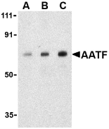
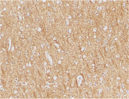
Left: Western blot analysis of AATF in human small intestine cell lysate with AATF antibody at (A) 0.5, (B) 1 and (C) 2 µg/ml. Below: Immunohistochemistry of AATF in human small intestine tissue with AATF antibody at 10 µg/ml.

Source : AATF antibody was raised against a 12 amino acid peptide from near the carboxy terminus of human AATF.

Purification : Immunoaffinity purified IgG

Clonality and Clone : This is a polyclonal antibody.

Host : AATF antibody was raised in rabbit.

Immunogen : Human AATF Peptide (Cat. No. 3039P)

Application : AATF antibody can be used for

Tested Application(s) : E, WB, IHC

Buffer : Antibody is supplied in PBS containing 0.02% sodium azide.

Blocking Peptide : Cat. No. 3039P - AATF Peptide

Long-Term Storage : AATF antibody can be stored at 4ºC, stable for one year. As with all antibodies care should be taken to avoid repeated freeze thaw cycles. Antibodies should not be exposed to prolonged high temperatures.

Positive Control
  1. Cat. No. 1308 - Human Small Intestine Tissue Lysate
Species Reactivity : H, M

GI Number : 7657013

Accession Number : NP_036270

Short Description : An anti-apoptotic transcription factor

References
  1. AATF antibody can be stored at 4ºC, stable for one year. Page G, Lodige I, Kogel D et al. AATF, a novel transcription factor that interacts with Dlk/ZIP kinase and interferes with apoptosis. FEBS Lett. 1999; 462:187-191.
  2. Kawai T, Matsumoto M, Takeda K, et al. ZIP kinase, a novel serine/threonine kinase which mediates apoptosis. Mol. Cell Biol. 1998; 18:1642-51.
  3. Lindfors K, Halttunen T, Huotari P, et al. Identification of novel transcription factor-like gene from human intestinal cells. Biochem. Biophys. Res. Commun. 2000; 276:660-6.
  4. Xie J and Guo Q. AATF protects neural cells against oxidative damage induced by amyloid b-peptide. Neurobiol. of Dis. 2004; 16:150-7.
For more detail regarding Peptide please visit here : Custom Peptide Synthesis

ATR Antibody

ATR Antibody

The Anthrax toxin receptor (ATR) was initially discovered as the tumor endothelial marker 8 (TEM8) (1). This protein, which exists in three isoforms (36, 40, and 60 kDa), is highly expressed in tumor vessels as well as in the vasculature of developing embryos, suggesting that it may normally play a role in angiogenesis. However, it also acts as the receptor for anthrax toxin (2). Following the binding of this protein by the protective antigen (PA) of anthrax, PA is cleaved and heptamerizes to form the binding site for both edema factor (EF) and lethal factor (LF) (3). This complex is then endocytosed by the cell; acidification in endosomes allows the release of EF and LF into the cytoplasm where they interfere with MAPK signaling and induce apoptosis (4).

Additional Names : ATR (IN), TEM8

Description
Left: Western blot analysis of ATR in HepG2 cell lysates with ATR antibody (IN) at (A) 0.5, (B) 1, and (C) 2 µg/ml. Below: Immunohistochemical staining of human brain tissue using ATR antibody at 2 µg/ml.


Source : ATR antibody was raised against a peptide corresponding to 13 amino acids near the center of human ATR.

Purification : Ion exchange chromatography purified

Clonality and Clone : This is a polyclonal antibody.

Host : ATR antibody was raised in rabbit.

Immunogen : Human ATR / TEM8 (Intermediate Domain) Peptide (Cat. No. 3117P)

Application : ATR antibody can be used for d

Tested Application(s) : E, WB, IHC

Buffer : Antibody is supplied in PBS containing 0.02% sodium azide.

Blocking Peptide : Cat. No. 3117P - ATR Peptide

Long-Term Storage : ATR antibody can be stored at 4ºC, stable for one year. As with all antibodies care should be taken to avoid repeated freeze thaw cycles. Antibodies should not be exposed to prolonged high temperatures.

Positive Control
  1. Cat. No. 1211 - HepG2 Cell Lysate
  2. Cat. No. 1303 - Human Brain Tissue Lysate
Species Reactivity : H, M, R

GI Number : 16933551

Accession Number : NP_444262

Short Description : (IN) Anthrax receptor

For more detail regarding Peptide please visit here : Custom Peptide Synthesis

Anthrax Protective Antigen Antibody

Anthrax Protective Antigen Antibody

Catalog# : 3413

Anthrax infection is initiated by the inhalation, ingestion, or cutaneous contact with Bacillus anthracis endospores. B. anthracis produces three polypeptides that comprise the anthrax toxin: protective antigen (PA), lethal factor (LF), and edema factor (EF) (1,2 for review). PA binds to two related proteins on the cell surface; these are termed tumor epithelial marker 8 (TEM8)/anthrax toxin receptor (ATR) (3) and capillary morphogenesis protein 2 (CMG2) (4), although it is still unclear which is physiologically relevant. Following PA binding to its receptor, PA is cleaved into two fragments by a furin-like protease. The bound fragment binds both LF and EF; the resulting complex is then endocytosed which allows the translocation of LF and EF into the cytoplasm (5). These toxins are usually sufficient to cause rapid cell death, and often the death of the organism.

Additional Names : Anthrax Protective Antigen (CT), Anthrax PA

Source : Anthrax protective antigen antibody was raised against a synthetic peptide corresponding to 14 amino acids at the carboxy-terminus of the Anthrax protective antigen protein.

Purification : Affinity chromatography purified via peptide column

Clonality and Clone : This is a polyclonal antibody.

Host : Anthrax Protective Antigen antibody was raised in rabbit.

Immunogen : Anthrax Protective Antigen (C-Terminus) Peptide (Cat. No. 3413P)

Application : Anthrax protective antigen ant

Tested Application(s) : E

Buffer : Antibody is supplied in PBS containing 0.02% sodium azide.

Blocking Peptide : Cat. No. 3413P - Anthrax Protective Antigen Peptide

Long-Term Storage : Anthrax Protective Antigen antibody can be stored at 4ºC, stable for one year. As with all antibodies care should be taken to avoid repeated freeze thaw cycles. Antibodies should not be exposed to prolonged high temperatures.

Species Reactivity : Bac

GI Number : 9280533

Accession Number : AAF86457

Short Description : (CT) Anthrax protective antigen

References
  1. Schwartz MN. Recognition and management of anthrax – an update. New Engl. J. Med. 2001; 345:1621-6.
  2. Moayeri M and Leppla SH. The roles of anthrax toxin in pathogenesis. Curr. Opin. Microbiol. 2004; 7:19-24.
  3. Bradley KA, Mogridge J, Mourez M, et al. Identification of the cellular receptor for anthrax toxin. Nature 2001; 414:225-9.
  4. Scobie HM, Rainey GJ, Bradley KA, et al. Human capillary morphogenesis protein 2 functions as an anthrax toxin receptor. Proc. Natl. Acad. Sci. USA 2003; 100:5170-4.
For more detail regarding Peptide please visit here : Custom Peptide Synthesis

Anthrax PA Antibody

Anthrax PA Antibody

Catalog# : 3411

Anthrax infection is initiated by the inhalation, ingestion, or cutaneous contact with Bacillus anthracis endospores. B. anthracis produces three polypeptides that comprise the anthrax toxin: protective antigen (PA), lethal factor (LF), and edema factor (EF) (1,2 for review). PA binds to two related proteins on the cell surface; these are termed tumor epithelial marker 8 (TEM8)/anthrax toxin receptor (ATR) (3) and capillary morphogenesis protein 2 (CMG2) (4), although it is still unclear which is physiologically relevant. Following PA binding to its receptor, PA is cleaved into two fragments by a furin-like protease. The bound fragment binds both LF and EF; the resulting complex is then endocytosed which allows the translocation of LF and EF into the cytoplasm (5). These toxins are usually sufficient to cause rapid cell death, and often the death of the organism.

Additional Names : Anthrax PA (IN), Anthrax PA

Source : Anthrax PA antibody was raised against a synthetic peptide corresponding to 16 amino acids in the middle of the Anthrax protective antigen protein.

Purification : Affinity chromatography purified via peptide column

Clonality and Clone : This is a polyclonal antibody.

Host : Anthrax PA antibody was raised in rabbit.

Immunogen : Human Anthrax PA (Intermediate Domain) Peptide (Cat. No. 3411P)

Application : Anthrax PA antibody can be use

Tested Application(s) : E

Buffer : Antibody is supplied in PBS containing 0.02% sodium azide.

Blocking Peptide : Cat. No. 3411P - Anthrax PA Peptide

Long-Term Storage : Anthrax PA antibody can be stored at 4ºC, stable for one year. As with all antibodies care should be taken to avoid repeated freeze thaw cycles. Antibodies should not be exposed to prolonged high temperatures.

Species Reactivity :Bac

GI Number : 9280533

Accession Number : AAF86457

Short Description : (IN) Anthrax protective antigen

References
  1. Schwartz MN. Recognition and management of anthrax – an update. New Engl. J. Med. 2001; 345:1621-6.
  2. Moayeri M and Leppla SH. The roles of anthrax toxin in pathogenesis. Curr. Opin. Microbiol. 2004; 7:19-24.
  3. Bradley KA, Mogridge J, Mourez M, et al. Identification of the cellular receptor for anthrax toxin. Nature 2001; 414:225-9.
  4. Scobie HM, Rainey GJ, Bradley KA, et al. Human capillary morphogenesis protein 2 functions as an anthrax toxin receptor. Proc. Natl. Acad. Sci. USA 2003; 100:5170-4.
For more detail regarding Peptide please visit here : Custom Peptide Synthesis

Anthrax Lethal Factor Antibody

Anthrax Lethal Factor Antibody

Catalog# : 3415

Anthrax infection is initiated by the inhalation, ingestion, or cutaneous contact with Bacillus anthracis endospores. B. anthracis produces three polypeptides that comprise the anthrax toxin: protective antigen (PA), lethal factor (LF), and edema factor (EF) (1,2 for review). PA binds to two related proteins on the cell surface; these are termed tumor epithelial marker 8 (TEM8)/anthrax toxin receptor (ATR) (3) and capillary morphogenesis protein 2 (CMG2) (4), although it is still unclear which is physiologically relevant. Following PA binding to its receptor, PA is cleaved into two fragments by a furin-like protease. The bound fragment binds both LF and EF; the resulting complex is then endocytosed which allows the translocation of LF and EF into the cytoplasm (5). LF is the primary toxin of anthrax and functions as a highly specific protease that cleaves members of the mitogen-activated protein kinase kinase (MAPKK) family near their amino terminus, interfering with MAPK signaling and inducing apoptosis (6).

Additional Names : Anthrax Lethal Factor (IN), Anthrax LF

Source : Anthrax lethal factor antibody was raised against a synthetic peptide corresponding to 16 amino acids in the middle of the Anthrax lethal factor protein.

Purification : Affinity chromatography purified via peptide column

Clonality and Clone : This is a polyclonal antibody.

Host : Anthrax Lethal Factor antibody was raised in rabbit.

Immunogen : Anthrax Lethal Factor (Intermediate Domain) Peptide (Cat. No. 3415P)

Application : Anthrax lethal factor antibody

Tested Application(s) : E

Buffer : Antibody is supplied in PBS containing 0.02% sodium azide.

Blocking Peptide : Cat. No. 3415P - Anthrax Lethal Factor Peptide

Long-Term Storage : Anthrax Lethal Factor antibody can be stored at 4ºC, stable for one year. As with all antibodies care should be taken to avoid repeated freeze thaw cycles. Antibodies should not be exposed to prolonged high temperatures.

Species Reactivity : Bac

GI Number : 50402185

Accession Number : P15917

Short Description : (IN) Anthrax lethal factor

References
  1. Schwartz MN. Recognition and management of anthrax – an update. New Engl. J. Med. 2001; 345:1621-6.
  2. Moayeri M and Leppla SH. The roles of anthrax toxin in pathogenesis. Curr. Opin. Microbiol. 2004; 7:19-24.
  3. Bradley KA, Mogridge J, Mourez M, et al. Identification of the cellular receptor for anthrax toxin. Nature 2001; 414:225-9.
  4. Scobie HM, Rainey GJ, Bradley KA, et al. Human capillary morphogenesis protein 2 functions as an anthrax toxin receptor. Proc. Natl. Acad. Sci. USA 2003; 100:5170-4.
For more detail regarding Peptide please visit here : Custom Peptide Synthesis

Anthrax Edema Factor Antibody

Anthrax Edema Factor Antibody

Catalog# : 3419

Anthrax infection is initiated by the inhalation, ingestion, or cutaneous contact with Bacillus anthracis endospores. B. anthracis produces three polypeptides that comprise the anthrax toxin: protective antigen (PA), lethal factor (LF), and edema factor (EF) (1,2 for review). PA binds to two related proteins on the cell surface; these are termed tumor epithelial marker 8 (TEM8)/anthrax toxin receptor (ATR) (3) and capillary morphogenesis protein 2 (CMG2) (4), although it is still unclear which is physiologically relevant. Following PA binding to its receptor, PA is cleaved into two fragments by a furin-like protease. The bound fragment binds both LF and EF; the resulting complex is then endocytosed which allows the translocation of LF and EF into the cytoplasm (5). EF is a calmodulin and Ca++-dependent adenylate cyclase responsible for the edema seen in the disease. It is thought to benefit the B. anthracis bacteria by inhibiting cells of the host immune system (reviewed in 6).

Additional Names : Anthrax Edema Factor, Anthrax EF

Source : Anthrax Edema Factor antibody was raised against a synthetic peptide corresponding to 16 amino acids near the carboxy terminus of the Anthrax edema factor protein.

Purification : Affinity chromatography purified via peptide column

Clonality and Clone : This is a polyclonal antibody.

Host : Anthrax Edema Factor antibody was raised in rabbit.

Immunogen : Anthrax Edema Factor Peptide (Cat. No. 3419P)

Application : Anthrax Edema Factor antibody

Tested Application(s) : E

Buffer : Antibody is supplied in PBS containing 0.02% sodium azide.

Blocking Peptide : Cat. No. 3419P - Anthrax Edema Factor Peptide

Long-Term Storage : Anthrax Edema Factor antibody can be stored at 4ºC, stable for one year. As with all antibodies care should be taken to avoid repeated freeze thaw cycles. Antibodies should not be exposed to prolonged high temperatures.

Species Reactivity : Bac

GI Number : 729244

Accession Number : P40136

Short Description : Anthrax edema factor

References
  1. Schwartz MN. Recognition and management of anthrax – an update. New Engl. J. Med. 2001; 345:1621-6.
  2. Moayeri M and Leppla SH. The roles of anthrax toxin in pathogenesis. Curr. Opin. Microbiol. 2004; 7:19-24.
  3. Bradley KA, Mogridge J, Mourez M, et al. Identification of the cellular receptor for anthrax toxin. Nature 2001;
  4. Scobie HM, Rainey GJ, Bradley KA, et al. Human capillary morphogenesis protein 2 functions as an anthrax toxin receptor. Proc. Natl. Acad. Sci. USA 2003; 100:5170-4.
For more detail regarding Peptide please visit here : Custom Peptide Synthesis

Abundant Serum Protien Depletion Kit

Abundant Serum Protien Depletion Kit

Catalog# : NB-17300
Price/Unit : $220.00
Unit : 25 preps

This Depletion Kit provides a fast and simple procedure for the effective depletion of major serum proteins including albumin , alpha -antitrypsin, transferin and haptoglobin from serum and plasma samples. The kit is unique in that it is based on an ion-exchange mechanism and not the use of specific antibodies. As a result, the kit can be used to deplete serum proteins from a wide variety of samples, including human and various animals.

Each spin column can deplete up to 500 µg of serum proteins. Albumin has been found to be depleted by 70%, transferrin and haptoglobin by 50% and alpha -antitrypsin by 90%. The complexity of the sample is thus greatly reduced, allowing for the detection of less abundant proteins present in the sample. Eluted samples are ready for use in various downstream applications including 2D gel electrophoresis, LC/MS and microarrays.

ProteoSpin™ Abundant Serum Protein Depletion Kit Benefits

  • Efficient removal of highly abundant proteins - The kit depletes 70% of albumin, 90% of alpha-antitrypsin, and 50% of transferrin and haptoglobin, enabling the visualization of low abundance proteins..
  • Fast and easy processing - Efficiently process up to 10 samples in less than 30 minutes using an easy-to-use protocol.
  • Does not rely on antibodies - Based on an ion exchange mechanism, the kit can be applied to serum and plasma samples from a range of sources, including human and various animals.
  • Complete Kit -All columns and solutions required are provided.
  • Low price per column - Compared to columns based on antibodies, these ion-exchange columns are much lower in price, yet perform to the same high standards.
  • Depletion of proteins and salts - Most proteins bind to the column, while salts and many of the major proteins are removed in the flowthrough.
  • Suitable for downstream applications - Final elution suitable for:
  • SDS-PAGE
  • 2D gel electrophoresis
  • LC/MS
  • Protein
ProteoSpin™ Abundant Serum Protein Depletion Kit Contents

Each ProteoSpin™ Abundant Serum Protein Depletion Kit provides essential components for use with the provided protocols.

  • Column Activation and Wash Buffer
  • Elution Buffer
  • Neutralizer
  • Mini Spin Columns
  • Elution Tubes
  • Short Protocol Card
  • Application Manual
Customer-supplied Reagents and Equipment

  • Benchtop microcentrifuge
  • Micropipettors

Figure 1. Abundant proteins are depleted from human serum to a large degree using the ProteoSpin™ Abundant Serum Protein Depletion Kit. Lane A represents the input human serum proteins, while Lanes C and D represent the elution after the abundant proteins were depleted with the kit. Lane B represents the binding flowthrough, and contains the proteins that have been depleted from the serum sample.

Figure 2. A 2D gel was run after the abundant serum proteins were depleted from human serum using the ProteoSpin™ Abundant Serum Protein Depletion Kit. The abundant proteins including albumin, alpha-antitrypsin, transferrin and haptoglobin have been removed from the sample, allowing the visualization and detection of the lower abundance proteins of interest.

Product Usage
For laboratory research use only, not for diagnostic use! Not for use in humans.

For more detail regarding Peptide please visit here : Custom Peptide Synthesis

HLA DNA typing – Disease Association

HLA or “human leukocyte antigen” system is the major histocompatibility complex (MHC) in humans, which includes several hundreds of genes located in a highly polymorphic region of chromosome 6 that encode for proteins critical for the immune system [1]. Historically, these proteins are called antigens because of their discovery as crucial factors in tissue rejection by the host, a critical matter of in organ transplantations. Thus, typing of the HLAs from donor and recipient, allows matching those with the highest similarities between their HLA genes or alleles to minimize the chances of rejection; an application that is still the most important for HLA typing. In fact, the HLA profile defines the cells from an individual or “self”, distinguishing them from those from other individuals showing a different HLA profile, “non-self”. As indicated, HLAs play a critical role in the immune system, particularly in antigen presentation, the process by which macrophages and dendritic cells or antigen presenting cells, process antigens for recognition by T cells. The two main HLA groups involved in antigen presentation are the Class I and Class II antigens that have different immunological roles.

HLA Class I antigens (A, B and C) transport and present peptides produced by degradation of proteins inside the cell, i.e. endogenous antigens such as viral antigens, to CD8+ T cells (killer T cells) that seek and destroy only those cells that in addition to the HLA Class I, carry also foreign or not-self antigens (T cell immunity). Under normal conditions, these killer T cells recognize but do not destroy those cells with the right “self” tag, i.e. HLA-I, unless they also carry a non-self antigen. Yet, under pathological conditions, like in certain autoimmune conditions, killer T cells can destroy cells carrying self-antigens, causing damage to the body.

In contrast to HLA-Class I, the role of HLA Class II antigens (DRB1, DRB3,4,5, DQA1/DQB1, DPB1) is to present the peptides from processed proteins that are produced outside the cell, exogenous antigens derived from sources like bacteria and protozoa, to helper T-cells to stimulate their multiplication. These helper T cells then stimulate B-cells to produce antibodies against the foreign antigens (humoral immunity) [2].

Although HLA typing is largely use to match organ donors and recipients, advances in the biomedical field has allowed the typing techniques to become more accurate and yield more detail information. Studies using this new information have made obvious the fact that there are significant correlations between certain HLA types and specific diseases. Consequently, HLA typing is becoming a tool to screen susceptibility to certain autoimmune diseases, as a factor affecting the prognosis of certain autoimmune and infectious diseases. However, the association between HLA type and most infectious diseases is not very clear because of the multiplicity of factors that can affect their outcome. Nevertheless, in some cases such a relation is obvious, a situation that may be helpful in establishing either the resistance or susceptibility to certain infectious diseases and even a prognosis of the disease progression [3]. Another area in which HLA typing is becoming quite important is cancer, a class of diseases where HLAs can play a role in increasing susceptibility and resistance, as well as in tumor cells evading immune surveillance. Moreover, in some cases, the HLA type shows a good correlation with the prognosis of the disease upon treatment. Here, we would discuss the different applications of HLA DNA-typing to autoimmune, infectious diseases and cancer.

Autoimmune diseases

As previously indicated, the immune system under normal conditions is usually tolerant to cells carrying “self” HLAs, i.e. it does not mount an immune response against them. Yet, in many cases, there is a mild low level of autoimmunity that presumably allows immune surveillance by the immune system and the destruction of newly formed cancer cells. Although apparently there are several genes suspected to be involved in autoimmunity, there is strong evidence that certain inherited HLAs are involved in certain specific autoimmune diseases. Because of their important role in the presentation of processed antigens to T cells, HLAs are in a unique position to modify the immune response and cause autoimmunity. HLAs can determine which antigenic peptides are bound for presentation to the immune system, thus it can present “self” peptides auto reactive T cells. In other cases, certain HLAs may bind peptides from infectious agents that share some similarities to the self-antigens to generate an immune response against self-antigens; this molecular mimicry often leads to autoimmunity. In fact, several HLAs, like HLA-B27, are associated with various autoimmune inflammatory diseases. Some of these autoimmune diseases, their association to specific HLAs, and the relative risk of acquiring the disease as a function of specific HLAs are briefly discuss here.

Rheumatoid Arthritis. An inflammatory disease that is the result of a complex immune cascade and that its susceptibility is dependent on the presence of HLA-DR4 and HLA-DRB1 [4]. Presence of these HLA types increases the risk factor of contracting the disease 7-fold when compared to that population not carrying these HLA genes. In Peruvian mestizo patients the HLA allele DRB1*1402 is associated with a significant risk of contracting the disease.

Post-infectious Arthritis. An inflammatory condition of the joints, triggered by gastrointestinal or genitourinary bacterial infections or by a viral infection. Although it is assume that the synovial fluid is sterile, studies using PCR had detected bacterial DNA in the fluid. It has been found that individuals that suffer of this disease carry the gene for HLA-B27 and that the risk factor increases 10 to 20-fold [5]. Diagnosis is usually made by examining the synovial fluid for the presence of pathogenic antigens or DNA. However¸ detection of the HLA-B27 gene should be an effective confirmatory test.

Type I Diabetes. A disease strongly linked to HLA genes, however, the relative importance of the different HLA genes involve is quite complex and changing with new discoveries in the area. Moreover, in addition to the HLA genes there are other unrelated genes that play a role in the susceptibility to contract the disease. The HLA-DR3 and DR4 genes have been identified as playing important roles and that the presence of any of these two genes increases the risk of contracting the disease 5-6-fold, while the presence of both genes increases the risk factor to 15 [6]. Yet, new research indicates that some HLA-DQ genes play a role in susceptibility to the disease that is even higher than that of the DR genes.

Autoimmune Hepatitis. An inflammation of the liver due to unknown causes that raises the possibility that the chronic hepatitis observed in many patients may be in many cases due to an autoimmune reaction. The incidence of autoimmune hepatitis in South America although not well established it is apparently significant. Like other autoimmune diseases, there are genetic factors that affect its occurrence, e.g. HLAs; however, different ethnic groups have different susceptibility alleles. For instance, in South America is HLA DRB1*1301 while in Europe is HLA DR3. These HLAs have significant differences in their sequences within the antigen binding grove of the HLA DR molecule [7]. This fact strongly suggests that perhaps an unidentified indigenous etiologic agent must be the cause of autoimmune hepatitis in South America.

Celiac Disease. This autoimmune disorder of the small intestine is triggered by gluten proteins that occur in genetically predisposed people. The condition causes chronic diarrhea, fatigue and other problems, with the only effective therapy being a gluten-free diet. Although for some time it was assumed that the disease was limited to Europeans, recently it has been established that this condition is worldwide spread. For instance, the prevalence of this condition in Peru is estimated to around 0.40% or 1 of every 250 people, a prevalence quite similar to that of the US and many European countries. Over 95% of the celiac patients have some isoform of HLA-DQ2 or DQ8, however, their frequency varies geographically around the world. In effect, in South America where DQ8 accounts for 90% of the phenotype, the dominant HLA associated with celiac disease is DQ8 [8].

There are other autoimmune conditions with a strong association with HLAs, such as: Ankylosing Spondylitis associated to HLA B27; Narcolepsy a neurological disease closely associated with the HLA-DQB1*0602, i.e. over 90% of the patients carry this gene, but 20% of the population carry this gene, a situation that indicates the role of other factors in triggering this disease; Primary Sjögren Syndrome, in South America the HLA-DRB1*0301-DQB1*0201 haplotype is associated with the disease and apparently plays a role in the production of autoantibodies [9].

Infectious diseases

HLAs’ crucial role in mounting an immune response is show by the fact, that in many vaccines the nature of an individual’s HLA profile decides the production of an effective or ineffective immune response. In many cases, the susceptibility, resistance and progression of an infectious disease show an association with the individual’s genetic HLA profile. Perhaps the best study diseases are those caused by Mycobacteria, i.e. M. tuberculosis and M. leprae, in both cases the form of disease is link to the HLA profile. In tuberculosis the severity of the disease is associated with HLA DR2, i.e. the DR2 patients have a strong antibody response but a low T cell immunity, the later being the protective response against tuberculosis [10]. Consequently, the HLA DR2 does not only increase the susceptibility to the disease, but it also affects its progression. Yet, it is apparent that besides the HLA component, there are also other factors deciding TB susceptibility. A brief review of some infections associated with certain HLAs is given here.

Malaria. The association between HLA class I-B53 and protection from severe malaria is well established. In fact, a nonapeptide from P. falciparum hepatic phase that is HLA-B53 restricted and recognized by cytotoxic T lymphocytes (CTL) has been identify, which provides the bases to explain the killing of infected cells by (CTLs) [11]. This finding supports the approach of using the hepatic phase of the parasite to develop an effective vaccine.

Hepatitis B and C. The hepatitis B virus (HBV) is worldwide the major cause of chronic hepatitis and hepatocellular carcinoma. HLA-Class II DR1*1301-02 has been identified as being associated with resistance to infection by HBV and protection against chronic hepatitis [12]. The same allele has been associated with protection against chronic hepatitis due to hepatitis C virus (HCV).

Epstein-Barr Virus (EBV). The etiological agent of infectious mononucleosis that infects B cells, in addition to CD21 needs HLA Class II-DR, to enter the cell. It has been show that HLA-DR can be substitute by HLA-DP or a subset of HLA-DQ as co-receptors for EBV. These findings raise the possibility that individuals expressing HLA-DR, -DP or –DQ, are susceptible to EBV infection [13].

Cancer

Like in the case of autoimmune diseases, there are clear associations between some cancer’s resistance or susceptibility and the classic HLA profile of an individual. Like in autoimmunity, the HLA profile is largely dependent on the ethnic group, particularly in the cases where cancer is trigger by an infectious agent, e.g. virus and bacteria. The role of HLA in resistance or susceptibility to cancer is not well understood, but it is possible that because of their role in immunity they influence the immune surveillance process. Nevertheless, independent of their role, it has been show that HLAs are associated with either susceptibility or resistance to certain cancers. In addition to the classical HLAs, a non-classic HLA, HLA-G, is becoming relevant to cancer development, as well as a new biomarker for tumor cells [14]. Following is a brief discussion of two cancers quite common in Peru where HLA can affect their development.

Breast cancer. There are many genes unrelated to HLAs that affect the susceptibility to breast cancer. However, in the US two HLA Class II genes, HLA DQB*03032 and HLA DRB1*11, have been identified that may have a protective role in human breast cancer in Caucasians [15].

Cervical cancer. This cancer is cause by infection with several strains of human papilloma virus(HPV) with HPV16 being one of the most oncogenic. HLA typing have shown that two alleles, DQB1*0602 and DRB1*1501, are associated with the presence of cancer in Hispanic patients [16]. Apparently, these two alleles also contribute to the inability to clear HPV infection. However, there are also HLA alleles, DRB1*1301 and *1302, that protect against cancer induced by HPV infection.

HLA-G.

The expression of this non-classic HLA Class I is under epigenetic control, i.e. when the DNA is methylated the expression of HLA-G is suppressed [17]. There is evidence that HLA-G promotes tolerance during pregnancy, however, this protein is also produce by many types of cancer cells. This situation suggests that HLA-G may play some role in tumor cells evading immune surveillance by inducing tolerance of the immune system. In addition to the cell-associated HLA-G, there is a secreted, circulating form, sHLA-G, that may suppress the immune regulation of cancer cells [18]. The production of HLA-G can be a good indicator for the clinical prognosis of different cancers. As a rule, HLA-G is always associated with malignant tumor cells and it never has been found in normal cells. Thus, HLA-G appears to be a promising biomarker to predict the clinical progression of different cancers.

REFERENCES

1. Gerlach John. Arch Pathol Lab Med, 2002, 126:281-284.

2. Mingari MC et al. Hum Immunnol. 2000;61:44-50

3. Klein J et al. N. Engl. J. Med. 2000, 343:782-786

4. Weyand CN et al. Arthritis Res. 2000, 2:212-214.

5. Amital H et al. Clin Exp Rheumatol. 2008, 26:S27-32.

6. Chiu FK et al. J Endocrinol Invest. 2004, 27:480-484.

7. De Silva S et al. Liver Int. 2006, 26:509-511.

8. Green PH et al. Lancet, 2003, 362:383-391.

9. Quelvenecc E et al. Clin Exp Rheumatol. 1998, 16:725-728

10. Selvaraj P. Ind J Tub. 2000; 47:133-138.

11. Sing N. et al. Emerging Infect Dis. 1997; 3:41-49

12. Ramezani A et al. J Gastroenterol Hepatol. 2008; 23:1716-1721

13. Haan KM et al. J Virol. 2000; 74:2451-2454

14. Ie-Ming S. Hum Immunol. 2007; 68:272-276

15. Chaudhuri S et al. Proc Nat Acad Sci USA 2000; 97:11451-11454

16. Garcia Corona C. et al. Arch Dermatol. 2004; 140: 1227-1231

17. Menendez L et al. Molecular Cancer 2008; 7:43

18. Rouas-Freiss N et al. Cancer Res. 2005; 65:10139-10144

Bio-Synthesis Inc, has been producing synthetic oligonucleotides and peptides for over 25 years; not only DNA, but RNA, other modified oligonucleotides, and more recently bio-conjugates, which have a number of applications on gene expression inhibition and related anti-sense studies. Also synthetic peptides and peptide antibodies for a number of biological relevant applications, in the areas of proteomics, epigenetics, immune regulation, post translational modifications, antisense , gene expression control, RNA interference just to mention a few.

For more detail regarding Peptide please visit here : Custom Peptide Synthesis

Monday, February 22, 2010

FRET Peptides

Definition
Fluorescence resonance energy transfer (FRET) is a distance-dependent interaction between the electronic excited states of two dye molecules in which excitation is transferred from a donor molecule to an acceptor molecule without emission of a photon1. Following are some of the standard dye combinations used: a)Fluorescein and Dabcyl or Dabsyl b) Fluorescein and Tamra c) Methoxy-coumarin-acetic-acid(MCA) and 2,4-Dinitrophenyl(DNP).

Discovery
The first internally quenched fluorescent peptide reported was a substrate for angiotensin I-converting enzyme (ACE), namely Abz-Gly-Phe(NO2)-Pro where the fluorescence of the N-terminal ortho-amino benzoic acid (Abz) was quenched by the p-nitro-phenylalanine [Phe(NO2)] group (Carmel and Yaron 1978) 2. A new generation of fluorescence-quenched substrates was developed by Chagas et al., (1991), using the FRET peptide concept described substrates for tissue and plasma kallikrein containing Abz as the fluorescent group and EDDnp (2,4-dinitrophenyl ethylenediamine) as the quencher group. The FRET peptides introduced by Chagas et al., (1991) was a breakthrough in the study of proteases’ specificity, and the synthesis of different Abzpeptidyl-EDDnp sequences provided the opportunity for us to study the activity of various endopeptidases such as human rennin3.

Structural Characteristics
FRET peptides are labelled with two dye molecules. These can be identical, but in most applications, different dyes are commonly used. FRET describes the transfer of energy from an initially excited donor (dye 1) to an acceptor (dye 2). Typically, this donor emits light at a wavelength λd that overlaps with the absorption wavelength λa of the acceptor. If donor and acceptor dye are in close proximity (10 – 100 Å),

this energy transfer happens in one of two ways, depending on the chemical structure of the acceptor:

a) the transferred energy is converted to molecular vibrations (acceptor is dark quencher)

b) the transferred energy is emitted as light with a longer wavelength (acceptor is fluorescent).

Mode of Action
In a FRET peptide fluorescent donor group attached to one of the amino acid residues of the peptide transfers energy to a quenching acceptor attached to another residue in the sequence following the resonance mechanism. This process occurs whenever the emission spectrum of the fluorophore overlaps with the absorption spectrum of the acceptor (Sapsford et al., 2006)4. The FRET peptides exhibit internal fluorescence quenching when intact, but cleavage of any peptide bond between the donor/acceptor pair liberates fluorescence that can be detected continuously, allowing a quantitative measurement of the enzyme activity.

Functions
Fluorescence Energy Resonance Transfer (FRET) peptides are an excellent alternative for enzyme kinetic studies and for analysis of the enzymatic activity in biological fluids, crude tissue extracts or on the surface of cells in culture.

FRET peptides are used as suitable substrates in enzyme studies, such as:
1. Functional characterization of peptidases / proteases / kinases / phosphatases.
2. Kinetic characterization of peptidases / proteases / kinases / phosphatases.
3. Screening and detection of new proteolytic enzymes.
4. They can be used for conformational investigation of peptide folding.

References
1.Hossain MA, Mihara H, Ueno A (2003). Fluorescence resonance energy transfer in a novel cyclodextrin-peptide conjugate for detecting steroid molecules. Bioorg Med Chem Lett., 13(24):4305-4308.
2.Carmel A, Yaron A (1978). An intramolecularly quenched fluorescent tripeptide as a fluorogenic substrate of angiotensin-I-converting enzyme and of bacterial dipeptidyl carboxypeptidase. Eur J Biochem., 87(2):265-273.
3.Chagas JR, Juliano L, Prado ES (1991). Intramolecularly quenched fluorogenic tetrapeptide substrates for tissue and plasma kallikreins. Anal Biochem., 192(2):419-425.
4.Sapsford KE, Berti L, Medintz IL (2006). Materials for fluorescence resonance energy transfer analysis: beyond traditional donor-acceptor combinations. Angew Chem Int Ed Engl., 45(28):4562-4589.

Please visit our Stumbleupon Profile Bio-Synthesis

For more detail regarding Peptide please visit here : Custom Peptide Synthesis