FFPE RNA/DNA Purification Kit
Catalog# : NB-25000
Price/Unit : $295.00
Unit : 50 preps
For the rapid isolation and purification of total RNA (including microRNA) and genomic DNA from FFPE tissue samples.
* Purify all sizes of RNA, from large mRNA and ribosomal RNA down to microRNA (miRNA) and small interfering RNA (siRNA)
* Preferential purification of RNA from other cellular components without the use of phenol or chloroform
* Purified RNA and genomic DNA are of the highest quality for use in numerous downstream applications
* Available in 96 well format
Bio-Synthesis's FFPE RNA/DNA Purification Kit provides a rapid method for the isolation and purification of total RNA (including microRNA) and genomic DNA from formalin-fixed paraffin-embedded (FFPE) tissue samples. Alternatively, the kit can be used to isolate total RNA alone or genomic DNA alone from FFPE tissue samples through varying the protease digestion time and performing optional RNase or DNAse digestions.
Using formalin to fix tissues leads to crosslinking of the nucleic acids and proteins, and the process of embedding the tissue samples can also lead to fragmentation of the nucleic acids over time. Bio-Synthesis's FFPE RNA/DNA Purification Kit provides conditions that allow for the partial reversing of the formalin modifications, resulting in a high quality and yield of nucleic acids.
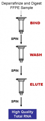
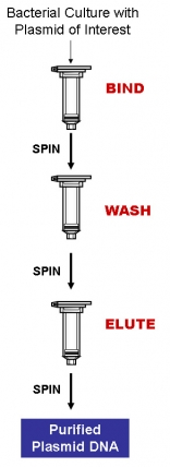
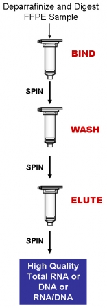
Purification is based on spin column chromatography using Bio-Synthesis's proprietary resin as the separation matrix. The process first involves deparaffinization of the FFPE samples through a series of xylene and ethanol washes. Next, the FFPE samples are digested with the provided Proteinase K and Digestion Buffer. Binding Solution and ethanol are then added to the lysate, and the solution is loaded onto a spin-column (BIND). Under these conditions only the RNA and/or DNA will bind while most of the contaminants will be removed in the flowthrough. At this point, any traces of genomic DNA can be digested allowing for pure RNA samples to be isolated. Alternatively, traces of the RNA may be digested at this point if a pure sample of genomic DNA is required instead. The bound nucleic acid is then washed (WASH) in order to remove any impurities. Lastly, the purified total nucleic acd is eluted in 50 µL of the provided Elution Buffer or water (ELUTE). Please see the procedure flowchart to the right.
This kit is also available in a 96-well format for high-throughput FFPE RNA/DNA purification. Purifictaion with the 96-well plate can be performed using either a vacuum manifold or centrifugation.
 FEATURES AND BENEFITS
* High quality and integrity of the isolated nucleic acid - Both the purified total RNA and/or genomic DNA are of the highest quality and integrity, and can be used in any sensitive downstream application.
* Isolate a diversity of RNA species - All RNA species can be isolated, from large mRNA and ribosomal RNA down to microRNA and siRNA.
* High yields - Bio-Synthesis's FFPE RNA/DNA Purification Kit allows for the purification of high yields of total RNA or DNA.
* Rapid procedure - Isolate total RNA and/or genomic DNA from FFPE tissue sections using a rapid spin column format or a high-throughput 96-well plate format.
* Versatile procedure - The kit allows for the purification of either pure total RNA, pure genomic DNA or both RNA and genomic DNA through the use of tailored protease digestion times.
APPLICATIONS
The purified RNA and genomic DNA are of the highest available quality from FFPE samples, and can be used in a number of downstream applications including:
* qPCR
* qRT-PCR
* Reverse transcription PCR
* Primer extension
* Mutation screening
* Expression array assays
* Microarray analyses
* Sequencing
* Southern blotting
* SNP analysis
* Next Generation Sequencing
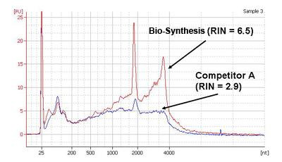
 Figure 1. High Quality and Yield of Total RNA. Total RNA was isolated from one slice of hamster FFPE kidney section (20 micron thickness) using Bio-Synthesis's FFPE RNA/DNA Purification Kit and a leading competitor’s kit. One microliter of the 50 µL purified RNA was then resolved on an Agilent 2100 BioAnalzyer using an RNA Nano 6000 chip. As it can be seen, Bio-Synthesis's not only isolated higher yields of total RNA, but the RNA was also of a higher quality as evidenced by the higher RIN values obtained with Bio-Synthesis's RNA.
FFPE RNA/DNA Purification Kit Contents
1. Digestion Buffer
2. Binding Solution
3. Enzyme Incubation Buffer
4. Wash Solution
5. Elution Buffer
6. Proteinase K
7. Spin Columns
8. Collection Tubes
9. Elution Tubes
10. Product Insert
Long-Term Storage : All solutions should be kept tightly sealed and stored at room temperature. The Proteinase K should be stored in aliquots at -20°C upon reconstitution. These reagents should remain stable for at least 1 year in their unopened containers.
Product Usage
For laboratory research use only, not for diagnostic use! Not for use in humans.
FFPE RNA Purification Kit
Catalog# : NB-25300
Price/Unit : $320.00
Unit : 50 preps
For the rapid and efficient extraction and purification of RNA (including microRNA) from FFPE samples
* Extract total RNA (including microRNA) from FFPE samples
* No phenol extraction step
* Includes DNAse for optional on-column DNA removal
* Isolated RNA is of the highest quality and integrity
Bio-Synthesis's FFPE RNA Purification Kit provides a rapid method for the isolation and purification of total RNA (including microRNA) from formalin-fixed paraffin-embedded (FFPE) tissue samples in as little as 1 hour. Using formalin to fix tissues leads to crosslinking of the RNA and proteins, and the process of embedding the tissue samples can also lead to fragmentation of the RNA over time. Bio-Synthesis's FFPE RNA Purification Kit provides conditions that allow for the partial reversing of the formalin modifications, resulting in a high quality and yield of RNA. The kit is able to purify all sizes of RNA, from large mRNA and ribosomal RNA down to microRNA (miRNA) and small interfering RNA (siRNA), depending on the age of the FFPE tissue as fragmentation of the RNA is known to occur over time.
Purification is based on spin column chromatography using Bio-Synthesis's proprietary resin as the separation matrix. The process first involves deparaffinization of the FFPE samples through a series of xylene and ethanol washes. Next, the FFPE samples are digested with the provided Proteinase K and Digestion Buffer. Binding Solution and ethanol are then added to the lysate, and the solution is loaded onto a spin-column (BIND). Under these conditions only the RNA will bind while most of the contaminants will be removed in the flowthrough. At this point, any remaining traces of genomic DNA can be digested using an optional protocol, allowing for pure RNA samples to be isolated. The bound RNA is then washed (WASH) in order to remove any impurities. Lastly, the purified total RNA is eluted in 50 µL of the provided Elution Buffer or water (ELUTE). Please see the procedure flowchart to the right.
 FEATURES AND BENEFITS
* High quality and integrity of the isolated RNA - The purified total RNA is of the highest quality and integrity, and can be used in any sensitive downstream applications.
* Isolate a diversity of RNA species - All RNA species can be isolated, from large mRNA and ribosomal RNA down to microRNA (miRNA) and small interfering RNA (siRNA).
* High yields - Bio-Synthesis's FFPE RNA Purification Kit allows for the purification of high yields of total RNA.
* No phenol:chloroform extractions - Total RNA is isolated from FFPE tissue samples without the use of harmful chemicals such as phenol or chloroform.
* Rapid procedure - Isolate total RNA from FFPE tissue sections using a rapid spin column format in as little as 1 hour
APPLICATIONS
Purified RNA is of the highest quality, and can be used in a number of downstream applications including:
* Bioanalyzer
* Quantitative, real-time RT-PCR for both large mRNA and small RNA including miRNA
* RT-PCR for both large mRNA and small RNA including miRNA
* Northern blotting
* RNase protection
* Primer extension
* Expression array assays
* Next Generation Sequencing
* microRNA Cloning
 Figure 1. High Quality and Yield of Total RNA. Bio-Synthesis's FFPE RNA Purification Kit isolates FFPE RNA that exceeds both yield and quality of competitors. Total RNA was isolated from one slice of hamster FFPE kidney section (20 micron thickness) using Bio-Synthesis's FFPE RNA Purification Kit and a leading competitor’s kit. One microliter of the 50 µL purified RNA was then resolved on an Agilent 2100 BioAnalzyer using an RNA Nano 6000 chip. As it can be seen, Bio-Synthesis's not only isolated higher yields of total RNA, but the RNA was also of a higher quality as evidenced by the higher RIN values obtained with Bio-Synthesis's RNA.
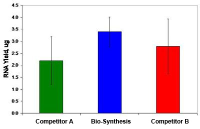
 Figure 2. Higher Yield of FFPE RNA Isolated by Bio-Synthesis's FFPE RNA Purification Kit. Bio-Synthesis's FFPE RNA Purification Kit isolates FFPE RNA that exceeds the yield of competitors. Total RNA was isolated from one slice of hamster FFPE kidney section (20 micron thickness) using Bio-Synthesis's FFPE RNA Purification Kit and two leading competitor’s kits. Eighteen isolations were performed for each product. The graph demonstrates the mean yield of RNA according to spectrophotometry for 18 sample replicates. Vertical bars represent the standard deviation. Bio-Synthesis's kit consistently purified total RNA with a higher yield than for those obtained using the market competitor's kits.
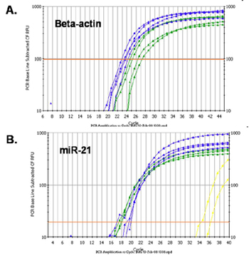
 Figure 3. High Yield of a Diversity of RNA Species. Bio-Synthesis's FFPE RNA Purification Kit effectively recovers all sizes of RNA from large mRNA to small RNA including microRNAs. Total RNA was isolated from one slice of hamster FFPE kidney section (20 micron thickness) using Bio-Synthesis's FFPE RNA Purification Kit and a leading competitor’s kit. Five microliters of the 50 µL purified RNA was then used as the template in a 20 µL reverse transcription reaction with oligo dT primer or miR-21 stem-loop reverse primer. Two microliters of the RT reaction was then used in a 15 µL qPCR reaction for detecting the beta-actin gene (Panel A) and for detecting miR-21(Panel B), respectively. In both graphs the blue lines correspond to Bio-Synthesis's isolated-RNA and the green lines correspond to competitor-isolated RNA. As it can be seen, Bio-Synthesis's kit isolated higher yields of RNA in both cases, as indicated by the lower Ct values of the blue lines. Also, Bio-Synthesis's kit successfully isolated not only large RNA (Panel A) but also microRNA (Panel B), indicating the diversity of RNA species isolated.
 FFPE RNA Purification Kit Contents - Spin Columns
1. Digestion Buffer
2. Binding Solution
3. Enzyme Incubation Buffer
4. Wash Solution
5. RNA Elution Buffer
6. Proteinase K
7. DNase I
8. Spin Columns
9. Collection Tubes
10. Elution Tubes
11. Product Insert
Long-Term Storage : All solutions should be kept tightly sealed and stored at room temperature. The DNAse I should be stored at -20°C upon arrival. The Proteinase K should be stored in aliquots at -20oC upon reconstitution. These reagents should remain stable for at least 1 year in their unopened containers.
Product Usage
For laboratory research use only, not for diagnostic use! Not for use in humans.
CleanAll RNA/DNA Clean-Up and Concentration Kit
Catalog# : NB-23800
Price/Unit : $240.00
Unit : 20 preps
For the rapid and efficient purification, cleanup and concentration of RNA or DNA
* Purifies all sizes of RNA, from large mRNA and ribosomal RNA down to microRNA (miRNA) and small interfering RNA (siRNA)
* RNA is preferentially purified from other reaction components such as proteins and nucleotides, without the use of phenol or chloroform
* Purifies all sizes of DNA, from small PCR products to plasmids to genomic DNA.
* Rapid and efficient spin-column format
Features and Benefits | Specifications
BSI's CleanAll RNA/DNA Clean-Up and Concentration Micro Kit provides a rapid method for the purification, cleanup and concentration of RNA or DNA from different isolation methods or upstream applications. The kit can be used as an alternative to organic extraction and ethanol precipitation to clean up various enzymatic reactions. The CleanAll Kit purifies RNA from phenol/guanidine-based protocols or from various upstream enzymatic reactions such as DNase treatment, labeling and in vitro transcription. Furthermore, endotoxins can be removed from previously purified RNA solutions. The kit can be used to clean up DNA from digestions, ligations, PCR reactions, labeling reactions, DNA modification reactions and staining. The kit also provides a protocol for the rapid removal of endotoxins from previously purified DNA down to 0.1 EU/µg DNA or less.
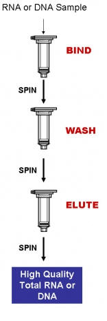
Purification is based on spin column chromatography using BSI’s proprietary resin as the separation matrix. Briefly, the samples or reaction containing RNA or DNA is mixed with Binding Solution and the nucleic acids are bound to BSI's column (BIND). Under these conditions only the nucleic acids will bind to BSI's resin while most of the contaminating proteins and nucleotides are removed in the flowthrough. The bound nucleic acid is then washed to remove any remaining impurities (WASH). Lastly, the purified total RNA or DNA is eluted into 50 µL of the provided Elution Buffer or water (ELUTE). Please see procedure flowchart to the right.

FEATURES AND BENEFITS
* Versatile performance - Both DNA and RNA can be cleaned and concentrated using the same kit. Clean-up nucleic acids from a variety of isolation methods and upstream enzymatic reactions.
* Fast and easy processing - Rapid spin-column format allows for the processing of 10 samples in 20 minutes.
* Suitable for all sizes of RNA - Purify all sizes of RNA, from large mRNA and ribosomal RNA down to microRNA (miRNA) and small interfering RNA (siRNA).
* Suitable for all sizes of DNA - Purify all sizes of DNA, from small PCR products, native or linearized plasmids, to genomic DNA. Using an alternative protocol, even oligonucleotides and smaller DNA fragments can be purified.
* Optional endotoxin removal - Optional protocol is provided for the rapid removal of endotoxins from previously purified RNA or DNA.
* No phenol:chlorofom extractions - BSI’s CleanAll kit purifies RNA without the use of harmful chemicals such as phenol or chloroform
APPLICATIONS AND DATA
The kit can be used to clean up RNA and DNA from upstream applications such as:
* Digestions
* Ligations
* PCR reactions
* Labeling reactions
* DNA modification reactions
* Staining
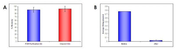
 Figure 1. High Recoveries and Efficient Endotoxin Removal. High recoveries of DNA can be seen when using the CleanAll kit, and these high recoveries are not compromised even during endotoxin removal. In Panel A, 1 µg of a PCR product was cleaned using the CleanAll RNA/DNA Clean-Up and Concentration Kit as well as BSI's PCR Purification Kit. More than 0.9 µg of the DNA was recovered from both kits with a comparable percent recovery of over 90%. In Panel B, 10 µg of plasmid DNA preparation was cleaned from endotoxin by using the CleanAll RNA/DNA Kit. Endotoxin levels of the plasmid DNA sample were reduced from 0.75 EU/µg plasmid DNA to less 0.05 EU/µg plasmid DNA. Approximately 95% of the endotoxin units in the DNA sample were removed using the kit, therefore the kit allows for high recoveries coupled with exceptional cleanup.

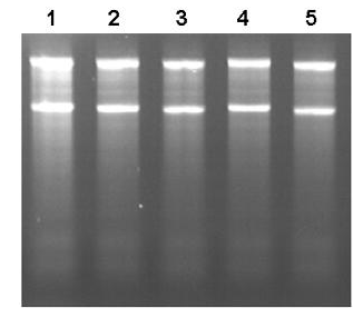
Figure 2: Clean-Up of RNA with High Recovery. BSI’s CleanAll RNA/DNA Clean-Up and Concentration Kit can be used to clean up various enzymatic reactions including DNase treatment. In Figure 2 above, Lane 1 is the RNA input, while lanes 2-5 contain the RNA that has been cleaned using BSI’s CleanAll Kit. It can be seen that in all cases the recovery is high, and the purified RNA is intact and of a high quality.
CleanAll RNA/DNA Clean-Up and Concentration Kit Components
1. Binding Solution
2. Wash Solution
3. Elution Buffer
4. Micro Spin Columns
5. Collection Tubes
6. Elution tubes
7. Product Insert
Long-Term Storage : All solutions should be kept tightly sealed and stored at room temperature. These reagents should remain stable for at least 1 year in their unopened containers.
Product Usage
For laboratory research use only, not for diagnostic use! Not for use in humans.
EndoG Monoclonal Antibody
Catalog# : PM-4581
The fragmentation of nuclear DNA is a hallmark of apoptotic cell death. The activities of caspase and nuclease are involved in the DNA fragmentation. Caspase-activated deoxyribonuclease (CAD), also termed DNA fragmentation factor (DFF40), is one such nuclease, and is capable of inducing DNA fragmentation and chromatin condensation after cleavage by caspase-3 of its inhibitor ICAD/DFF45. Caspase and CAD independent DNA fragmentation also exists. Recent studies demonstrated that another nuclease, endonuclease G (EndoG), is specifically activated by apoptotic stimuli and is able to induce nucleosomal fragmentation of DNA independently of caspase and DFF/CAD. EndoG is a mitochondrion-specific nuclease that translocates to the nucleus and cleaves chromatin DNA during apoptosis. The homologue of mammalian EndoG is the first mitochondrial protein identified to be involved in apoptosis in C. elegans. EndoG also cleaves DNA in vitro.
Additional Names : EndoG (7F2D7): Endonuclease G
 Source : Mouse monoclonal EndoG antibody was raised against a recombinant protein corresponding to amino acids 51 – 140 of human EndoG.
Source : Mouse monoclonal EndoG antibody was raised against a recombinant protein corresponding to amino acids 51 – 140 of human EndoG.
Purification : Immunoaffinity chromotography purified IgG
Clonality and Clone : This is a monoclonal antibody. (Clone 7F2D7)
Host : EndoG monoclonal antibody was raised in mouse. Please use anti-rabbit secondary antibodies.
Immunogen : Recombinant protein corresponding to amino acids 51 – 140 of human EndoG.
Application : EndoG monoclonal antibody can be used for detection of EndoG by Western blot at 5 – 10 µg/ml.
Tested Application(s) : E, WB
Buffer : Antibody is supplied in PBS containing 0.02% sodium azide.
Long-Term Storage : EndoG monoclonal antibody can be stored at 4ºC, stable for one year.
Positive Control
1.Cat. No. 1211 - HepG2 Cell Lysate
2.Cat. No. 95-103 - EndoG Recombinant Protein
Species Reactivity :H, M, R
Short Description : Endonclease G
References
1.Li LY, Luo X, Wang X. Endonuclease G is an apoptotic DNase when released from mitochondria. Nature 2001; 412:95-9.
2.Parrish J, Li L, Klotz K, et al. Mitochondrial endonuclease G is important for apoptosis in C. elegans. Nature 2001; 412:90-4
3.Hengartner MO. Apoptosis. DNA destroyers. Nature 2001; 412:27, 29.
4.Widlak P, Li LY, Wang X, et al. Action of recombinant human apoptotic endonuclease G on naked DNA and chromatin substrates: cooperation with exonuclease and DNase I. J. Biol. Chem. 2001; 276:48404-9.
EndoG Monoclonal Antibody
Catalog# : PM-4579
The fragmentation of nuclear DNA is a hallmark of apoptotic cell death. The activities of caspase and nuclease are involved in the DNA fragmentation. Caspase-activated deoxyribonuclease (CAD), also termed DNA fragmentation factor (DFF40), is one such nuclease, and is capable of inducing DNA fragmentation and chromatin condensation after cleavage by caspase-3 of its inhibitor ICAD/DFF45. Caspase and CAD independent DNA fragmentation also exists. Recent studies demonstrated that another nuclease, endonuclease G (EndoG), is specifically activated by apoptotic stimuli and is able to induce nucleosomal fragmentation of DNA independently of caspase and DFF/CAD. EndoG is a mitochondrion-specific nuclease that translocates to the nucleus and cleaves chromatin DNA during apoptosis. The homologue of mammalian EndoG is the first mitochondrial protein identified to be involved in apoptosis in C. elegans. EndoG also cleaves DNA in vitro.
Additional Names : EndoG (7G1G10): Endonuclease G
 Description
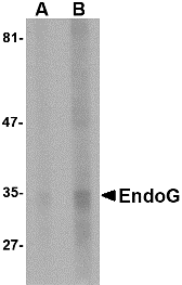
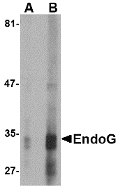
Left: Western blot analysis of EndoG expression in HepG2 cell lysate with EndoG antibody at (A) 5 and (B) 10 µg/ml.
Source : Mouse monoclonal EndoG antibody was raised against a recombinant protein corresponding to amino acids 51 – 140 of human EndoG.
Purification : Immunoaffinity chromotography purified IgG
Clonality and Clone : This is a monoclonal antibody. (Clone 7G1G10)
Host : EndoG monoclonal antibody was raised in mouse. Please use anti-rabbit secondary antibodies.
Immunogen : Recombinant protein corresponding to amino acids 51 – 140 of human EndoG.
Application : EndoG monoclonal antibody can be used for detection of EndoG by Western blot at 5 – 10 µg/ml.
Tested Application(s) : E, WB
Buffer : Antibody is supplied in PBS containing 0.02% sodium azide.
Long-Term Storage : EndoG monoclonal antibody can be stored at 4ºC, stable for one year.
Positive Control
1.Cat. No. 1211 - HepG2 Cell Lysate
2.Cat. No. 95-103 - EndoG Recombinant Protein
Species Reactivity :H, R
Short Description : Endonclease G
References
1.Li LY, Luo X, Wang X. Endonuclease G is an apoptotic DNase when released from mitochondria. Nature 2001; 412:95-9.
2.Parrish J, Li L, Klotz K, et al. Mitochondrial endonuclease G is important for apoptosis in C. elegans. Nature 2001; 412:90-4
3.Hengartner MO. Apoptosis. DNA destroyers. Nature 2001; 412:27, 29.
4.Widlak P, Li LY, Wang X, et al. Action of recombinant human apoptotic endonuclease G on naked DNA and chromatin substrates: cooperation with exonuclease and DNase I. J. Biol. Chem. 2001; 276:48404-9.
EndoG Monoclonal Antibody
Catalog# : PM-4577
The fragmentation of nuclear DNA is a hallmark of apoptotic cell death. The activities of caspase and nuclease are involved in the DNA fragmentation. Caspase-activated deoxyribonuclease (CAD), also termed DNA fragmentation factor (DFF40), is one such nuclease, and is capable of inducing DNA fragmentation and chromatin condensation after cleavage by caspase-3 of its inhibitor ICAD/DFF45. Caspase and CAD independent DNA fragmentation also exists. Recent studies demonstrated that another nuclease, endonuclease G (EndoG), is specifically activated by apoptotic stimuli and is able to induce nucleosomal fragmentation of DNA independently of caspase and DFF/CAD. EndoG is a mitochondrion-specific nuclease that translocates to the nucleus and cleaves chromatin DNA during apoptosis. The homologue of mammalian EndoG is the first mitochondrial protein identified to be involved in apoptosis in C. elegans. EndoG also cleaves DNA in vitro.
Additional Names : EndoG (7G1C10): Endonuclease G
 Description
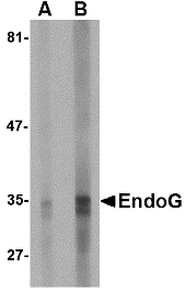
Left: Western blot analysis of EndoG expression in HepG2 cell lysate with EndoG antibody at (A) 2.5 and (B) 5 µg/ml.
Source : Mouse monoclonal EndoG antibody was raised against a recombinant protein corresponding to amino acids 51 – 140 of human EndoG.
Purification : Immunoaffinity chromotography purified IgG
Clonality and Clone : This is a monoclonal antibody. (Clone 7G1C10)
Host : EndoG monoclonal antibody was raised in mouse.Please use anti-rabbit secondary antibodies.
Immunogen : Recombinant protein corresponding to amino acids 51 – 140 of human EndoG.
Application : EndoG monoclonal antibody can be used for detection of EndoG by Western blot at 2.5 – 5 µg/ml.
Tested Application(s) : E, WB
Buffer : Antibody is supplied in PBS containing 0.02% sodium azide.
Long-Term Storage : EndoG monoclonal antibody can be stored at 4ºC, stable for one year.
Positive Control
1.Cat. No. 1211 - HepG2 Cell Lysate
2.Cat. No. 95-103 - EndoG Recombinant Protein
Species Reactivity :H, M, R
Short Description : Endonclease G
References
1.Li LY, Luo X, Wang X. Endonuclease G is an apoptotic DNase when released from mitochondria. Nature 2001; 412:95-9.
2.Parrish J, Li L, Klotz K, et al. Mitochondrial endonuclease G is important for apoptosis in C. elegans. Nature 2001; 412:90-4
3.Hengartner MO. Apoptosis. DNA destroyers. Nature 2001; 412:27, 29.
4.Widlak P, Li LY, Wang X, et al. Action of recombinant human apoptotic endonuclease G on naked DNA and chromatin substrates: cooperation with exonuclease and DNase I. J. Biol. Chem. 2001; 276:48404-9.
EndoG Antibody
Catalog# : 3035
The fragmentation of nuclear DNA is a hallmark of apoptotic cell death. The activities of caspase and nuclease are involved in the DNA fragmentation. Caspase-activated deoxyribonuclease (CAD), also termed DNA fragmentation factor (DFF40), is one such nuclease, and is capable of inducing DNA fragmentation and chromatin condensation after cleavage by caspase-3 of its inhibitor ICAD/DFF45. Caspase and CAD independent DNA fragmentation also exists. Recent studies demonstrated that another nuclease, endonuclease G (endoG), is specifically activated by apoptotic stimuli and is able to induce nucleosomal fragmentation of DNA independently of caspase and DFF/CAD (1,2). EndoG is a mitochondrion-specific nuclease that translocates to the nucleus and cleaves chromatin DNA during apoptosis. The homologue of mammalian EndoG is the first mitochondrial protein identified to be involved in apoptosis in C. elegans (2). EndooG also cleaves DNA in vitro (4).
Additional Names : EndoG
 Description
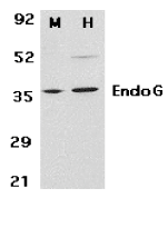
Left: Western blot analysis of EndoG in mouse (M) 3T3 and human (H) HepG2 cell lysates with EndoG antibody at 2 µg/ml.
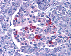
Below: Immunohistochemistry of EndoG in human pancreas with EndoG antibody at 15 µg/ml.
Other Product Images
 Source : EndoG antibody was raised with a synthetic peptide corresponding to 15 amino acids near the amino terminus of human EndoG.
Purification : Affinity chromatography purified via peptide column
Clonality and Clone : This is a polyclonal antibody.
Host : EndoG antibody was raised in rabbit. Please use anti-rabbit secondary antibodies.
Immunogen : Human EndoG Peptide (Cat. No. 3035P)
Application : EndoG antibody can be used for detection of EndoG by Western blot at 1 to 2 µg/ml.HepG2 and 3T3 cell lysates can be used as positive control and a 35 kDa band can be detected. It is human, mouse and rat reactive.
Tested Application(s) : E, WB, IHC
Buffer : Antibody is supplied in PBS containing 0.02% sodium azide.
Blocking Peptide : Cat. No. 3035P - EndoG Peptide
Long-Term Storage : EndoG antibody can be stored at 4ºC, stable for one year. As with all antibodies care should be taken to avoid repeated freeze thaw cycles. Antibodies should not be exposed to prolonged high temperatures.
Positive Control
1.Cat. No. 1211 - HepG2 Cell Lysate
2.Cat. No. 1212 - 3T3 Whole Cell Lysate
3.Cat. No. 1307 - Human Pancreas Tissue Lysate
Species Reactivity :H, M, R
GI Number : 53759134
Accession Number : NP_004426
Short Description : Endonuclease G
References
1.Li LY, Luo X, Wang X. Endonuclease G is an apoptotic DNase when released from mitochondria. Nature. 2001;412(6842):95-9.
2.Parrish J, Li L, Klotz K, Ledwich D, Wang X, Xue D. Mitochondrial endonuclease G is important for apoptosis in C. elegans. Nature. 2001;412(6842):90-4
3.Hengartner MO. Apoptosis. DNA destroyers. Nature. 2001;412(6842):27, 29.
4.Widlak P, Li LY, Wang X, Garrard WT. Action of recombinant human apoptotic endonuclease G on naked DNA and chromatin substrates: cooperation with exonuclease and DNase I. J Biol Chem. 2001;276(51):48404-9.
DRAM Antibody
Catalog# : 4035
Damage-regulated autophagy modulator (DRAM) is a p53 target gene encoding a lysosomal protein that induces autophagy, a process that degrades cytosolic proteins and organelles. It has been suggested that activation of DRAM by p53 is simultaneous to the activation by p53 of one or more proapoptotic genes such as PUMA, Bax, etc., and that the signaling pathways regulated by these genes together promote a full cell death response. By itself, DRAM cannot induce apoptosis, but the fact that it is inactivated in certain cancers highlights the importance of DRAM and suggests that autophagy may play a more important role in cancer than initially suspected. At least two different isoforms of DRAM are known to exist.
Additional Names : DRAM (NT), Damage-regulated autophagy modulator
 Description
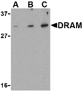
Left: Western blot analysis of DRAM in K562 cell lysate with DRAM antibody at (A) 0.5, (B) 1 and (C) 2 µg/ml.
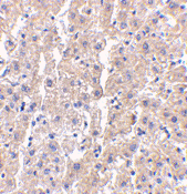
Below: Immunohistochemistry of DRAM in human liver tissue with DRAM antibody at 2.5 µg/ml.
Other Product Images
 Source : DRAM antibody was raised against a 16 amino acid peptide from near the amino terminus of human DRAM.
Purification : Affinity chromatography purified via peptide column
Clonality and Clone : This is a polyclonal antibody.
Host : DRAM antibody was raised in rabbit. Please use anti-rabbit secondary antibodies.
Application : DRAM antibody can be used for detection of DRAM by Western blot at 0.5 – 1 µg/ml.
Tested Application(s) : E, WB, IHC
Buffer : Antibody is supplied in PBS containing 0.02% sodium azide.
Blocking Peptide : Cat.No. 4035P - DRAM Peptide
Long-Term Storage : DRAM antibody can be stored at 4ºC, stable for one year. As with all antibodies care should be taken to avoid repeated freeze thaw cycles. Antibodies should not be exposed to prolonged high temperatures.
Positive Control
1.Cat. No. 1204 - K562 Cell Lysate
2.Cat. No. 1304 - Human Liver Tissue Lysate
Species Reactivity :H, M, R
GI Number : 22450862
Accession Number : AAH18435
Short Description : (NT) Damage-regulated autophagy modulator
References
1.Crighton D, Wilkinson S, O’Prey J, et al. DRAM, a p53-induced modulator of autophagy, is critical for apoptosis. Cell 2006; 126:121-34.
2.Gozuacik D and Kimchi A. Autophagy as a cell death and tumor suppressor mechanism. Oncogene. 2004; 23:2891-906.
3.Crighton D, Wilkinson S, and Ryan KM. DRAM links autophagy to p53 and programmed cell death. Autophagy 2007; 3:72-4. (07-01D)
Low Copy Plasmid DNA MiniPrep
Catalog# : NB-17800
Price/Unit : $110.00
Unit : 25 preps
For the rapid purification of low copy plasmids
* High yield of low copy plasmid DNA
* Alcohol-free protocol
* Fast and easy processing
Bio-Synthesis's Low Copy Plasmid MiniPrep Kit is designed for the rapid preparation of low copy number plasmid DNA from small batch cultures of E. coli. The kit is able to purify 0.5 - 1 µg of low copy number plasmid DNA from 3 mL of bacterial culture. Purification of plasmids up to 14,000 bp in size have been verified.
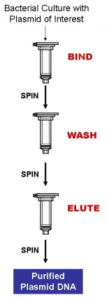
Purification is based on spin column chromatography using Bio-Synthesis's proprietary resin as the separation matrix. Briefly, the bacteria harbouring the plasmid is pelleted and lysed, and the released plasmid DNA is bound to Bio-Synthesis's column in the presence of Binding Solution (BIND). Under these conditions only the DNA will bind to Bio-Synthesis's resin while most of the contaminating RNA and cellular proteinaceous components are removed in the flowthrough. The bound DNA is then washed to remove any remaining impurities (WASH). Lastly, the purified DNA is eluted into 30 - 50 µL of the provided Elution Buffer or water (ELUTE). Please see procedure flowchart to the right.
The kit purifies DNA without alcohols, a significant improvement over other techniques to achieve full compatibility with alcohol-sensitive applications. The purified DNA is free of genomic DNA, RNA and cellular proteins and is suitable for many downstream applications.
 FEATURES AND BENEFITS
* Fast and easy processing - Rapid spin-column format allows for the processing of multiple samples in 30 minutes.
* High yield - 0.5 to 1 µg of low copy number plasmid DNA can be purified from 3 mL of bacterial culture
* Recovered plasmid DNA is suitable for downstream applications - Purified plasmid DNA is fully compatible with restriction enzyme digestions, and manual or automated sequencing.
* Alcohol-free protocol - No alcohols are used during the purification process, thus the DNA is compatible with alcohol sensitive downstream applications.
APPLICATIONS
* Restriction digests
* Sequencing
* Transformation
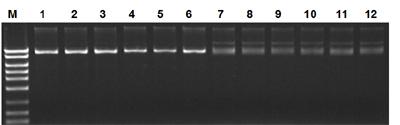
 Figure 1. Isolation of High Quality DNA from Low Copy Number Plasmids. The Low Copy Number Plasmid MiniPrep Kit was used to isolate plasmid DNA from 3 mL cultures. Lanes 1-6 are pSYX20 DNA (10-20 copies per cell) while lanes 7-12 are pSC101DNA (2-3 copies per cell). Five microlitres of the total elutions were loaded onto a 1X TAE, 1% agarose gel, with Lanes 1-3 and 7-9 corresponding to 30 µL elutions, and Lanes 4-6 and 10-12 corresponding to 50 µL elutions. In all cases, the DNA is of a high quality, indicated by the presence of sharp bands and no degradation of the isolated DNA. Lane M is Bio-Synthesis's MidRanger 1kb DNA Ladder. Quantification of the DNA is presented in Table 1.
LowCopy Plasmid DNA MiniPrep Kit Contents
1.Resuspension Buffer
2.Lysis Solution
3.Binding Solution
4.RNAse A
5.Wash Solution
6.Elution Buffer
7.Micro Spin Columns
8.Collection Tubes
9.Elution Tubes
10.Product Insert
Long-Term Storage : All solutions should be kept tightly sealed and stored at room temperature. These reagents should remain stable for at least 1 year in their unopened containers. The Resuspension Buffer should be stored at 4oC upon addition of RNAse A enzyme.
Product Usage
For laboratory research use only, not for diagnostic use! Not for use in humans.
BAC DNA MiniPrep
Catalog# : NB-18000
Price/Unit : $110.00
Unit : 25 preps
For the rapid preparation of BAC DNA
* High DNA recovery
* Alcohol-free protocol
* Convenient spin column protocol
Bio-Synthesis's BAC DNA MiniPrep Kit is designed for the rapid preparation of Bacterial Artificial Chromosomes (BACs) and other large DNA constructs from small batch cultures of Escherichia coli. The BAC DNA MiniPrep Kit typically recovers between 0.6 and 1 µg of DNA from 3 mL of bacterial culture. Purification of constructs up to 130 kbp in size have been verified.
Purification is based on spin column chromatography using Bio-Synthesis's proprietary resin as the separation matrix. Briefly, the bacteria harbouring the plasmid is pelleted and lysed, and the released plasmid DNA is bound to Bio-Synthesis's column in the presence of Binding Solution (BIND). Under these conditions only the DNA will bind to Bio-Synthesis's resin while most of the contaminating RNA and cellular proteinaceous components are removed in the flowthrough. The bound DNA is then washed to remove any remaining impurities (WASH). Lastly, the purified DNA is eluted into 30 - 50 µL of the provided Elution Buffer or water (ELUTE). Please see procedure flowchart to the right.
Bio-Synthesis's BAC DNA Miniprep Kit purifies DNA without alcohols, a significant improvement over other techniques to achieve full compatibility with alcohol-sensitive applications. The purified DNA is free of genomic DNA, RNA and cellular proteins and is suitable for many downstream applications.
 FEATURES AND BENEFITS
* High Recovery - 0.6 to 1 µg of BAC DNA can be purified from 3.0 mL of bacterial culture.
* Alcohol-free protocol - No alcohols are used durig the purification process, thus the DNA is compatible with alcohol sensitive downstream applications.
* Fast and easy processing - Rapid spin-column format allows for the processing of multiple samples in 1 hour.
* Recovered DNA is suitable for downstream applications - Purified BAC DNA is fully compatible with restriction enzyme digestions and sequencing.
* High binding capacity of columns - The binding capacity of the columns in the BAC DNA MiniPrep Kit is 25 µg
APPLICATIONS
Purified BAC DNA is ready for a range of downstream applications including:
* sequencing
* transformation
* restriction digestion
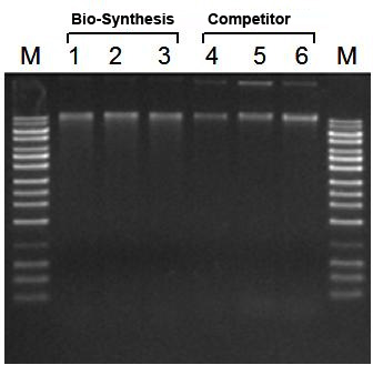
 Figure 1. Higher DNA Yield than Competitor. BAC DNA from 3 mL overnight cultures was isolated with the Bio-Synthesis BAC MiniPrep Kit (Lanes 1-3) and with a competitor’s kit (Lanes 4-6). Ten microliters of 50 µL of recovered DNA was loaded in each lane of a 1X TAE, 0.9% agarose gel. Lane M is Bio-Synthesis's UltraRanger 1kb DNA Ladder. Note the quality, consistency and yield of the DNA isolated using Bio-Synthesis's kit (Lanes 1-3).
 BAC DNA MiniPrep Kit Contents
1.Resuspension Buffer
2.Lysis Solution
3.Neutralizaation Solution
4.Binding Solution
5.RNAse A
6.RNase T1
7.Wash Solution
8.Elution Buffer
9.Micro Spin Columns
10.Collection Tubes
11.Elution Tubes
12.Product Insert
Long-Term Storage : All solutions should be kept tightly sealed and stored at room temperature. The RNAse A and RNAse T1 should be stored at -20°C upon arrival. Once RNAse A has been added to the Resuspension Buffer, however, the solution should be stored at 4°C. All the reagents should remain stable for at least 1 year in their unopened containers.
Product Usage
For laboratory research use only, not for diagnostic use! Not for use in humans.
Water RNA/DNA Purification Kit - 0.45 µm
Catalog# : NB-26450
Price/Unit : $420.00
Unit : 25 preps
For convenient purification of RNA and DNA from microorganisms in water samples
* Isolate total DNA and RNA from all microorganisms found in water, including bacteria, fungi and algae
* RNA and DNA are both column purified simultaneously using the same column
* Elution contains concentrated DNA and RNA without the need for further precipitation
* Complete RNA (including microRNA) without phenol
* Isolated RNA and DNA are of high quality and integrity for all downstream applications
* Available in 0.45 µm and 0.22 µm filter formats
Bio-Synthesis's Water RNA/DNA Purification Kit provides a convenient and rapid method for the detection of microorganisms from environmental water samples. The kit allows for the rapid isolation and purification of total RNA and DNA simultaneously from the microorganisms found in different types of water samples. The total RNA and DNA (including genomic DNA) are isolated from all the microorganisms found in the water, including bacteria, fungi and algae without the use of any inhibitory organic substances. The kit purifies genomic DNA, and all sizes of RNA, from large mRNA and ribosomal RNA down to microRNA (miRNA) and small interfering RNA (siRNA). The purified RNA and DNA are highly concentrated, and can be used directly in a number of downstream applications including real time PCR, reverse transcription PCR, Northern blotting, Southern blotting and sequencing reactions.
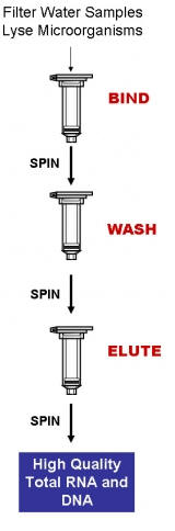
Purification is based on spin column chromatography using Bio-Synthesis's proprietary resin as the separation matrix. Briefly, the microorganisms present in the water sample are collected using either a 0.22 µm Filter Column or a 0.45 µm Filter Column. The captured microorganisms are then lysed using a provided Bead Tube and Lysis Solution. Next, ethanol is added to the lysate and it is loaded onto a spin column (BIND). Under these conditions only the RNA and DNA will bind to Bio-Synthesis's resin, while the proteins and other contaminants are removed in the flowthrough. Next, an optional step can be carried out in which the genomic DNA can be digested allowing for a more pure RNA sample to be isolated. Alternatively, the RNA can be digested resulting in a more pure genomic DNA sample. The bound nucleic acid is then washed to remove any impurities (WASH). Lastly, the purified RNA and/or DNA is eluted with 50 µL of the provided Elution Buffer or water (ELUTE). Please see the procedure flowchart to the right.
 FEATURES AND BENEFITS
* Rapid isolation of DNA/RNA from microorganisms in non-turbid water samples –Only three buffers are required to complete isolation of total DNA and RNA from all microorganisms found in water, including bacteria, fungi and algae.
* Complete column purification - The RNA and DNA are both column purified simultaneously using the same column.
* Fast and easy processing - Rapid spin column format allows for the isolation of both RNA and DNA in under 45 minutes.
* Isolate highly concentrated RNA and DNA - The purified RNA and DNA is eluted in low volumes, allowing for the direct use of the nucleic acid in downstream applications
* Isolate a diversity of RNA species - All sizes of RNA are isolated, from large mRNA down to microRNA, without the use of phenol or chloroform
APPLICATIONS
The purified RNA and DNA are of the highest quality and can be used in a number of downstream applications including:
* Quantitative, real-time PCR
* Quantitative, real-time RT-PCR
* Conventional PCR
* Reverse transcription PCR
* Northern blotting
* Southern blotting
* Sequencing
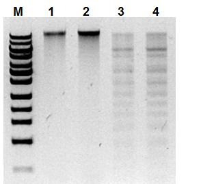
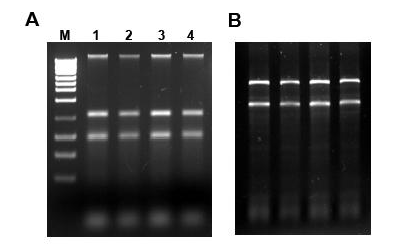
 Figure 1. High Yield and Purity of RNA and DNA. Total RNA and DNA were simultaneously isolated from 50 mL of water sample containing 107 cfu/mL E.coli using Bio-Synthesis's Water RNA/DNA Purification Kit and subsequently run on gels for visual analysis. Panel A shows 10 µL aliquots (no RNase treatment) of the 50 µL elutions run on a 1% TAE agarose gel. Genomic DNA and 16S and 23S rRNA bands were visable. Panel B shows 5 µL aliquots (on-column DNase was applied) of the elution run on a 1.5% formaldehyde agarose gel. 16S and 23S rDNA was seen without DNA contamination. From observing the gels it can be seen that the kit allows for the isolation and purification of high yields of concentrated and high quality RNA and DNA.
 Water RNA/DNA Purification Kit - 0.45 µm Components
1.Lysis Solution
2.Nucleic Acid Wash Solution
3.Enzyme Incubation Buffer
4.Nucleic Acid Elution Buffer
5.Mini Spin Columns
6.Filter Columns
7.Bead Tubes
8.Collection Tubes
9.Elution tubes
10.Product Insert
Long-Term Storage : All solutions should be kept tightly sealed and stored at room temperature. These reagents should remain stable for at least 1 year in their unopened containers.
Product Usage
For laboratory research use only, not for diagnostic use! Not for use in humans.
Water RNA/DNA Purification Kit - 0.22 µm
Catalog# : NB-26400
Price/Unit : $440.00
Unit : 25 preps
Product Usage
For laboratory research use only, not for diagnostic use! Not for use in humans.
Water RNA/DNA Purification Kit - 0.22 µm
Catalog# : NB-26400
Price/Unit : $440.00
Unit : 25 preps
Product Usage
For laboratory research use only, not for diagnostic use! Not for use in humans.
Plant/Fungi RNA Total Purification Kit
Catalog# : NB-25800
Price/Unit : $295.00
Unit : 50 preps
For the rapid purification of total RNA (including microRNA) from plants and fungi
* Extract total RNA, including viral RNA, from plant or fungal species
* Robust lysis buffer is well-suited to even challenging samples such as pine needle or grape leaf
* Isolate total RNA (including microRNA) without phenol
* Liquid nitrogen not needed for homogenization
* Isolated RNA is of high quality, integrity and diversity
Bio-Synthesis's Plant/Fungi Total RNA Purification Kit provides a rapid method for the isolation and purification of total RNA, including viral RNA, from a wide range of plant and filamentous fungal species. Total RNA can be purified from fresh or frozen plant tissues, plant cells or filamentous fungi samples using this kit. All sizes of RNA are purified, from large mRNA and ribosomal RNA down to microRNA (miRNA) and small interfering RNA (siRNA). The procedure is rapid and convenient, as it does not rely on the use of liquid nitrogen in order to homogenize the samples.
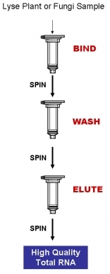
Purification is based on spin column chromatography using Bio-Synthesis's proprietary resin as the separation matrix. Briefly, the cells or tissue are macerated in a mortar with the provided Lysis Solution. Alternatively, liquid nitrogen can be used to homogenize the sample. The lysate is then spun to pellet any debris, ethanol is added to the clarified lysate, and the solution is loaded onto a spin-column (BIND). Under these conditions only the RNA will bind to Bio-Synthesis's resin while most of the DNA and proteins are removed in the flowthrough. The bound RNA is then washed in order to remove any remaining impurities (WASH). Lastly, the purified total RNA is eluted into 50 µL of the supplied Elution Buffer or water (ELUTE). Please see the procedure flowchart to the right.
The RNA is preferentially purified from other cellular components, such as proteins, without the use of phenol or chloroform. The purified RNA is of the highest integrity, and can be used in a number of downstream applications including real time PCR, reverse transcription PCR, Northern blotting, RNase protection and primer extension, and expression array assays. This kit is available in two convenient sizes - 50 preps and 96 preps.
 FEATURES AND BENEFITS
* No liquid nitrogen required for homogenization - Liquid nitrogen is not required for homogenization of samples, making RNA purification rapid and convenient.
* Isolate RNA from a wide range of samples - Total RNA can be isolated from a wide range of plant and filamentous fungi samples.
* Isolate a diversity of RNA species - All sizes of RNA are isolated from large mRNA down to microRNA, without the use of phenol or chloroform.
* Isolate total RNA, including viral RNA - RNA samples can be used for the detection of viral pathogens, as viral RNA is isolated with the total RNA.
* High yield of RNA - High yields of purified RNA can be isolated with this kit.
* No phenol:chloroform extractions - Total RNA is isolated without the use of harmful chemicals such as phenol or chloroform. RNA is of the highest quality and can be used in a number of downstream applications.
* Available in 2 sizes - This kits is available as both a 50 prep kit and a 96 prep kit.
APPLICATIONS
Purified RNA is of the highest quality and can be used in a number of downstream applications, including:
* qRT-PCR
* Reverse transcription PCR
* Northern blotting
* RNAse protection
* Primer extension
* Expression array assays
* Next Generation Sequencing

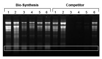
Figure 1. Isolation of High Quality RNA, even from Difficult Samples. Total RNA was isolated from 50 mg samples of apple (Lanes 1), peach (Lanes 2), grape (Lanes 3), pine needle (Lanes 4), strawberry (Lanes 5) and pear (Lanes 6) using Bio-Synthesis's kit and a competitors kit. Five µL of total RNA from the 50 µL of elution was loaded on 1X MOPS 1.0 % Formaldehyde-Agarose RNA gel for analysis. Bio-Synthesis's kit allowed for the isolation of high quality RNA from all the samples, including the difficult samples, while the competitor failed to isolate RNA from grape, pine needles and strawberry. Furthermore, only Bio-Synthesis's kit was able to isolate the small RNA species (white box).
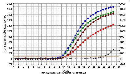
 Figure 2. Detection of EF1-α using one step RT-qPCR from Challenging Plant Samples. Total RNA was isolated from 50 mg samples of apple (red), peach (green), pine needle (blue) and grape leaves (burgundy) using Bio-Synthesis's Plant/Fungi Total RNA Purification Kit. Three µL of the eluted 50 µL of RNA was used in an RT-qPCR reaction for the detection of EF1-α. EF1-α was detected from all samples, indicating that the RNA is of high quality and that the Plant/Fungi RNA Purification kit is highly sensitive for total RNA isolation.
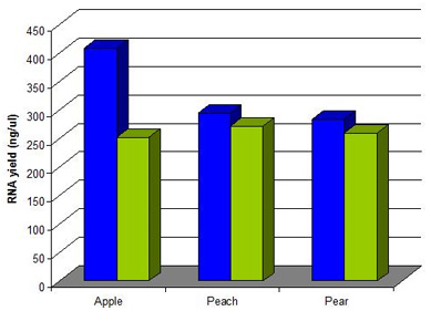
 Figure 3. High Yield of Plant RNA Samples isolated from Apple, Peach and Pear. Total RNA was isolated using Bio-Synthesis's kit (Blue bars) and competitor’s kit (Green bars) from 50 mg of apple, peach and pear according to the provided manual. In each case the RNA was eluted into 50 µL. RNA yield was measured by spectrophotometer in triplicate, and the average concentration was used for data. Bio-Synthesis's kit resulted in higher yields of total RNA, including miRNA. Total processing time was less with Bio-Synthesis's kit than competitor’s kit.
 Plant/Fungi RNA Purification Kit Components
1.Lysis Solution
2.Wash Solution
3.Elution Buffer
4.Mini Spin Columns
5.Collection Tubes
6.Elution tubes
7.Product Insert
Long-Term Storage : All solutions should be kept tightly sealed and stored at room temperature. These reagents should remain stable for at least 1 year in their unopened containers.
Product Usage
For laboratory research use only, not for diagnostic use! Not for use in humans.
DR4 Antibody
Catalog# : 1139
Apoptosis, or programmed cell death, occurs during normal cellular differentiation and development of multicellular organisms. Apoptosis is induced by certain cytokines including TNF and Fas ligand in the TNF family through their death domain containing receptors, TNFR1 and Fas. A novel death domain containing receptor was recently identified and designated DR4 (for death receptor 4). The ligand for this novel death receptor has been identified and termed TRAIL2,3, which is a new member in the TNF family. DR4 is also called TRAIL receptor-1 (TRAIL-R1). DR4 is expressed in most of human tissues including spleen, peripheral blood leukocytes, small intestine and thymus. Like TNFR1, Fas and DR3, DR4 mediates apoptosis and NF-kappaB activation in many tissues and cells.
Additional Names : DR4 (CT), TRAIL-R1
 Description
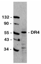
Left: Western blot analysis of DR4 in HeLa total cell lysate with DR4 antibody at 1:500 dilution.
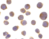
Below: Immunocytochemistry of DR4 in Jurkat cells with DR4 antibody at 10 µg/ml.
Other Product Images

Source : DR4 antibody was raised against a peptide corresponding to amino acids near the carboxy terminus of human DR4 protein.
Purification : Antibody is DEAE purified
Clonality and Clone : This is a polyclonal antibody.
Host : DR4 antibody was raised in rabbit. Please use anti-rabbit secondary antibodies.
Immunogen : Human DR4 (C-Terminus) Peptide (Cat. No. 1139P)
Application : DR4 antibody can be used for detection of DR4 expression by Western blot at 1:500 to 1:1000 dilution. A 57 kDa band should be detected.
Tested Application(s) : E, WB, ICC
Buffer : Antibody is supplied in PBS containing 0.02% sodium azide.
Blocking Peptide : Cat. No. 1139P - DR4 Peptide
Long-Term Storage : DR4 antibody can be stored at 4ºC, stable for one year. As with all antibodies care should be taken to avoid repeated freeze thaw cycles. Antibodies should not be exposed to prolonged high temperatures.
Positive Control
1.Cat. No. 1201 - HeLa Cell Lysate
Species Reactivity :H
GI Number : 1945072
Accession Number : AAC51226
Short Description : (CT) Death Receptor for TRAIL
References
1.Pan G; O'Rourke K; Chinnaiyan AM; O'Rourke K; Gentz R; Ebner R; Ni J; Dixit VM. The receptor for the cytotoxic ligand TRAIL. Science; 1997;276:111-113
2.Wiley SR, Schooley K, Smolak PJ, Din WS, Huang CP, Nicholl JK, Sutherland GR, Smith TD, Rauch C, Smith CA, et al. Identification and characterization of a new member of the TNF family that induces apoptosis. Immunity 1995;3:673-682
3.Pitti RM; Marsters SA; Ruppert S; Donahue CJ; Moore A; Ashkenazi A. Induction of apoptosis by Apo-2 ligand, a new member of the tumor necrosis factor cytokine family. J. Biol. Chem. 1996;271:12687-90
4.Schneider P, Thome M, Burns K, Bodmer JL, Hofmann K, Kataoka T, Holler N, Tschopp J. TRAIL receptors 1 (DR4) and 2 (DR5) signal FADD-dependent apoptosis and activate NF-kappaB. Immunity 1997;7:831-836 (RD1299)
DR4 Antibody
Catalog# : 1167
Apoptosis, or programmed cell death, occurs during normal cellular differentiation and development of multicellular organisms. Apoptosis is induced by certain cytokines including TNF and Fas ligand in the TNF family through their death domain containing receptors, TNFR1 and Fas. A novel death domain containing receptor was recently identified and designated DR4 (for death receptor 4)1. The ligand for this novel death receptor has been identified and termed TRAIL2,3, which is a new member in the TNF family. DR4 is also called TRAIL receptor-1 (TRAIL-R1)4. DR4 is expressed in most of human tissues including spleen, peripheral blood leukocytes, small intestine and thymus. Like TNFR1, Fas and DR3, DR4 mediates apoptosis and NF-kappaB activation in many tissues and cells.
Additional Names : DR4 (NT), TRAIL-R1
 Description
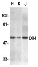
Left: Western blot analysis of DR4 in HeLa (H), K562 (K), and Jurkat (J) whole cell lysate with DR4 antibody at 1:500 dilution.
Source : DR4 antibody was raised against a peptide corresponding to amino acid 1 to 20 of human DR4 mature protein.
Purification : Antibody is DEAE purified
Clonality and Clone : This is a polyclonal antibody.
Host : DR4 antibody was raised in rabbit. Please use anti-rabbit secondary antibodies.
Immunogen : Human DR4 (N-Terminus) Peptide (Cat. No. 1167P)
Application : DR4 antibody can be used for detection of DR4 by Western blot at 1:500 to 1:1000 dilution. A 57 kDa band can be detected. DR4 antibody has no cross reaction to DR5.
Tested Application(s) : E, WB
Buffer : Antibody is supplied in PBS containing 0.02% sodium azide.
Blocking Peptide : Cat. No. 1167P - DR4 Peptide
Long-Term Storage : DR4 antibody can be stored at 4ºC, stable for one year. As with all antibodies care should be taken to avoid repeated freeze thaw cycles. Antibodies should not be exposed to prolonged high temperatures.
Positive Control
1.Cat. No. 1201 - HeLa Cell Lysate
2.Cat. No. 1205 Jurkat Whole Cell Lysate
Species Reactivity :H
GI Number : 1945072
Accession Number : AAC51226
Short Description : (NT) Death Receptor for TRAIL
References
1.Pan G; O'Rourke K; Chinnaiyan AM; O'Rourke K; Gentz R; Ebner R; Ni J; Dixit VM. The receptor for the cytotoxic ligand TRAIL. Science; 1997;276:111-113
2.Wiley SR, Schooley K, Smolak PJ, Din WS, Huang CP, Nicholl JK, Sutherland GR, Smith TD, Rauch C, Smith CA, et al. Identification and characterization of a new member of the TNF family that induces apoptosis. Immunity 1995;3:673-682
3.Pitti RM; Marsters SA; Ruppert S; Donahue CJ; Moore A; Ashkenazi A. Induction of apoptosis by Apo-2 ligand, a new member of the tumor necrosis factor cytokine family. J. Biol. Chem. 1996;271:12687-90
4.Schneider P, Thome M, Burns K, Bodmer JL, Hofmann K, Kataoka T, Holler N, Tschopp J. TRAIL receptors 1 (DR4) and 2 (DR5) signal FADD-dependent apoptosis and activate NF-kappaB. Immunity 1997;7:831-836 (RD1299)
DR3 Antibody
Catalog# : 1120
Apoptosis, or programmed cell death, occurs during normal cellular differentiation and development of multicellular organisms. Apoptosis is induced by certain cytokines including TNF and Fas ligand of the TNF family through their death domain containing receptors, TNFR1 and Fas. A novel cell death receptor was recently identified by several groups independently and designated DR3, Wsl-1, Apo-3, TRAMP and LARD1-5. The ligand for this novel cell death receptor has not yet been defined. DR3 is highly expressed in the tissues enriched in lymphocytes including PBL, thymus and spleen. Like TNFR1, DR3 induces apoptosis and NF-kappaB activation.
Additional Names : DR3 (CT), Wsl-1, Apo-3
 Description
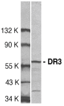
Left: Western blot analysis of DR3 in Jurkat total cell lysate with DR3 antibody at 1:500 dilution.
Source : DR3 antibody was raised against a peptide corresponding to amino acids 398 to 417 of human DR3.
Purification : Antibody is DEAE purified
Clonality and Clone : This is a polyclonal antibody.
Host : DR3 antibody was raised in rabbit. Please use anti-rabbit secondary antibodies.
Immunogen : Human DR3 (C-Terminus) Peptide (Cat. No. 1120P)
Application : DR3 antibody can be used for Western blot at 1:500 to 1:1000 dilution. A 59 kDa band should be detected and has no cross reaction to other death receptors.
Tested Application(s) : E, WB
Buffer : Antibody is supplied in PBS containing 0.02% sodium azide.
Blocking Peptide : Cat. No. 1120P - DR3 Peptide
Long-Term Storage : DR3 antibody can be stored at 4ºC, stable for one year. As with all antibodies care should be taken to avoid repeated freeze thaw cycles. Antibodies should not be exposed to prolonged high temperatures.
Positive Control
1.Cat. No. 1205 - Jurkat Cell Lysate
Species Reactivity :H, M, R
GI Number : 37181738
Accession Number : AAQ88676
Short Description : (CT) Cell Death Receptor
References
1.Chinnaiyan AM; O'Rourke K; Yu GL; Lyons RH; Garg M; Duan DR; Xing L; Gentz R; Ni J; Dixit VM. Science, 1996;274:990-
2.Kitson J; Raven T; Jiang YP; Goeddel DV; Giles KM; Pun KT; Grinham CJ; Brown R; Farrow SN. Nature, 1996;384:372-5.
3.Marsters SA; Sheridan JP; Donahue CJ; Pitti RM; Gray CL; Goddard AD; Bauer KD; Ashkenazi A. Curr Biol, 1996;6:1669-76.
4.Bodmer JL; Burns K; Schneider P; Hofmann K; Steiner V; Thome M; Bornand T; Hahne M; Schroter M; Becker K; et al. Immunity, 1997;6:79-88.
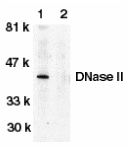
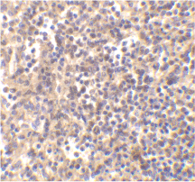
DNase II AntibodyCatalog# : 2059Apoptosis is characterized by several morphological nuclear changes including chromatin condensation and nuclear fragmentation. These changes are triggered by the activation of members of caspase family, caspase activated DNase, and several novel proteins including AIF and Acinus . DNase II causes both chromatin condensation and DNA fragmentation . The genes encoding human (2-4), porcine (4), and murine (5) DNase II have been cloned. The DNase II gene encodes a 40 kDa proenzyme. The mature enzyme consists of two non-identical subunits, the 32 kDa (a) and 12 kDa (b) chains, generated by proteolytic processing. Overexpression of DNase II induces chromatin condensation (3). DNase II is ubiquitously expressed in human tissues.Additional Names : DNase II DescriptionLeft: Western blot analysis of DNase II in human spleen tissue lysate in the absence (lane 1) or presence (lane 2) of blocking peptide with DNase antibody II at 1:500 dilution.Below: Immunohistochemistry of DNaseII in human spleen tissue with DNaseII antibody at 5 µg/ml.Other Product Images
 Source : DNase II antibody was raised against a peptide corresponding to amino acids 347 to 360 of human DNase II precursor (2-4) .Purification : Affinity chromatography purified via peptide columnClonality and Clone : This is a polyclonal antibody.Host : DNase II antibody was raised in rabbit. Please use anti-rabbit secondary antibodies.Immunogen : Human DNase II (C-Terminus) Peptide (Cat. No. 2059P)Application : DNase II antibody can be used for detection of DNase II expression by Western blot at 1:500 to 1:1000 dilution. An approximate 40 kDa band can be detected, which represents the pro-enzyme of DNase II.Tested Application(s) : E, WB, IHCBuffer : Antibody is supplied in PBS containing 0.02% sodium azide.Blocking Peptide : Cat. No. 2059P - DNase II PeptideLong-Term Storage : DNase II antibody can be stored at 4ºC, stable for one year. As with all antibodies care should be taken to avoid repeated freeze thaw cycles. Antibodies should not be exposed to prolonged high temperatures.Positive Control1.Cat. No. 1306 - Human Spleen Tissue Lysate2.Cat. No. 1208 - THP-1 Whole Cell LysateSpecies Reactivity :HGI Number : 2921838Accession Number : AF047016Short Description : DNase involved in apoptosisReferences 1.Zamzami N, Kroemer G. Condensed matter in cell death. Nature 1999;401:127-82.Yasuda T, Takeshita H, Iida R, Nakajima T, Hosomi O, Nakashima Y, Kishi K. Molecular cloning of the cDNA encoding human deoxyribonuclease II. J Biol Chem 1998;273:2610-63.Krieser RJ, Eastman A. The cloning and expression of human deoxyribonuclease II. A possible role in apoptosis. J Biol Chem 1998;273:30909-144.Shiokawa D, Tanuma S. Cloning of cDNAs encoding porcine and human DNase II. Biochem Biophys Res Commun 1998;247:864-9

 FEATURES AND BENEFITS
 Figure 1. High Quality and Yield of Total RNA. Total RNA was isolated from one slice of hamster FFPE kidney section (20 micron thickness) using Bio-Synthesis's FFPE RNA/DNA Purification Kit and a leading competitor’s kit. One microliter of the 50 µL purified RNA was then resolved on an Agilent 2100 BioAnalzyer using an RNA Nano 6000 chip. As it can be seen, Bio-Synthesis's not only isolated higher yields of total RNA, but the RNA was also of a higher quality as evidenced by the higher RIN values obtained with Bio-Synthesis's RNA.