Showing posts with label pna-oligo. Show all posts

Tuesday, July 13, 2010

DR3 Antibody

DR3 Antibody

Catalog# : 1166

Apoptosis, or programmed cell death, occurs during normal cellular differentiation and development of multicellular organisms. Apoptosis is induced by certain cytokines including TNF and Fas ligand of the TNF family through their death domain containing receptors, TNFR1 and Fas. A novel cell death receptor was recently identified by several groups independently and designated DR3, Wsl-1, Apo-3, TRAMP and LARD1-5. The ligand for this novel death receptor has been defined as TWEAK, also termed Apo3L. DR3 is highly expressed in the tissues enriched in lymphocytes including PBL, thymus and spleen. Like TNFR1, DR3 induces apoptosis and NF-kappaB activation.

Additional Names : DR3 (NT), Wsl-1, Apo-3
Description
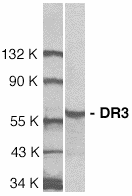
Left: Western blot analysis of DR3 in Jurkat total cell lysate with DR3 antibody at 1:1000 dilution.


Source : DR3 antibody was raised against a peptide corresponding to amino acids 25 to 40 of human DR3 precusor.

Purification : Antibody is DEAE purified

Clonality and Clone : This is a polyclonal antibody.

Host : DR3 antibody was raised in rabbit. Please use anti-rabbit secondary antibodies.

Immunogen : Human DR3 (N-Terminus) Peptide (Cat. No. 1166P)

Application : DR3 antibody can be used for detection of DR3 by Western blot at 1:500 to 1:1000 dilution. A 59 kDa band can be detected. DR3 has no cross reaction to other death receptors.

Tested Application(s) : E, WB

Buffer : Antibody is supplied in PBS containing 0.02% sodium azide.

Blocking Peptide : Cat. No. 1166P - DR3 Peptide

Long-Term Storage : DR3 antibody can be stored at 4ºC, stable for one year. As with all antibodies care should be taken to avoid repeated freeze thaw cycles. Antibodies should not be exposed to prolonged high temperatures.

Positive Control
1.Cat. No. 1205 - Jurkat Cell Lysate

Species Reactivity :H, M

GI Number : 37181738

Accession Number : AAQ88676

Short Description : (NT) Cell Death Receptor

References
1.Chinnaiyan AM; O'Rourke K; Yu GL; Lyons RH; Garg M; Duan DR; Xing L; Gentz R; Ni J; Dixit VM. Science, 1996;274:990-2.
2.Kitson J; Raven T; Jiang YP; Goeddel DV; Giles KM; Pun KT; Grinham CJ; Brown R; Farrow SN. Nature, 1996;384:372-5.
3.Marsters SA; Sheridan JP; Donahue CJ; Pitti RM; Gray CL; Goddard AD; Bauer KD; Ashkenazi A. Curr Biol, 1996;6:1669-76.
4.Bodmer JL; Burns K; Schneider P; Hofmann K; Steiner V; Thome M; Bornand T; Hahne M; Schroter M; Becker K; et al. Immunity, 1997;6:79-88.

DNase II Antibody

DNase II Antibody

Catalog# : 2059

Apoptosis is characterized by several morphological nuclear changes including chromatin condensation and nuclear fragmentation. These changes are triggered by the activation of members of caspase family, caspase activated DNase, and several novel proteins including AIF and Acinus . DNase II causes both chromatin condensation and DNA fragmentation . The genes encoding human (2-4), porcine (4), and murine (5) DNase II have been cloned. The DNase II gene encodes a 40 kDa proenzyme. The mature enzyme consists of two non-identical subunits, the 32 kDa (a) and 12 kDa (b) chains, generated by proteolytic processing. Overexpression of DNase II induces chromatin condensation (3). DNase II is ubiquitously expressed in human tissues.

Additional Names : DNase II
Description
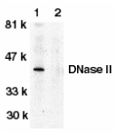
Left: Western blot analysis of DNase II in human spleen tissue lysate in the absence (lane 1) or presence (lane 2) of blocking peptide with DNase antibody II at 1:500 dilution.

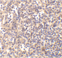
Below: Immunohistochemistry of DNaseII in human spleen tissue with DNaseII antibody at 5 µg/ml.

Other Product Images

Source : DNase II antibody was raised against a peptide corresponding to amino acids 347 to 360 of human DNase II precursor (2-4) .

Purification : Affinity chromatography purified via peptide column

Clonality and Clone : This is a polyclonal antibody.

Host : DNase II antibody was raised in rabbit. Please use anti-rabbit secondary antibodies.

Immunogen : Human DNase II (C-Terminus) Peptide (Cat. No. 2059P)

Application : DNase II antibody can be used for detection of DNase II expression by Western blot at 1:500 to 1:1000 dilution. An approximate 40 kDa band can be detected, which represents the pro-enzyme of DNase II.

Tested Application(s) : E, WB, IHC

Buffer : Antibody is supplied in PBS containing 0.02% sodium azide.

Blocking Peptide : Cat. No. 2059P - DNase II Peptide

Long-Term Storage : DNase II antibody can be stored at 4ºC, stable for one year. As with all antibodies care should be taken to avoid repeated freeze thaw cycles. Antibodies should not be exposed to prolonged high temperatures.

Positive Control
1.Cat. No. 1306 - Human Spleen Tissue Lysate
2.Cat. No. 1208 - THP-1 Whole Cell Lysate

Species Reactivity :H

GI Number : 2921838

Accession Number : AF047016

Short Description : DNase involved in apoptosis

References
1.Zamzami N, Kroemer G. Condensed matter in cell death. Nature 1999;401:127-8
2.Yasuda T, Takeshita H, Iida R, Nakajima T, Hosomi O, Nakashima Y, Kishi K. Molecular cloning of the cDNA encoding human deoxyribonuclease II. J Biol Chem 1998;273:2610-6
3.Krieser RJ, Eastman A. The cloning and expression of human deoxyribonuclease II. A possible role in apoptosis. J Biol Chem 1998;273:30909-14
4.Shiokawa D, Tanuma S. Cloning of cDNAs encoding porcine and human DNase II. Biochem Biophys Res Commun 1998;247:864-9

Sunday, July 11, 2010

Plant RNA/DNA Purification Kit

Plant RNA/DNA Purification Kit

Alternate Name:
Catalog# : NB-24400
Price/Unit : $240.00
Unit : 50 preps

For simultaneous isolation of total RNA and DNA from the same plant sample

No phenol extraction
High quality DNA and RNA are purified simultaneously using the same spin column
No need to split the lysate
All sizes of RNA are recovered, including microRNA

Bio-Synthesis's Plant RNA/DNA Purification Kit provides a rapid method for the isolation and purification of total RNA and genomic DNA simultaneously from a single sample of plants. The total RNA and genomic DNA are both column purified in under 30 minutes using a single column. It is often necessary to isolate total RNA and genomic DNA from a single plant sample, such as for studies of gene expression, mutant or transgenic plant characterization, and host plant-pathogen characterization. Traditionally the RNA and DNA would be isolated from different aliquots of a sample, however this novel technology will allow for their simultaneous isolation from the same sample. This will not only save time, but will also be of a great benefit when isolating RNA and DNA from precious, difficult to obtain or very small samples. Furthermore, gene expression analysis will be more reliable since the RNA and DNA are derived from the same sample, therefore eliminating inconsistent results.

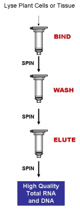
Purification is based on spin column chromatography using Bio-Synthesis's proprietary resin as the separation matrix. Briefly, the cells or tissue of interest are lysed with the provided Lysis Solution, ethanol is then added to the lysate, and the solution is loaded onto a spin-column (BIND). Under these conditions only the RNA and DNA will bind to the column while the proteins are removed in the flowthrough. Next, an optional step can be carried out in which the genomic DNA can be digested allowing for a more pure RNA sample to be isolated. Alternatively, traces of the RNA may be digested at this point if a pure sample of genomic DNA is required instead. The bound nucleic acids are then washed in order to remove any remaining impurities (WASH). Lastly, the purified nucleic acids are eluted into the provided Elution Buffer or water (ELUTE). Please see the procedure flowchart to the right.

FEATURES AND BENEFITS

  • Complete column purification - The RNA and DNA are both column purified simultaneously using the same column.
  • Reduce variability - RNA and DNA are isolated from a single plant sample with no splitting of the lysate, thus reducing inconsistent results and variability.
  • Isolate from small samples - Simultaneous isolation of RNA and DNA from a single sample. Ideal for precious, difficult to obtain or small samples.
  • Isolate a diversity of RNA species - All sizes of RNA are isolated, from large mRNA down to microRNA, without the use of phenol or chloroform.
  • Isolate DNA-free plant RNA or RNA-free plant DNA - Optional protocols for on-column DNase or RNase digestion are provided if the user wishes to isolate pure, DNA-free RNA or pure, RNA-free DNA..

APPLICATIONS

Purified RNA and DNA are of the highest quality and can be used in a number of downstream applications including:

  • Real-time PCR
  • Southern blotting
  • SNP analysis
  • Sequencing
  • AFLP
  • RFLP
  • RAPD
  • Microsatellite analysis
  • qRT-PCR
  • Primer extension
  • Expression array assays
  • Next Generation Sequencing

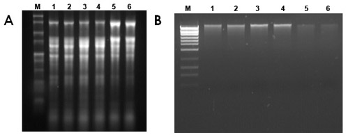
Figure 1. Isolation of Total RNA and Genomic DNA from Tobacco, Tomato and Peach Leaf Tissue. Total RNA and genomic DNA were isolated from 50 mg of tobacco leaf, 50 mg of tomato leaf and 50 mg of peach leaf using Bio-Synthesis's Plant RNA/DNA Purification Kit. Panel A is a 1X MOPS 1.5% agarose gel showing the total RNA that was isolated after the optional on-column DNase digestion. Five µL of total RNA from each 150 µL elution was mixed with 2x RNA loading dye and denatured at 70ºC for 10 minutes and loaded onto the gel. Lane M is Bio-Synthesis's 1 kb RNA Ladder, Lanes 1 and 2 contain RNA isolated from tobacco cells, Lanes 3 and 4 contain RNA isolated from tomato cells, and Lanes 5 and 6 contain RNA isolated from peach cells. Panel B is a 1.5% agarose gel containing the genomic DNA that was isolated after the optional on-column RNase digestion, and in each case 10 µL of the 150 µL elution was loaded. Lane M is Bio-Synthesis's HighRanger 1kb DNA Ladder, Lanes 1 and 2 contain the tobacco DNA, Lanes 3 and 4 contain the tomato DNA, and Lanes 5 and 6 contain the peach DNA. The RNA and DNA are intact and of the highest quality, and can be used in a number of different downstream applications.

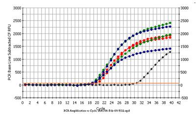
Figure 2. Detection of 18s rDNA by Real-time PCR (SYBR Green). Total DNA and RNA from 50 mg of tobacco, tomato and peach were simultaneously isolated with Bio-Synthesis's Plant RNA/DNA Purification Kit (with the optional on-column RNase treatment). Next, 2µL of DNA from each 150 µL elution was mixed in 20 µL of PCR reaction and used in a real-time PCR (95ºC for 3min and 40 cycles at 95 ºC for 15 sec. and 60 ºC for 30 sec.). DNA from tobacco (green), tomato (red) and peach (blue) was successfully amplified, indicating the high quality of the DNA for downstream application. Primer dimer (black) also was shown.

Plant RNA / DNA Purification Kit Contents

1.Lysis Solution
2.Nucleic Acid Wash Solution
3.Nucleic Acid Elution Buffer
4.Enzyme Incubation Buffer
5.Mini spin columns
6.Collection Tubes
7.Elution Tubes
8.Product Insert

Long-Term Storage : All solutions should be kept tightly sealed and stored at room temperature. These reagents should remain stable for at least 1 year in their unopened containers.
Product Usage
For laboratory research use only, not for diagnostic use! Not for use in humans.

dNTP Mix

dNTP Mix (25 mM)

Alternate Name:
Catalog# : NB-28105
Price/Unit : $297.00
Unit : 1 ml
For Robust and Consistent PCR and RT-PCR Amplification

Available as dNTP Set (100 mM each of dATP, dCTP, dGTP, dTTP)
Available as dNTP Mixes at 2 different concentrations (10 mM each and 25 mM each)
Highly pure
Suitable for many different PCR applications

BSI’s dNTP Set (Cat# NB-28104) contains one tube each of dATP, dCTP, dGTP and dTTP at a concentration of 100 mM, supplied in water.

BSI’s dNTP Mixes contain dATP, dCTP, dGTP and dTTP at a concentration of either 10 mM each (Cat # NB-28106) or 25 mM each (Cat # NB-28105) supplied in water.

BSI’s highly pure dNTP Set and dNTP Mixes can be used in all standard and sensitive PCR applications.

APPLICATIONS
  • Standard PCR
  • Multiplex PCR
  • Genotyping
  • Methylation-Sensitive PCR (MSP)
  • Nested PCR
  • One-Step RT-PCR
  • Two-Step RT-PCR
  • Multiplex RT-PCR
Quality Assurance

  • dNTP solutions are certified free of nucleases and phosphatases.
  • Long-Term Storage : dNTP Set and dNTP Mixes should be stored at –20°C. Avoid repeated freeze-thawing.
  • Product Usage
  • For laboratory research use only, not for diagnostic use! Not for use in humans.

dNTP Mix

dNTP Mix (10 mM)

Catalog# : NB-28106
Price/Unit : $178.00
Unit : 1 ml
For Robust and Consistent PCR and RT-PCR Amplification

Available as dNTP Set (100 mM each of dATP, dCTP, dGTP, dTTP)
Available as dNTP Mixes at 2 different concentrations (10 mM each and 25 mM each)
Highly pure
Suitable for many different PCR applications

BSI’s dNTP Set (Cat# NB-28104) contains one tube each of dATP, dCTP, dGTP and dTTP at a concentration of 100 mM, supplied in water.

BSI’s dNTP Mixes contain dATP, dCTP, dGTP and dTTP at a concentration of either 10 mM each (Cat # NB-28106) or 25 mM each (Cat # NB-28105) supplied in water.

BSI’s highly pure dNTP Set and dNTP Mixes can be used in all standard and sensitive PCR applications.

APPLICATIONS
  • Standard PCR
  • Multiplex PCR
  • Genotyping
  • Methylation-Sensitive PCR (MSP)
  • Nested PCR
  • One-Step RT-PCR
  • Two-Step RT-PCR
  • Multiplex RT-PCR
Quality Assurance

  • dNTP solutions are certified free of nucleases and phosphatases.
  • Long-Term Storage : dNTP Set and dNTP Mixes should be stored at –20°C. Avoid repeated freeze-thawing.
  • Product Usage
  • For laboratory research use only, not for diagnostic use! Not for use in humans.

Taq Polymerase (with 10X Taq Reaction Buffer)

Taq Polymerase (with 10X Taq Reaction Buffer)

Alternate Name:
Catalog# : NB-28088
Price/Unit : $205.00
Unit : 1000 units (5 U/ul)

Recombinant source
  • Functionality preserved at wide range of temperatures
  • Highly reliable and reproducible reactions
  • Supplied with detergent-free Reaction Buffer for compatibility with microarray and DHPL applications
  • Compatible with the use of modified nucleotides (dUTP, dITP, biotin-, digoxigenin- and fluorescently-labeled nucleotides)
  • Available in 2 different concentrations: 5 units/µL and 1 unit/µL

Taq DNA Polymerase is a highly thermostable DNA polymerase that possesses a 5´→ 3´ polymerase activity and a very low 5´→ 3´ exonuclease activity. The enzyme is supplied with detergent-free 10X Taq Reaction Buffer, making it compatible with microarray and DHPLC applications.

Bio-Synthesis's Taq DNA Polymerase with 10X Taq Reaction Buffer is available in 2 convenient sizes:

−1000 units at a concentration of 5000 units/mL (Cat # 28088)

Enzyme Source:
An E. coli strain with a cloned Taq DNA Polymerase gene from Thermus aquaticus YT-1

APPLICATIONS
  • Routine PCR
  • Preparation of PCR products for cloning
  • Primer extension
  • Microarray analysis
  • DHPLC
  • DNA labelling
  • DNA sequencing
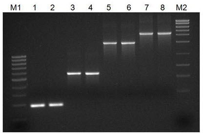
Figure 1. Successful Amplification of Different Sized Amplicons. Bio-Synthesis's Taq DNA Polymerase was used in a series of PCR reactions to amplify a number of different sized amplicons from the adenovirus E2 region. Lanes 1 and 2 correspond to a 189 bp product, Lanes 3 and 4 correspond to a 565 bp product, Lanes 5 and 6 correspond to a 1501 bp product, and Lanes 7 and 8 correspond to a 2202 bp product. Bio-Synthesis's Taq DNA Polymerase was able to amplify all the different sized products with a high sensitivity and yield. Lane M1 is Bio-Synthesis's PCR Sizer 100 bp DNA Ladder and Lane M2 is Bio-Synthesis's MidRanger 1kb DNA Ladder.

Reagents supplied - Cat# 28088:

1000 units of Taq DNA Polymerase (5000 units/mL)
4 x 1.25 mL Taq Reaction Buffer (10X)

Reaction Conditions:
1X Taq Reaction Buffer (10 mM Tris-HCl, 50 mM KCl, 1.5 mM MgCl2, PCR stabilizer, pH 8.3 @ 25°C)

Storage Buffer:
  • 50 mM Tris-HCl pH 8 at 25°C, 50 mM KCl, 50mM NaCl, 0.5 mM Dithiothreitol, 0.1 mM EDTA, 50% Glycerol, 1% Triton X-100
  • Long-Term Storage : -20°C
  • Product Usage
  • For laboratory research use only, not for diagnostic use! Not for use in humans.

Tuesday, July 6, 2010

DEDAF Antibody

DEDAF Antibody

Catalog# : 2227

Apoptosis is related to many diseases and induced by a family of cell death receptors and their ligands. Cell death signals are transduced by death domain (DD) death effector domain (DED), and caspase recruitment domain (CARD) containing molecules. Several molecules including caspases and adaptor FADD contain DEDs. A novel protein that interacts with DED of caspase-8 and 10, and FADD was identified recently and designated DEDAF for DED associated factor . DEDAF is identical to the transcriptional repressor RYBP (2). DEDAF/RYBP is expressed in multiple tissues and cell lines. DEDAF interacts with FADD and augments the formation of CD95/FADD/capase-8 complexes at the cell membrane, and interacts with DED-containing DNA biding protein (DEDD) in the nucleus indicating it is involved in the regulation of both cytoplasmic and nuclear events of apoptosis.

Additional Names : DEDAF
Description
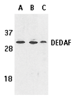
Left: Western blot analysis of DEDAF expression in human A549 (lane A), HepG2 (lane B), and mouse 3T3 (lane C) cell lysates with DEDAF antibody at 1 µg /ml.

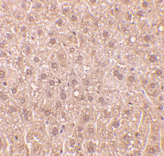
Below: Immunohistochemistry of DEDAF in mouse liver tissue with DEDAF antibody at 10 µg/ml.

Other Product Images

Source : DEDAF antibody was raised against a synthetic peptide corresponding to amino acids 215 to 228 of human DEDAF. The sequence is identical to that of mouse origin.

Purification : Affinity chromatography purified via peptide column

Clonality and Clone : This is a polyclonal antibody.

Host : DEDAF antibody was raised in rabbit. Please use anti-rabbit secondary antibodies.

Immunogen : Human DEDAF Peptide (Cat. No. 2227P)

Application : DEDAF can be used for detection of DEDAF by Western blot at 0.5 to 1 µg/ml.A band at approximately 32 kDa can be detected.

Tested Application(s) : E, WB, IHC

Buffer : Antibody is supplied in PBS containing 0.02% sodium azide.

Blocking Peptide : Cat. No. 2227P - DEDAF Peptide

Long-Term Storage : DEDAF antibody can be stored at 4ºC, stable for one year. As with all antibodies care should be taken to avoid repeated freeze thaw cycles. Antibodies should not be exposed to prolonged high temperatures.

Positive Control
1.Cat. No. 1203 - A549 Cell Lysate
2.Cat. No. 1211 - HepG2 Whole Cell Lysate
3.Cat. No. 1404 - Mouse Liver Tissue Lysate

Species Reactivity :H, M, R

GI Number : 5802963

Accession Number : AF179286

Short Description : A novel regulator of apoptosis

References

1.Zheng L, Schickling O, Peter ME, Lenardo MJ. The death effector domain-associated factor (DEDAF) plays distinct regulatory roles in the nucleus and cytoplasm. J Biol Chem. 2001;276(34):31945-52.
2.Garcia E, Marcos-Gutierrez C, del Mar Lorente M, Moreno JC, Vidal M. RYBP, a new repressor protein that interacts with components of the mammalian Polycomb complex, and with the transcription factor YY1. EMBO J. 1999;18(12):3404-18. (RD0201)

Monday, June 28, 2010

DFF40 Antibody

DFF40 Antibody

Catalog# : 2155

Apoptosis is related to many diseases and induced by a family of cell death receptors and their ligands. Cell death signals are transduced by death domain containing adapter molecules and members of the caspase family of proteases. These death signals finally cause the degradation of chromosomal DNA by activated DNase. A mouse DNase that causes DNA fragmentation was identified recently and designated CAD for caspase activated deoxyribonuclease (1,2). The human homologue of mouse CAD was more recently identified by three groups independently and termed CPAN, DFF40, and human CAD, respectively, (3-5). DFF45/ICAD is the inhibitory protein of DFF40/CAD (1,2,6) and forms complex with DFF40/CAD. Upon cleavage of DFF45/ICAD by activated caspase, DFF40/CAD is released and activated and eventually causes the degradation of DNA in the nuclei. Activation of DFF40/CAD, which causes DNA degradation, is the hallmark of apoptotic cell death.

Additional Names : DFF40 (IN), CAD

Description
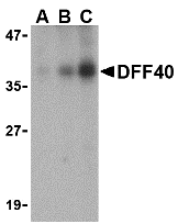
Left: Western blot analysis of DFF40 in Jurkat cell lysate with DFF40 antibody at (A) 0.5, (B) 1 and (C) 2 µg/ml.

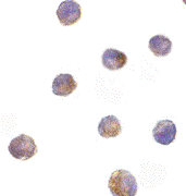
Below: Immunocytochemistry of DFF40 in K562 cells with DFF40 antibody at 10 µg/ml.

Other Product Images
Source : DFF40 antibody was raised against a peptide corresponding to amino acids 203 to 218 of human DFF40 .

Purification : Affinity chromatography purified via peptide column

Clonality and Clone : This is a polyclonal antibody.

Host : DFF40 antibody was raised in rabbit. Please use anti-rabbit secondary antibodies.

Immunogen : Human DFF40 (Intermediate Domain) Peptide (Cat. No. 2155P)

Application : DFF40 antibody can be used for detection of DFF40 /CAD by Western blot at 1:500 dilution. A 40 kDa band can be detected. It is human, mouse, and rat reactive,

Tested Application(s) : E, WB, ICC

Buffer : Antibody is supplied in PBS containing 0.02% sodium azide.

Blocking Peptide : Cat. No. 2155P - DFF40 Peptide

Long-Term Storage : DFF40 antibody can be stored at 4ºC, stable for one year. As with all antibodies care should be taken to avoid repeated freeze thaw cycles. Antibodies should not be exposed to prolonged high temperatures.

Positive Control
1.Cat. No. 1204 - K562 Cell Lysate
2.Cat. No. 1205 Jurkat Whole Cell Lysate

Species Reactivity :H, M, R

GI Number : 4758150

Accession Number : NP_004393

Short Description : (IN) Caspase Activated DNase

References

1.Enari M, Sakahira H, Yokoyama H, Okawa K, Iwamatsu A, Nagata S. A caspase-activated DNase that degrades DNA during apoptosis, and its inhibitor ICAD. Nature 1998;391:43-50
2.Sakahira H, Enari M, Nagata S. Cleavage of CAD inhibitor in CAD activation and DNA degradation during apoptosis. Nature 1998;391:96-99
3.Halenbeck R, MacDonald H, Roulston A, Chen TT, Conroy L, Williams LT. CPAN, a human nuclease regulated by the caspase-sensitive inhibitor DFF45. Curr Biol 1998;8:537-40
4.Liu X, Li P, Widlak P, Zou H, Luo X, Garrard WT, Wang X The 40-kDa subunit of DNA fragmentation factor induces DNA fragmentation and chromatin condensation during apoptosis. Proc Natl Acad Sci USA 1998;95:8461-6

Sunday, June 27, 2010

DEDD2 Antibody

DEDD2 Antibody

Catalog# : 3071

Apoptotic signals are often triggered by cell surface death receptors through protein-protein interactions mediated by conserved domains such as the death effector domain (1). A novel death effector domain (DED)-containing protein, DEDD2, has been recently identified and its over-expression in transfected cells induces moderate apoptosis and results in substantial sensitization to apoptosis induced by Fas, TRAIL, and FADD (2). More recently, work has shown that DEDD2 can bind caspase-8 and -10 in addition to FLIP but not FADD (2,3). Like the related protein DEDD, DEDD2 translocates from the cytosol to the nucleus upon induction of apoptosis, and it has been suggested that DEDD2 may target caspase-8 to the nucleus and that DEDD2 thus plays a critical role in death receptor-induced apoptosis (3). At least two alternatively spliced transcript variants encoding distinct isoforms have been found for DEDD2.

Additional Names : DEDD2, DNA-binding death effector domain-containing protein 2, FLAME-3

Description
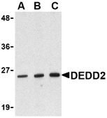
Left: Western blot analysis of DEDD2 in RAW264.7 cell lysate with DEDD2 antibody at (A) 0.5, (B) 1 and (C) 2 μg/ml.


Source : DEDD2 antibody was raised against a peptide corresponding to 11 amino acids near the amino-terminus of human DEDD2.

Purification : Immunoaffinity chromatography purified IgG

Clonality and Clone : This is a polyclonal antibody.

Host : DEDD2 antibody was raised in rabbit. Please use anti-rabbit secondary antibodies.

Immunogen : Human DEDD2 Peptide (Cat. No. 3071P)

Application : DEDD2 antibody can be used for detection of DEDD2 by Western blot at 0.5 to 1 µg/ml.

Tested Application(s) : E, WB

Buffer : Antibody is supplied in PBS containing 0.02% sodium azide.

Blocking Peptide : Cat. No. 3071P - DEDD2 Peptide

Long-Term Storage : DEDD2 antibody can be stored at 4ºC, stable for one year. As with all antibodies care should be taken to avoid repeated freeze thaw cycles. Antibodies should not be exposed to prolonged high temperatures.

Positive Control
1.Cat. No. 1283 - RAW264.7 Cell Lysate

Species Reactivity :H, M, R

GI Number : 19923050

Accession Number : NP_579874

Short Description : A novel death effector domain containing protein

References
1.Tibbetts MD, Zheng L, and Lenardo MJ. The death effector domain protein family: regulators of cellular homeostasis. Nat. Immunol. 2003; 4:404-9.
2.Roth W, Stenner-Liewen F, Pawlowski K, et al. Identification and characterization of DEDD2, a death effector domain-containing protein. J. Biol. Chem. 2002; 277:7501-8.
3.Alcivar A, Hu S, Tang J, et al. DEDD and DEDD2 associate with caspase-8/10 and signal cell death. Oncogene 2003; 22:291-7.

DEDAF Antibody

DEDAF Antibody

Catalog# : 2227

Apoptosis is related to many diseases and induced by a family of cell death receptors and their ligands. Cell death signals are transduced by death domain (DD) death effector domain (DED), and caspase recruitment domain (CARD) containing molecules. Several molecules including caspases and adaptor FADD contain DEDs. A novel protein that interacts with DED of caspase-8 and 10, and FADD was identified recently and designated DEDAF for DED associated factor . DEDAF is identical to the transcriptional repressor RYBP (2). DEDAF/RYBP is expressed in multiple tissues and cell lines. DEDAF interacts with FADD and augments the formation of CD95/FADD/capase-8 complexes at the cell membrane, and interacts with DED-containing DNA biding protein (DEDD) in the nucleus indicating it is involved in the regulation of both cytoplasmic and nuclear events of apoptosis.

Additional Names : DEDAF
Description
Left: Western blot analysis of DEDAF expression in human A549 (lane A), HepG2 (lane B), and mouse 3T3 (lane C) cell lysates with DEDAF antibody at 1 µg /ml.

Below: Immunohistochemistry of DEDAF in mouse liver tissue with DEDAF antibody at 10 µg/ml.

Other Product Images

Source : DEDAF antibody was raised against a synthetic peptide corresponding to amino acids 215 to 228 of human DEDAF. The sequence is identical to that of mouse origin.

Purification : Affinity chromatography purified via peptide column

Clonality and Clone : This is a polyclonal antibody.

Host : DEDAF antibody was raised in rabbit. Please use anti-rabbit secondary antibodies.

Immunogen : Human DEDAF Peptide (Cat. No. 2227P)

Application : DEDAF can be used for detection of DEDAF by Western blot at 0.5 to 1 µg/ml.A band at approximately 32 kDa can be detected.

Tested Application(s) : E, WB, IHC

Buffer : Antibody is supplied in PBS containing 0.02% sodium azide.

Blocking Peptide : Cat. No. 2227P - DEDAF Peptide

Long-Term Storage : DEDAF antibody can be stored at 4ºC, stable for one year. As with all antibodies care should be taken to avoid repeated freeze thaw cycles. Antibodies should not be exposed to prolonged high temperatures.

Positive Control
1.Cat. No. 1203 - A549 Cell Lysate
2.Cat. No. 1211 - HepG2 Whole Cell Lysate
3.Cat. No. 1404 - Mouse Liver Tissue Lysate

Species Reactivity :H, M, R

GI Number : 5802963

Accession Number : AF179286

Short Description : A novel regulator of apoptosis

References
1.Zheng L, Schickling O, Peter ME, Lenardo MJ. The death effector domain-associated factor (DEDAF) plays distinct regulatory roles in the nucleus and cytoplasm. J Biol Chem. 2001;276(34):31945-52.
2.Garcia E, Marcos-Gutierrez C, del Mar Lorente M, Moreno JC, Vidal M. RYBP, a new repressor protein that interacts with components of the mammalian Polycomb complex, and with the transcription factor YY1. EMBO J. 1999;18(12):3404-18. (RD0201)

DcR3 Antibody

DcR3 Antibody

Catalog# : 2140

Apoptosis is induced by certain cytokines including TNF and Fas ligand in the TNF family through their death domain containing receptors. Several novel members in the TNFR family including DR3, DR4, DR5, and DR6 were recently discovered and function as cell death receptors. Two decoy receptors, DcR1 and DcR2, were recently identified to compete with DR4 and DR5 for their ligand TRAIL binding. A novel decoy receptor was more recently discovered and designated DcR3 and TR6, respectively, (1,2). Unlike DcR1 and DcR2, DcR3 is a soluble rather than a membrane associated molecule. DcR3 binds to FasL and LIGHT and inhibits FasL and LIGHT induced apoptosis (1,2). DcR3 transcript is expressed in a number of lung and colon carcinomas and in some normal tissues.

Additional Names : DcR3, DCR3, TR6

Description
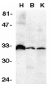
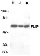
Left: Western blot analysis of DcR3 in human heart (H), brain (B), and kidney (K) tissue lysates with DcR3 antibody at 1:500 dilution.

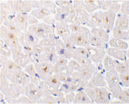
Below: Immunohistochemistry of DcR3 in human heart tissue with DcR3 antibody at 1 µg/ml.

Other Product Images
Source : DcR3 antibody was raised against a peptide corresponding to amino acids near the carboxy terminus of human DcR3 precursor.

Purification : Ion exchange chromatography purified

Clonality and Clone : This is a polyclonal antibody.

Host : DcR3 antibody was raised in rabbit. Please use anti-rabbit secondary antibodies.

Immunogen : Human DcR3 (N-Terminus) Peptide (Cat. No. 2140P)

Application : DcR3 antibody can be used for detection of DcR3 expression by Western blot at 1:500 to 1:1000 dilution. An approximately 33 kDa band can be detected.

Tested Application(s) : E, WB, IHC

Buffer : Antibody is supplied in PBS containing 0.02% sodium azide.

Blocking Peptide : Cat. No. 2140P - DcR3 Peptide

Long-Term Storage : DcR3 antibody can be stored at 4ºC, stable for one year. As with all antibodies care should be taken to avoid repeated freeze thaw cycles. Antibodies should not be exposed to prolonged high temperatures.

Positive Control
1.Cat. No. 1301 - Human Heart Tissue Lysate

Species Reactivity :H, M, R

GI Number : 4106877

Accession Number : AF104419

Short Description : Decoy Receptor for FASL and LIGHT

References
1.Pitti RM, Marsters SA, Lawrence DA, Roy M, Kischkel FC, Dowd P, Huang A, Donahue CJ, Sherwood SW, Baldwin DT, Godowski PJ, Wood WI, Gurney AL, Hillan KJ, Cohen RL, Goddard AD, Botstein D, Ashkenazi A. Genomic amplification of a decoy receptor for Fas ligand in lung and colon cancer. Nature 1998;396:699-703
2.Yu KY, Kwon B, Ni J, Zhai Y, Ebner R, Kwon BS. A newly identified member of tumor necrosis factor receptor superfamily (TR6) suppresses LIGHT-mediated apoptosis. J Biol Chem 1999;274:13733-6 (RD1299)

Sunday, June 13, 2010

Daxx Antibody

Daxx Antibody

Catalog# : 1163

Apoptosis, or programmed cell death, occurs during normal cellular differentiation and development of multicellular organisms. Apoptosis is induced by certain cytokines including TNF and Fas ligand of the TNF family through their death domain containing receptors, TNFR1 and Fas. Cell death signals are transduced by death domain (DD)- containing adapter molecules and members of the ICE/CED-3 protease family. A novel DD-containing molecule was recently cloned from mouse, human and monkey and designated Daxx. Daxx binds specifically to the Fas death domain and enhances Fas induced apoptosis and activates the Jun N-terminal kinase (JNK) pathway. Daxx is widely expressed in fetal and adult human and mouse tissues indicating its important function in Fas signaling pathways.

Additional Names : Daxx (CT), Daxx

Description
Left: Western blot analysis of Daxx in HeLa total cell lysate with Daxx antibody at 1 mg/ml.

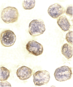
Below: Immunocytochemistry of DAXX in HeLa cells with DAXX antibody at 10 µg/ml.

Other Product Imagesc


Source : Daxx antibody was raised against a peptide corresponding to amino acids near the carboxy-terminus of human Daxx.

Purification : Antibody is DEAE purified

Clonality and Clone : This is a polyclonal antibody.

Host : Daxx antibody was raised in rabbit. Please use anti-rabbit secondary antibodies.


Immunogen : Human Daxx (C-Terminus) Peptide (Cat. No. 1163P)
Application : Daxx antibody can be used for detection of Daxx by Western blot at 1 – 2 µg/ml.A 120 kDa major band can be detected.

Tested Application(s) : E, WB, ICC

Buffer : Antibody is supplied in PBS containing 0.02% sodium azide.

Blocking Peptide : Cat. No. 1163P - Daxx Peptide

Long-Term Storage : Daxx antibody can be stored at 4ºC, stable for one year. As with all antibodies care should be taken to avoid repeated freeze thaw cycles. Antibodies should not be exposed to prolonged high temperatures.

Positive Control
1.Cat. No. 1201 - HeLa Cell Lysate

Species Reactivity :H, M

GI Number : 48146287

Accession Number : CAG33366

Short Description : (CT) Adapter Molecule

References
1.Yang X, Khosravi-Far R, Chang HY, Baltimore D. Daxx, a novel Fas-binding protein that activates JNK and apoptosis. Cell 1997;89:1067-1076
2.Kiriakidou M, Driscoll DA, Lopez-Guisa JM, Strauss JF 3rd. Cloning and expression of primate Daxx cDNAs and mapping of the human gene to chromosome 6p21.3 in the MHC region. DNA Cell Biol 1997;16:1289-1298 (RD1299)

DAPK2 Antibody

DAPK2 Antibody

Catalog# : 2323

Apoptosis is mediated by death domain containing adapter molecules and a caspase family of proteases. Certain serine/threonine protein kinases, such as RIP and DAP kinase, are mediators of apoptosis. DAP kinase (DAPK) is pro-apoptotic calcium-regulated serine/threonine kinase containing death domain. Ectopic expression of DAPK induces cell death and suppresses oncogenic transformation. DAPK mediates IFNg induced apoptosis. A novel DAP kinase-related protein was recently identified and designated DAPK2 and DRP-1 (1, 2). Ectopicly expressed DAPK2 induced apoptosis in various types of cells (1,2). DAPK has high sequence homology to ZIP kinase and DRAK1/2, and they represent a novel family of serine/threonine kinases, which mediates apoptosis through their catalytic activities. The messenger RNA of DAPK2 is expressed in multiple human tissues (1).

Additional Names : DAPK2

Description
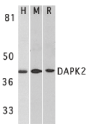
Left: Western blot analysis of DAPK2 in A431 (H), mouse spleen (M), and rat kidney (R) lysates with DAPK2 antibody at 1 µg/ml.

Below: Immunohistochemistry of DAPK2 in mouse spleen cells with DAPK2 antibody at 2 µg/ml.

Other Product Images


Source : DAPK2 antibody was raised against a peptide corresponding to amino acids near the carboxy terminus of human DAPK2. The sequence of this antigenic peptide is identical to the corresponding amino acids of mouse origin (1,2).

Purification : Affinity chromatography purified via peptide column

Clonality and Clone : This is a polyclonal antibody.

Host : DAPK2 antibody was raised in rabbit. Please use anti-rabbit secondary antibodies.

Immunogen : Human DAP kinase 2 Peptide (Cat. No. 2323P)

Application : DAPK2 antibody can be used for detection of DAPK2 by Western blot at 0.5 to 1 µg/ml.An approximately 42 kDa band can be detected. DAPK2 has no cross responses to DAPK1.

Tested Application(s) : E, WB, IHC

Buffer : Antibody is supplied in PBS containing 0.02% sodium azide.

Blocking Peptide : Cat. No. 2323P - DAPK2 Peptide

Long-Term Storage : DAPK2 antibody can be stored at 4ºC, stable for one year. As with all antibodies care should be taken to avoid repeated freeze thaw cycles. Antibodies should not be exposed to prolonged high temperatures.

Positive Control
1.Cat. No. 1202 - A431 Cell Lysate
2.Cat. No. 1406 - Mouse Spleen Tissue Lysate
3.Cat. No. 1465 - Rat Kidney Lysate

Species Reactivity :H, M, R

GI Number : 6521210

Accession Number : BAA88063

Short Description : Death-Associated Protein kinase 2

References
1.Kawai T, Nomura F, Hoshino K, Copeland NG, Gilbert DJ, Jenkins NA, Akira S. Death-associated protein kinase 2 is a new calcium/calmodulin-dependent protein kinase that signals apoptosis through its catalytic activity. Oncogene 1999;18(23):3471-80
2.Inbal B, Shani G, Cohen O, Kissil JL, Kimchi A. Death-associated protein kinase-related protein 1, a novel serine/threonine kinase involved in apoptosis. Mol Cell Biol 2000;20(3):1044-54 (WD0101)

Friday, June 11, 2010

CIDE-B Antibody

CIDE-B Antibody

Catalog# : 2321

Apoptosis is related to many diseases and induced by a family of cell death receptors and their ligands. Cell death signals are transduced by death domain containing adapter molecules and members of the caspase family of proteases. These death signals finally cause the degradation of chromosomal DNA by activated DNase. DFF45/ICAD has been identified as inhibitor of caspase activated DNase DFF40/CAD. DFF45 related proteins CIDE-A and CIDE-B (for cell death-inducing DFF-like effector A and B) were recently identified. CIDE contains a new type of domain termed CIDE-N, which has high homology with the regulatory domains of DFF45/ICAD and DFF40/CAD. Expression of CIDE-B induces apoptosis, which is inhibited by DFF45. CIDE-B is a DFF45-inhibitable effector that promotes cell death and DNA fragmentation. CIDE-B is expressed mainly in liver and at lower levels in spleen, kidney, peripheral blood lymphocytes and bone marrow. CIDE-B antibody has no cross activity to CIDE-A.

Additional Names : CIDE-B (IN), Cell death-inducing DFF-like effector B

Description
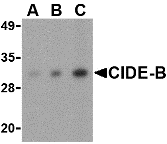
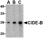
Left: Western blot analysis of CIDE-B in mouse small intestine tissue lysate with CIDE-B antibody at (A) 0.5, (B) 1 and (C) 2 µg/ml.

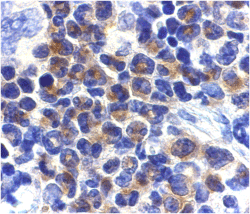
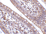
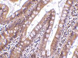
Below: Immunohistochemistry of CIDE-B in human small intestine tissue with CIDE-B antibody at 5 µg/ml.

Other Product Images
Source : CIDE-B antibody was raised against a peptide corresponding to 14 amino acids near the center of human CIDE-B.

Purification : Affinity chromatography purified via peptide column

Clonality and Clone : This is a polyclonal antibody.

Host : CIDE-B antibody was raised in rabbit. Please use anti-rabbit secondary antibodies.

Application : CIDE-B antibody can be used for detection of CIDE-B by Western blot at 0.5 – 1 µg/ml.

Tested Application(s) : E, WB, IHC

Buffer : Antibody is supplied in PBS containing 0.02% sodium azide.

Blocking Peptide : Cat.No. 2321P - CIDE-B Peptide

Long-Term Storage : CIDE-B antibody can be stored at 4ºC, stable for one year. As with all antibodies care should be taken to avoid repeated freeze thaw cycles. Antibodies should not be exposed to prolonged high temperatures.

Positive Control
1.Cat. No. 1408 - Mouse Small Intestine Tissue Lysate
2.Cat. No. 1308 - Human Small Intestine Tissue Lysate

Species Reactivity :H, M, R

GI Number : 6746627

Accession Number : AAF27658

Short Description : (IN) Cell death-inducing DFF-like effector B

References
1.Inohara N, Koseki T, Chen S,et al. CIDE, a novel family of cell death activators with homology to the 45 kDa subunit of the DNA fragmentation factor. EMBO J. 1998; 17:2526-33.
2.Inohara N, Koseki T, Chen S, et al. Identification of regulatory and catalytic domains in the apoptosis nuclease DFF40/CAD. J. Biol. Chem. 1999; 274:270-4.

Thursday, June 10, 2010

CIDE-B Antibody

CIDE-B Antibody

Catalog# : 2319

Apoptosis is related to many diseases and induced by a family of cell death receptors and their ligands. Cell death signals are transduced by death domain containing adapter molecules and members of the caspase family of proteases. These death signals finally cause the degradation of chromosomal DNA by activated DNase. DFF45/ICAD has been identified as inhibitor of caspase activated DNase DFF40/CAD. DFF45 related proteins CIDE-A and CIDE-B (for cell death-inducing DFF-like effector A and B) were recently identified. CIDE contains a new type of domain termed CIDE-N, which has high homology with the regulatory domains of DFF45/ICAD and DFF40/CAD (1,2). Expression of CIDE-B induces apoptosis, which is inhibited by DFF45. CIDE-B is a DFF45-inhibitable effector that promotes cell death and DNA fragmentation. CIDE-B is expressed mainly in liver and at lower levels in spleen, kidney, peripheral blood lymphocytes and bone marrow. CIDE-B antibody has no cross activity to CIDE-A.

Additional Names : CIDE-B (C2), Cell death-inducing DFF-like effector B

Description
Left: Western blot analysis of CIDE-B in mouse small intestine tissue lysate with CIDE-B antibody at (A) 0.5, (B) 1 and (C) 2 µg/ml.

Below: Immunohistochemistry of CIDE-B in human small intestine tissue with CIDE-B antibody at 5 µg/ml.

Other Product Images
Source : CIDE-B antibody was raised against a peptide corresponding to 15 amino acids near the carboxy terminus of human CIDE-B.

Purification : Affinity chromatography purified via peptide column

Clonality and Clone : This is a polyclonal antibody.

Host : CIDE-B antibody was raised in rabbit. Please use anti-rabbit secondary antibodies.

Application : CIDE-B antibody can be used for detection of CIDE-B by Western blot at 0.5 – 1 µg/ml.

Tested Application(s) : E, WB, IHC

Buffer : Antibody is supplied in PBS containing 0.02% sodium azide.

Blocking Peptide : Cat.No. 2319P - CIDE-B Peptide

Long-Term Storage : CIDE-B antibody can be stored at 4ºC, stable for one year. As with all antibodies care should be taken to avoid repeated freeze thaw cycles. Antibodies should not be exposed to prolonged high temperatures.

Positive Control
1.Cat. No. 1408 - Mouse Small Intestine Tissue Lysate
2.Cat. No. 1308 - Human Small Intestine Tissue Lysate

Species Reactivity :H, M, R

GI Number : 6746627

Accession Number : AAF27658

Short Description : (C2) Cell death-inducing DFF-like effector B

References
1.Inohara N, Koseki T, Chen S,et al. CIDE, a novel family of cell death activators with homology to the 45 kDa subunit of the DNA fragmentation factor. EMBO J. 1998; 17:2526-33
2.Inohara N, Koseki T, Chen S, et al. Identification of regulatory and catalytic domains in the apoptosis nuclease DFF40/CAD. J. Biol. Chem. 1999; 274:270-4.

CIDE-A Antibody

CIDE-A Antibody

Catalog# : 2089

Apoptosis is related to many diseases and induced by a family of cell death receptors and their ligands. Cell death signals are transduced by death domain containing adapter molecules and members of the caspase family of proteases. These death signals finally cause the degradation of chromosomal DNA by activated DNase. DFF45/ICAD has been identified as inhibitor of caspase activated DNase DFF40/CAD. DFF45 related proteins CIDE-A and CIDE-B (for cell death-inducing DFF-like effector A and B) were recently identified (1). CIDE contains a new type of domain termed CIDE-N, which has high homology with the regulatory domains of DFF45/ICAD and DFF40/CAD (1,2). Expression of CIDE-A induces DNA fragmentation and activates apoptosis, which is inhibited by DFF45. CIDE-A is a DFF45-inhibitable effector that promotes cell death and DNA fragmentation. CIDE-A is expressed in many tissues.

Additional Names : CIDE-A (CT), CIDE-A
Description
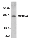
Left: Western blot analysis of CIDE-A in mouse heart tissue lysate with CIDE-A antibody at 1:500 dilution.

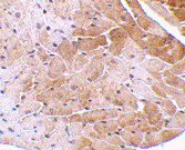
Below: Immunohistochemistry of CIDE-A in mouse heart tissue with CIDE-A antibody at 5 µg/ml.

Other Product Images
Source : CIDE-A antibody was raised against a peptide
corresponding to amino acids 200 to 214 of mouse CIDE-A.

Purification : Affinity chromatography purified via peptide column

Clonality and Clone : This is a polyclonal antibody.

Host : CIDE-A antibody was raised in rabbit. Please use anti-rabbit secondary antibodies.

Immunogen : Mouse mCIDE-A (C-Terminus) Peptide (Cat. No. 2089P)

Application : CIDE-A antibody can be used for detection of CIDE-A by Western blot at 1:500 to 1:1000 dilution. An approximately 25 kDa band can be detected. It has no cross activity to CIDE-B.

Tested Application(s) : E, WB, IHC

Buffer : Antibody is supplied in PBS containing 0.02% sodium azide.

Blocking Peptide : Cat. No. 2089P - CIDE-A Peptide

Long-Term Storage : CIDE-A antibody can be stored at 4ºC, stable for one year. As with all antibodies care should be taken to avoid repeated freeze thaw cycles. Antibodies should not be exposed to prolonged high temperatures.

Positive Control
1.Cat. No. 1401 - Mouse Heart Tissue Lysate

Species Reactivity :M

GI Number : 3114592

Accession Number : AAC34985

Short Description : (CT) Cell death-Inducing DFF-like Effector

References
1.Inohara N, Koseki T, Chen S, Wu X, Nunez G. CIDE, a novel family of cell death activators with homology to the 45 kDa subunit of the DNA fragmentation factor. EMBO J 1998;17:2526-33
2.Inohara N, Koseki T, Chen S, Benedict MA, Nunez G. Identification of regulatory and catalytic domains in the apoptosis nuclease DFF40/CAD. J Biol Chem 1999 ;274:270-4 (RD1299)

Wednesday, April 14, 2010

2X PCR Master Mix

2X PCR Master Mix

Catalog# : NB-28009

Price/Unit : $339.00

Unit : 500 reactions

2x PCR Master Mix
  • Convenient ready-to-use solution
  • High sensitivity and yield
  • Robust amplification
  • Available in 3 convenient sizes: 100, 200 and 500 reactions (20 µL vol)
Features and Benefits | Specifications

BSI's 2X PCR Master Mix is a ready-to-use solution that contains components required for PCR amplification including Taq DNA polymerase, dNTPs, reaction buffer, MgCl2, KCl and a PCR enhancer/stabilizer. The user needs only to add template, the primer set and water to the master mix in order to set up the PCR reaction. This convenient 2X PCR Master Mix reduces the time required to set up PCR reactions and reduces the possibility of contamination, particularly when preparing large numbers of reactions. The optimized master mix allows for robust amplification of DNA templates with high yields of PCR products.

Taq DNA Polymerase is a highly thermostable DNA polymerase that possesses a 5´→ 3´ polymerase activity and a very low 5´→ 3´ exonuclease activity. The source of Taq included with BSI’s 2X PCR Master Mix is an E. coli strain with a cloned Taq DNA Polymerase gene from Thermus aquaticus YT-1.

BSI’s 2X PCR Master Mix is available in 3 convenient sizes:

Cat # NB-28007 - 100 reactions (sufficient for 100 reactions x 20 µL reaction volume)
Cat # NB-28008 - 200 reactions (sufficient for 200 reactions x 20 µL reaction volume)
Cat # NB-28009 – 500 reactions (sufficient for 500 reactions x 20 µL reaction volume)

FEATURES AND BENEFITS
  • Convenient - With the ready-to-use Master Mix, the user needs only to add template, the primer set and water to the master mix in order to set up the PCR reaction.
  • Time Savings - Set up PCR reactions in a shorter time, as less pipetting steps are required.
  • Cost Efficient - No need to buy separate enzymes, dNTPs and buffers. All are included with the ready-to-use Master Mix.
  • High Sensitivity and Yield – the optimized Master Mix allows for highly sensitive amplifications with high yields of PCR products.
  • Robust Amplification - BSI’s 2X PCR Master Mix is capable of amplifying difficult templates with a high degree of reproducibility.
APPLICATIONS AND DATA
  • Routine PCR amplification of DNA templates
  • PCR for post reverse transcription step
  • Multiple band detection or genotyping
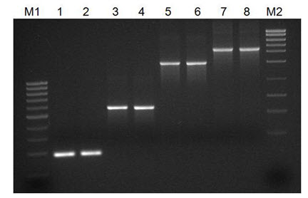
Figure 1. Successful Amplification of Different Sized Amplicons. BSI’s 2X PCR Master Mix was used in a series of PCR reactions to amplify a number of different sized amplicons from the adenovirus E2 region. Lanes 1 and 2 correspond to a 189 bp product, Lanes 3 and 4 correspond to a 565 bp product, Lanes 5 and 6 correspond to a 1501 bp product, and Lanes 7 and 8 correspond to a 2202 bp product. BSI’s 2X PCR Master Mix was able to amplify all the different sized products with a high sensitivity and yield. Lane M1 is BSI’s PCR Sizer 100 bp DNA Ladder (Cat #NB-11400) and Lane M2 is BSI’s MidRanger 1kb DNA Ladder (Cat # NB-11700).

SPECIFICATIONS

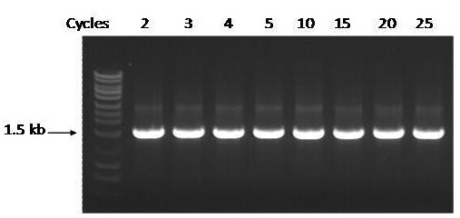
Figure 1. Highly Stable after Multiple Freeze-Thaw Cycles The stability of BSI’s 2X Master Mix was tested by subjected the mix to multiple freezing and thawing cycles, followed by using the Master Mix to amplify the 16s rDNA (1.5 kb) of Staphylococcus aureus gDNA. BSI’s 2X Master Mix showed robustness and outstanding stability from multiple freezing and thawing steps at -20ºC. M: FastRunner DNA Ladder (Cat#NB-12800). Ten μL of each PCR reaction was loaded on a 1% TAE agarose gel.

Reagents Supplied - Cat # 28007

2X PCR Master Mix (1 Vial, 100 Reactions) Sufficient reagent for 100 x 20 µL reactions

Reagents Supplied – Cat # 28008

2X PCR Master Mix (2 Vials, 100 Reactions each) Sufficient reagent for 200 x 20 µL reactions

Reagents Supplied – Cat # 28009

2X PCR Master Mix (5 Vials, 100 Reactions each) Sufficient reagent for 500 x 20 µL reactions

Storage Conditions and Product Stability
BSI’s 2X Master Mix should be stored at -20ºC. For everyday use an aliquot can be stored at 4ºC for up to three months. The Master Mix is stable for multiple freeze-thaw cycles (see Figure 2). When stored at the proper temperature this reagent is stable for at least 1 year.

Long-Term Storage : BSI’s 2X Master Mix should be stored at -20ºC. For everyday use an aliquot can be stored at 4ºC for up to three months. The Master Mix is stable for multiple freeze-thaw cycles (see Figure 2). When stored at the proper temperature this reagent is stable for at least 1 year.

Product Usage
For laboratory research use only, not for diagnostic use! Not for use in humans.

For more detail regarding Peptide please visit here : Custom Peptide Synthesis

2X PCR Master Mix

2X PCR Master Mix

Catalog# : NB-28007

Price/Unit : $117.80

Unit : 100 reactions

2x PCR Master Mix
  • Convenient ready-to-use solution
  • High sensitivity and yield
  • Robust amplification
  • Available in 3 convenient sizes: 100, 200 and 500 reactions (20 µL vol)
Features and Benefits | Specifications

BSI's 2X PCR Master Mix is a ready-to-use solution that contains components required for PCR amplification including Taq DNA polymerase, dNTPs, reaction buffer, MgCl2, KCl and a PCR enhancer/stabilizer. The user needs only to add template, the primer set and water to the master mix in order to set up the PCR reaction. This convenient 2X PCR Master Mix reduces the time required to set up PCR reactions and reduces the possibility of contamination, particularly when preparing large numbers of reactions. The optimized master mix allows for robust amplification of DNA templates with high yields of PCR products.

Taq DNA Polymerase is a highly thermostable DNA polymerase that possesses a 5´→ 3´ polymerase activity and a very low 5´→ 3´ exonuclease activity. The source of Taq included with BSI’s 2X PCR Master Mix is an E. coli strain with a cloned Taq DNA Polymerase gene from Thermus aquaticus YT-1.

BSI’s 2X PCR Master Mix is available in 3 convenient sizes:

Cat # NB-28007 - 100 reactions (sufficient for 100 reactions x 20 µL reaction volume)
Cat # NB-28008 - 200 reactions (sufficient for 200 reactions x 20 µL reaction volume)
Cat # NB-28009 – 500 reactions (sufficient for 500 reactions x 20 µL reaction volume)

FEATURES AND BENEFITS
  • Convenient - With the ready-to-use Master Mix, the user needs only to add template, the primer set and water to the master mix in order to set up the PCR reaction.
  • Time Savings - Set up PCR reactions in a shorter time, as less pipetting steps are required.
  • Cost Efficient - No need to buy separate enzymes, dNTPs and buffers. All are included with the ready-to-use Master Mix.
  • High Sensitivity and Yield – the optimized Master Mix allows for highly sensitive amplifications with high yields of PCR products.
  • Robust Amplification - BSI’s 2X PCR Master Mix is capable of amplifying difficult templates with a high degree of reproducibility.
APPLICATIONS AND DATA
  • Routine PCR amplification of DNA templates
  • PCR for post reverse transcription step
  • Multiple band detection or genotyping
Figure 1. Successful Amplification of Different Sized Amplicons. BSI’s 2X PCR Master Mix was used in a series of PCR reactions to amplify a number of different sized amplicons from the adenovirus E2 region. Lanes 1 and 2 correspond to a 189 bp product, Lanes 3 and 4 correspond to a 565 bp product, Lanes 5 and 6 correspond to a 1501 bp product, and Lanes 7 and 8 correspond to a 2202 bp product. BSI’s 2X PCR Master Mix was able to amplify all the different sized products with a high sensitivity and yield. Lane M1 is BSI’s PCR Sizer 100 bp DNA Ladder (Cat #NB-11400) and Lane M2 is BSI’s MidRanger 1kb DNA Ladder (Cat # NB-11700).

SPECIFICATIONS
Figure 1. Highly Stable after Multiple Freeze-Thaw Cycles The stability of BSI’s 2X Master Mix was tested by subjected the mix to multiple freezing and thawing cycles, followed by using the Master Mix to amplify the 16s rDNA (1.5 kb) of Staphylococcus aureus gDNA. BSI’s 2X Master Mix showed robustness and outstanding stability from multiple freezing and thawing steps at -20ºC. M: FastRunner DNA Ladder (Cat#NB-12800). Ten μL of each PCR reaction was loaded on a 1% TAE agarose gel.

Reagents Supplied - Cat # 28007

2X PCR Master Mix (1 Vial, 100 Reactions) Sufficient reagent for 100 x 20 µL reactions

Reagents Supplied – Cat # 28008

2X PCR Master Mix (2 Vials, 100 Reactions each) Sufficient reagent for 200 x 20 µL reactions

Reagents Supplied – Cat # 28009

2X PCR Master Mix (5 Vials, 100 Reactions each) Sufficient reagent for 500 x 20 µL reactions

Storage Conditions and Product Stability
BSI’s 2X Master Mix should be stored at -20ºC. For everyday use an aliquot can be stored at 4ºC for up to three months. The Master Mix is stable for multiple freeze-thaw cycles (see Figure 2). When stored at the proper temperature this reagent is stable for at least 1 year.

Long-Term Storage : BSI’s 2X Master Mix should be stored at -20ºC. For everyday use an aliquot can be stored at 4ºC for up to three months. The Master Mix is stable for multiple freeze-thaw cycles (see Figure 2). When stored at the proper temperature this reagent is stable for at least 1 year.

Product Usage
For laboratory research use only, not for diagnostic use! Not for use in humans.

For more detail regarding Peptide please visit here : Custom Peptide Synthesis

2X One-Step RT-PCR Master Mix

2X One-Step RT-PCR Master Mix

Catalog# : NB-28113

Price/Unit : $360.00

Unit : 100 reactions

2x One-Step RT-PCR Master Mix
  • Convenient ready-to-use solution
  • High sensitivity and yield
  • Robust amplification
  • Available in 3 convenient sizes: 100, 200 and 500 reactions (20 µL vol)

Features and Benefits | Specifications

BSI’s 2X One-Step RT-PCR Master Mix is a ready-to-use solution that contains components required for for RT-PCR amplification of RNA templates. The mix includes M-MuLV reverse transcriptase, Taq DNA polymerase, dNTPs, reaction buffer, MgCl2, KCl, and a PCR enhancer/stabilizer. The user needs only to add template, the primer set and water to the Master Mix to set up the RT-PCR reaction. This convenient 2X One-Step RT-PCR Master Mix reduces the time required to set up PCR reactions and reduces the possibility of contamination, particularly when preparing large numbers of reactions. The optimized master mix allows for robust amplification of RNA temples with high yields of PCR products.

Taq DNA Polymerase is a highly thermostable DNA polymerase that possesses a 5´→ 3´ polymerase activity and a very low 5´→ 3´ exonuclease activity. The source of Taq included with BSI’s 2X One-Step RT-PCR Master Mix is an E. coli strain with a cloned Taq DNA Polymerase gene from Thermus aquaticus YT-1. M-MuLV Reverse Transcriptase is an RNA-directed DNA polymerase that can synthesize a complementary DNA strand initiating from a primer using either RNA (cDNA synthesis) or single-stranded DNA as a template. The source of the Reverse Transcriptase included with BSI’s 2X One-Setp RT-PCR Master Mix is an E. coli strain with a cloned reverse transcriptase gene from M-MuLV.

BSI’s 2X One-Step RT-PCR Master Mix is available in 3 convenient sizes:

Cat # NB-28113 - 100 reactions (sufficient for 100 reactions x 20 µL reaction volume)
Cat # NB-28114 - 200 reactions (sufficient for 200 reactions x 20 µL reaction volume)
Cat # NB-28115 – 500 reactions (sufficient for 500 reactions x 20 µL reaction volume)

FEATURES AND BENEFITS
  • Convenient - With the ready-to-use Master Mix, the user needs only to add template, the primer set and water to the master mix in order to set up the RT-PCR reaction.
  • Time Savings - Set up RT-PCR reactions in a shorter time, as less pipetting steps are required.
  • Cost Efficient - No need to buy separate enzymes, dNTPs and buffers. All are included with the ready-to-use Master Mix.
  • High Sensitivity and yield – the optimized Master Mix allows for highly sensitive amplifications with high yields of PCR products.
  • Robust Amplification - BSI’s 2X One-Step RT-PCR Master Mix is capable of amplifying difficult templates with a high degree of reproducibility.
APPLICATIONS AND DATA

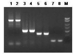
Figure 1. Successful Amplification of Different Sized Amplicons. BSI’s 2X One-Step RT-PCR Master Mix was used in RT-PCR reactions to amplify a number of different amplicons. Lanes 1 and 2 correspond to the E. coli 16s rRNA gene (large amplicon), which is 1.5 kb in size. Lanes 3 and 4 correspond to the E. coli 16s rRNA gene (small amplicon), which is 550 bp in size. Lanes 5 and 6 correspond to the human S15 gene, which is 360 bp in size. Lanes 7 and 8 correspond to the human 5S rRNA gene, which is 120 bp in size. BSI’s 2X One-Step RT-PCR Master Mix was able to amplify all the different sized products with a high sensitivity and yield.

SPECIFICATIONS
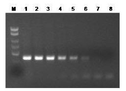
Figure 2. Robust Amplification from Different Amounts of RNA Input. BSI’s 2X One Step RT-PCR Master Mix was used to amplify the human S15 transcripts from a dilution series of HeLa total RNA isolated using BSI’s Total RNA Purification Kit. The 361 bp amplicon was robustly amplified from a broad range of RNA inputs, from the ng range down to fg range.

Reagents supplied – Cat # NB-28113

2X One-Step RT-PCR Master Mix (1 Vials, 100 Reactions) - Sufficient reagent for 100 x 20 µL reactions

Reagents supplied – Cat # NB-28114

2X One-Step RT-PCR Master Mix (2 Vials, 100 Reactions each) - Sufficient reagent for 200 x 20 µL reactions

Reagents supplied – Cat # NB-28115

2X One-Step RT-PCR Master Mix (5 Vials, 100 Reactions each) - Sufficient reagent for 500 x 20 µL reactions

Storage Conditions and Product Stability
2X One-Step RT-PCR Master Mix should be stored at -20°C. For everyday use an aliquot can be stored at 4°C for up to 3 months. Repeated freeze-thaw cycles are not recommended. When stored at the proper temperature this reagent is stable for at least 1 year.

Long-Term Storage : 2X One-Step RT-PCR Master Mix should be stored at -20°C. For everyday use an aliquot can be stored at 4°C for up to 3 months. Repeated freeze-thaw cycles are not recommended. When stored at the proper temperature this reagent is stable for at least 1 year.

Product Usage
For laboratory research use only, not for diagnostic use! Not for use in humans.

For more detail regarding Peptides please visit here : Custom Peptide Synthesis