CCR8 Antibody
Catalog# : 2097
CCR8 is one of the chemokine receptors that are required as coreceptors for HIV infection. The genes encoding human and murine CCR8 were cloned and designated TER1, CKR-L1, and ChemR1. The encoded seven transmembrane protein was identified as the receptor for human CC chemokine I-309 and renamed CCR8. Recently, CCR8 was found to serve as a coreceptor for diverse T-cell tropic, dual-tropic and macrophage-tropic HIV-1 strains. CCR8 mediates CC chemokine I-309 induced monocyte chemoattraction and HIV-1 envelope fusion and virus infection, which can be prevented by the CCR8 ligand I-309. CCR8 is expressed in spleen, thymus and T lymphoblastic cell lines.
Additional Names : CCR8 (EL), CCR8
 Description
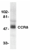
Left: Western blot analysis of CCR8 in human spleen lysate with CCR8 antibody at 1:500 dilution.
Source : CCR8 antibody was raised against a peptide corresponding to amino acids 183 to 201 of human CCR8, which locate in the second extracellular loop.
Purification : Affinity chromatography purified via peptide column
Clonality and Clone : This is a polyclonal antibody.
Host : CCR8 antibody was raised in rabbit. Please use anti-rabbit secondary antibodies.
Immunogen : Human CCR8 (Extracellular Loop) Peptide (Cat. No. 2097P)
Application : CCR8 antibody can be used for detection of CCR8 by Western blot at 1:500 to 1:1000 dilution. An approximately 50 kDa band can be detected.
Tested Application(s) : E, WB
Buffer : Antibody is supplied in PBS containing 0.02% sodium azide.
Blocking Peptide : Cat. No. 2097P - CCR8 Peptide
Long-Term Storage : CCR8 antibody can be stored at 4ºC, stable for one year. As with all antibodies care should be taken to avoid repeated freeze thaw cycles. Antibodies should not be exposed to prolonged high temperatures.
Positive Control
1. Cat. No. 1306 - Human Spleen Tissue Lysate
Species Reactivity :H
GI Number : 2231165
Accession Number : U45983
Short Description : (EL) HIV & chemokine receptor
References
1. Napolitano M, Zingoni A, Bernardini G, Spinetti G, Nista A, Storlazzi CT, Rocchi M, Santoni A. Molecular cloning of TER1, a chemokine receptor-like gene expressed by lymphoid tissues. J Immunol 1996;157:2759-63
2. Zaballos A, Varona R, Gutierrez J, Lind P, Marquez G. Molecular cloning and RNA expression of two new human chemokine receptor-like genes. Biochem Biophys Res Commun 1996;227:846-53
3. Samson M, Stordeur P, Labbe O, Soularue P, Vassart G, Parmentier M. Molecular cloning and chromosomal mapping of a novel human gene, ChemR1, expressed in T lymphocytes and polymorphonuclear cells and encoding a putative chemokine receptor. Eur J Immunol 1996;26:3021-8
4. Goya I, Gutierrez J, Varona R, Kremer L, Zaballos A, Marquez G. Identification of CCR8 as the specific receptor for the human beta-chemokine I-309: cloning and molecular characterization of murine CCR8 as the receptor for TCA-3. J Immunol 1998;160:1975-81
The availability of synthetic nucleic acids oligonucleotides and peptides, areas where BioSynthesis has extensive experience, has promoted the development of conjugates of these molecules cross-linked to compounds such as lipids, carbohydrates and small molecules (drugs) to yield products with distinct properties.
Lipid-oligonucleotide Conjugates
The main objective of cross-linking a lipid moiety to an oligonucleotide, either an oligodeoxynucleotide (ODN) or oligoribonucleotide (ORN), is to increase the hydrophobic character of the latter and its lipid-solubility. This way a conjugate would pass across the highly lipophilic cell membrane and into the cytosol, a process called transduction. Yet, depending on the lipid’s nature these conjugates may have also some other new biological properties.
Peptide-oligonucleotide Conjugates
Peptide-oligonucleotide conjugates have two moieties: a peptide that is covalently linked to an oligonucleotide: either an oligodeoxyribonucleotide (ODN) or an oligoribonucleotide (ORN). Alternatively, an oligonucleotide can be replaced with a bridged nucleic acid (BNA) that mimics the biological activity of that oligonucleotide. The oligonucleotide can interact with specific cell receptors to stimulate or inhibit biological responses, e.g. inhibition of a gene expression.
Carbohydrate-oligonucleotide Conjugates
These conjugates can have the carbohydrate moiety linked directly to the oligonucleotide or can be linked to a lipid or a peptide/protein. Due to the diversity of carbohydrate receptors present on the cell’s surface, oligosaccharides are useful for targeting conjugates to specific cells. Conjugation of antisense ODNs to a neoglycoprotein in which the protein has several mannose-6-phosphate residues, is readily internalized by cells having a receptor for this ligand on their surfaces.
CCR5 Antibody
Catalog# : 1112
Human immunodeficiency virus (HIV) and related virus require coreceptors, in addition to CD4, to infect target cells. Some G protein-coupled receptors including CCR5, CXCR4, CCR3, CCR2b and CCR8 in the chemokine receptor family, and four new human molecules GPR15, STRL33, GPR1 and V28 were recently identified as HIV coreceptors. Among them, CCR5 (CC-CKR-5) is a principal coreceptor for macrophage- and dual-tropic HIV-1 strains fusion and entry of human white blood cells. CCR5 is required for the infection by HIV-1, HIV-2, and SIV. The b-chemokines RANTES, MIP-1alpha and MIP-1beta are the ligands for CCR5 and prevent infection by M-tropic HIV-1. CXC5 associates with the surface CD4-gp120 of HIV complex and leads to membrane fusion and virus entry of target cells. The amino-terminal domain and the extracellular loops of CCR5 serve as HIV biding sites. CCR5 messenger RNA is expressed in lymphoid organs and monocytes.
Additional Names : CCR5 (NT), CCR5
 Description
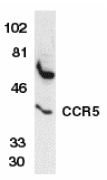
Left: Western blot analysis of CCR5 in THP-1 whole cell lysate with CCR5 antibody at 1:1000 dilution.
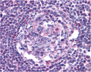
Below: Immunohistochemistry of CCR5 in human lymph node tissue with CCR5 antibody at 20 µg/ml.
Other Product Images
 Source : CCR5 antibody was raised against a peptide corresponding to amino acids 6 to 20 of human CCR5 .
Purification : Antibody is DEAE purified
Clonality and Clone : This is a polyclonal antibody.
Host : CCR5 antibody was raised in rabbit. Please use anti-rabbit secondary antibodies.
Immunogen : Human CCR5 (N-Terminus) Peptide (Cat. No. 1112P)
Application : CCR5 antibody can be used for Western blot at 1:1000 to 1:2000 dilution. An approximate 40 kDa band can be detected. CCR5 antibody can also detect CCR5 by immunohistochemistry at 20 µg/ml.
Tested Application(s) : E, WB, IHC
Buffer : Antibody is supplied in PBS containing 0.02% sodium azide.
Blocking Peptide : Cat. No. 1112P - CCR5 Peptide
Long-Term Storage : CCR5 antibody can be stored at 4ºC, stable for one year. As with all antibodies care should be taken to avoid repeated freeze thaw cycles. Antibodies should not be exposed to prolonged high temperatures.
Positive Control
1. Cat. No. 1208 - THP-1 Cell Lysate
2. Cat. No. 1369 - Human Lymph Node Tissue Lysate
Species Reactivity :H
GI Number : 2104520
Accession Number : AAB57793
Short Description : (NT) HIV & chemokine receptor
References
1. Dimitrov, DS. Cell 1997;91:721-30
2. Littman, DR. Cell 1998;93:677-80
3. Deng, H, et al. Nature 1996;381:661-6
4. Dragic, T, et al. Nature 1996;381:667-73
CCR3 Antibody
Catalog# : 1109
Human immunodeficiency virus (HIV) and related virus require coreceptors to infect target cells. Some G protein-coupled receptors including CCR5, CXCR4, CCR3, CCR2b, CCR8, GPR15, STRL33, and CX3CR1 in the chemokine receptor family were recently identified as HIV coreceptors. CCR5, CXCR4 and CCR3 are the principal receptors for HIV fusion and entry of target cells (1-4). CCR3 facilitates infection by a subset of virus. CCR3 and CCR5 promote efficient infection of microglia, the major target cells in the CNS (3,4). High levels of CCR3 and CXCR4 expression were found on the neurons from both the central and peripheral nervous systems (5). The CCR3 ligand, eotaxin, and an anti-CCR3 antibody inhibited HIV infection of microglia. These results indicate CCR3 plays an important role in HIV infection of CNS.
Additional Names : CCR3 (NT), CC chemokine receptor 3, eosinophil eotaxin receptor
 Description
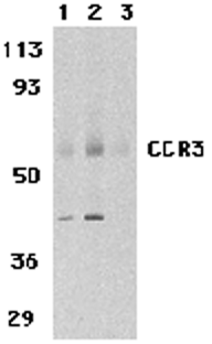
Left: Western blot analysis of CCR3 in human spleen tissue lysates with CCR3 antibody at 1 (lane 1) and 2 µg/ml (lane 2), and 2 µg/ml in the presence of blocking peptide (lane 3).
Source : CCR3 antibody was raised against a 14 amino acid peptide from near the amino terminus of human CCR3.
Purification : Immunoaffinity purified IgG
Clonality and Clone : This is a polyclonal antibody.
Host : CCR3 antibody was raised in rabbit. Please use anti-rabbit secondary antibodies.
Immunogen : Human CCR3 (N-Terminus) Peptide (Cat. No. 1109P)
Application : CCR3 antibody can be used for the detection of CCR3 by Western blot at 1 – 2 µg/ml.
Tested Application(s) : E, WB
Buffer : Antibody is supplied in PBS containing 0.02% sodium azide.
Blocking Peptide : Cat. No. 1109P - CCR3 Peptide
Long-Term Storage : CCR3 antibody can be stored at 4ºC, stable for one year. As with all antibodies care should be taken to avoid repeated freeze thaw cycles. Antibodies should not be exposed to prolonged high temperatures.
Positive Control
1. Cat. No. 1306 - Human Spleen Tissue Lysate
Species Reactivity :H, M, R
GI Number : 30581170
Accession Number : NP_847899
Short Description : (NT) HIV and chemokine receptor
References
1. Feng Y, Broder CC, Kennedy PE, et al. HIV-1 entry cofactor: functional cDNA cloning of a seven-transmembrane, G protein-coupled receptor. Science 1996; 272:872-7.
2. Deng H, Liu R, Ellmeier W, et al. Identification of a major co-receptor for primary isolates of HIV-1. Nature 1996; 381:661-6.
3. Choe H, Farzan M, Sun Y, et al. The beta-chemokine receptors CCR3 and CCR5 facilitate infection by primary HIV-1 isolates. Cell 1996; 85:1135-48.
4. He J, Chen Y, Farzan M, et al. CCR3 and CCR5 are co-receptors for HIV-1 infection of microglia. Nature 1997; 385:645-9.
CCR3 Antibody
Catalog# : 1105
Human immunodeficiency virus (HIV) and related virus require coreceptors to infect target cells. Some G protein-coupled receptors including CCR5, CXCR4, CCR3, CCR2b, CCR8, GPR15, STRL33, and CX3CR1 in the chemokine receptor family were recently identified as HIV coreceptors. CCR5, CXCR4 and CCR3 are the principal receptors for HIV fusion and entry of target cells. CCR3 facilitates infection by a subset of virus. CCR3 and CCR5 promote efficient infection of microglia, the major target cells in the CNS (3,4). High levels of CCR3 and CXCR4 expression were found on the neurons from both the central and peripheral nervous systems. The CCR3 ligand, eotaxin, and an anti-CCR3 antibody inhibited HIV infection of microglia. These results indicate CCR3 plays an important role in HIV infection of CNS.
Additional Names : CCR3 (CT), CC chemokine receptor 3, eosinophil eotaxin receptor
 Description
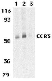
Left: Western blot analysis of CCR3 in human spleen tissue lysates with CCR3 antibody at 1 (lane 1) and 2 µg/ml (lane 2), and 2 µg/ml in the presence of blocking peptide (lane 3).
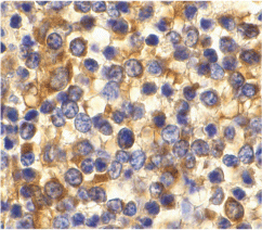
Below: Immunohistochemistry of CCR3 in human spleen tissue with CCR3 antibody at 10 µg/ml.
Other Product Images
 Source : CCR3 antibody was raised against a 14 amino acid peptide from near the carboxy terminus of human CCR3.
Purification : Immunoaffinity purified IgG
Clonality and Clone : This is a polyclonal antibody.
Host : CCR3 antibody was raised in rabbit. Please use anti-rabbit secondary antibodies.
Immunogen : Human CCR3 (C-Terminus) Peptide (Cat. No. 1105P)
Application : CCR3 antibody can be used for the detection of CCR3 by Western blot at 1 – 2 µg/ml.
Tested Application(s) : E, WB, IHC
Buffer : Antibody is supplied in PBS containing 0.02% sodium azide.
Blocking Peptide : Cat. No. 1105P - CCR3 Peptide
Long-Term Storage : CCR3 antibody can be stored at 4ºC, stable for one year. As with all antibodies care should be taken to avoid repeated freeze thaw cycles. Antibodies should not be exposed to prolonged high temperatures.
Positive Control
1. Cat. No. 1306 - Human Spleen Tissue Lysate
Species Reactivity :H, M, R
GI Number : 30581170
Accession Number : NP_847899
Short Description : (CT) HIV and chemokine receptor
References
1. Feng Y, Broder CC, Kennedy PE, et al. HIV-1 entry cofactor: functional cDNA cloning of a seven-transmembrane, G protein-coupled receptor. Science 1996; 272:872-7.
2. Deng H, Liu R, Ellmeier W, et al. Identification of a major co-receptor for primary isolates of HIV-1. Nature 1996; 381:661-6.
3. Choe H, Farzan M, Sun Y, et al. The beta-chemokine receptors CCR3 and CCR5 facilitate infection by primary HIV-1 isolates. Cell 1996; 85:1135-48.
4. He J, Chen Y, Farzan M, et al. CCR3 and CCR5 are co-receptors for HIV-1 infection of microglia. Nature 1997; 385:645-9.
Bonzo Antibody
Catalog# : 2209
Human immunodeficiency virus (HIV) and simian immunodeficiency virus (SIV) require coreceptors, in addition to CD4, to infect target cells. Some G protein-coupled receptors including CCR5, CXCR4, CCR3, and CCR2b in the chemokine receptor family have been identified as HIV coreceptors. An orphan G protein-coupled receptor was recently cloned and designated Bonzo, STRL33 and TYMSTR, and identified as HIV and SIV coreceptor (1-4). Bonzo/STRL33 serves as coreceptor for SIV, HIV-2 and HIV-1. The messenger RNA of Bonzo/STRL33 is expressed in lymphoid tissues and activated peripheral blood lymphocytes.
Additional Names : Bonzo (NT2), STRL33
 Description
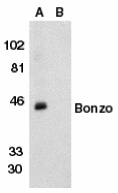
Left: Western blot analysis of Bonzo in human spleen tissue lysate in the absence (lane A) or presence (lane B) of peptide with Bonzo antibody (NT2) at 1:500 dilution.
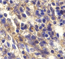
Below: Immunohistochemistry of Bonzo in human spleen cells with Bonzo antibody at 20 µg/ml.
Other Product Images
 Source : Bonzo antibody was raised against a peptide corresponding to amino acids near the amino terminus of human Bonzo/STRL33. The sequence of this peptide differs from those of African green monkey and pig-tailed macaque by one or two amino acids, respectively,.
Purification : Affinity chromatography purified via peptide column
Clonality and Clone : This is a polyclonal antibody.
Host : Bonzo antibody was raised in rabbit. Please use anti-rabbit secondary antibodies.
Immunogen : Human Bonzo (N-Terminus 2) Peptide (Cat. No. 2209P)
Application : Bonzo antibody can be used for detection of Bonzo by Western blot at 1:500 dilution.
Tested Application(s) : E, WB, IHC
Buffer : Antibody is supplied in PBS containing 0.02% sodium azide.
Blocking Peptide : Cat. No. 2209P - Bonzo Peptide
Long-Term Storage : Bonzo antibody can be stored at 4ºC, stable for one year. As with all antibodies care should be taken to avoid repeated freeze thaw cycles. Antibodies should not be exposed to prolonged high temperatures.
Positive Control
1. Cat. No. 1306 - Human Spleen Tissue Lysate
Species Reactivity :H
GI Number : 2209284
Accession Number : U73529
Short Description : (NT2) HIV coreceptor
References
1. Deng HK, Unutmaz D, KewalRamani VN, Littman DR. Expression cloning of new receptors used by simian and human immunodeficiency viruses. Nature 1997;388:296-300
2. Liao F, Alkhatib G, Peden KW, Sharma G, Berger EA, Farber JM. STRL33, A novel chemokine receptor-like protein, functions as a fusion cofactor for both macrophage-tropic and T cell line-tropic HIV-1. J Exp Med 1997;185:2015-23
3. Alkhatib G, Liao F, Berger EA, Farber JM, Peden KW. A new SIV co-receptor, STRL33. Nature 1997;388:238
4. Loetscher M, Amara A, Oberlin E, Brass N, Legler D, Loetscher P, D'Apuzzo M, Meese E, Rousset D, Virelizier JL, Baggiolini M, Arenzana-Seisdedos F, Moser B. TYMSTR, a putative chemokine receptor selectively expressed in activated T cells, exhibits HIV-1 coreceptor function. Curr Biol 1997;7:652-60 (RD0400)
ZBRK1 Antibody
Catalog# : 4817
ZBRK1 is a member of the Kruppel-associated box-zinc finger protein (KRAB-ZFP) protein family of transcriptional repressors. ZBRK1 has been shown to not only be up-regulated in senescent fibroblasts but also interacts with BRCA1, a transcriptional regulator with tumor suppressor activity. ZBRK1 binds to a specific sequence within the GADD45 gene, supporting the assembly of a nuclear complex containing BRCA1 and repressing its transcription. Recent experiments demonstrate that ZBRK1 can homo-oligomerize in vitro and in vivo through a C-terminal transcriptional repression domain (CTRD), and this oligomerization facilitates the ZBRK1-directed transcriptional repression through ZBRK1 response elements. These results suggest that higher order ZBRK1 oligomers may assemble through both protein-DNA and CTRD-dependent protein-protein interactions.
Additional Names : ZBRK1, Zinc finger protein 350, ZNF350
 Description
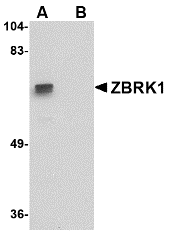
Left: Western blot analysis of ZBRK1 in A-20 lysate with ZBRK1 antibody at 1 µg/ml in (A) the absence and (B) the presence of blocking peptide.
Source : ZBRK1 antibody was raised against a 20 amino acid peptide near the carboxy terminus of the human ZBRK1.
Purification : Affinity chromatography purified via peptide column
Clonality and Clone : This is a polyclonal antibody.
Host : ZBRK1 antibody was raised in rabbit. Please use anti-rabbit secondary antibodies.
Application : ZBRK1 antibody can be used for detection of ZBRK1 by Western blot at 1 – 2 µg/ml.
Tested Application(s) : E, WB
Buffer : Antibody is supplied in PBS containing 0.02% sodium azide.
Blocking Peptide : Cat.No. 4817P - ZBRK1 Peptide
Long-Term Storage : ZBRK1 antibody can be stored at 4ºC, stable for one year. As with all antibodies care should be taken to avoid repeated freeze thaw cycles. Antibodies should not be exposed to prolonged high temperatures.
Positive Control
1. Cat. No. 1288 - A20 Cell Lysate
Species Reactivity :H, M, R
GI Number : 59710107
Accession Number : NP_067645
Short Description : Zinc finger protein 350
References
1. Zheng L, Pan H, Li S, et al. Sequence-specific transcription corepressor function for BRCA1 through a novel Zinc finger protein, ZBRK1. Mol. Cell 2000; 6:757-68.
2. Collins T, Stone JR, and Williams AJ. All in the family: the BTB/POZ, KRAB, and SCAN domains. Mol. Cell. Biol. 2001; 21:3609-15.
3. Tan W, Kim S, and Boyer TG. Tetrameric oligomerization mediates transcriptional repression by the BRCA1-dependent Kruppel-associated Box-Zinc finger protein ZBRK1. J. Biol. Chem. 2004; 279:55153-60.
Peptide sequence selection is likely the most difficult step in the development of anti-peptide antibodies. Although the design of peptides for use as antigens is not exact, The following summary highlights some important points that should be considered in the design of a peptide. If you would like BSI to help you in designing your peptide, click here to request for sequence analysis or visit our Gizmo for online interative analysis. Remember! You design. We can Help!
1. Examine N– and C-terminus of the protein. If they are hydrophilic, they often constitute an excellent choice.
2. If both N– and C-terminus are suitable, choose internal hydrophilic region of the protein. Hydrophilic sequences have the best chances to reside at the surface of the protein and therefore to be accessible to the antibodies.
3. Avoid possible glycosylation or phosphorylation sites
4. Avoid a series of hydrophobic amino acids such as alanine, tryptophan etc.
5. Short synthetic peptides are quite flexible and are unable to mimic alpha helical sequences. However, run algorithms for predicting secondary structures and avoid regions which are strongly predicted alpha helical prior to peptide selection.
6. A peptide range between 10-20 amino acids is recommended. Shorter sequences have fewer chances to include good peptides, longer peptides tend to make stable secondary structures which are not necessarily the same in the protein of interest. Longer peptides also increase the chances of cross-reactions with other proteins.
7. A BLAST homology searches of the selected peptides against proteins of the organism obtained from in order to exclude unwanted cross-reactions to other proteins as much as possible. Less than 4-5 identical amino acids in a continuous stretch are required to exclude a cross-reaction.
8. Carrier protein coupling strategy needs to be taken into account. Alternatively, use the MAP strategy for synthesis of the peptide (not suitable for peptides from the C-terminus).
-Peptides from the C-terminus of the protein should be linked to the carrier through the N-terminus.
-Peptides from the N-terminus of the protein should be linked to the carrier by their C-terminus.
-Internal peptides should be coupled at the less hydrophilic end.
-N-terminal coupling is specific only if the peptide does not contain lysine residue.
-C-terminal coupling is specific only if the peptide does not contain aspartyl or glutamyl residues.
-If no specific coupling can be done, add cysteine at one end of the peptide. The only restriction is there should not be any other cysteines in the sequence.
9. Conjugation of a peptide to a carrier protein containing multiple epitopes, such as Keyhole-Limpet Hemocyanin (KLH) or Bovine Serum Albumin (BSA), are most common. KLH is preferred since it is more antigenic in the majority of animals. BSA is often used as a blocking reagent in assays, thus an anti-peptide antibody raised against a BSA conjugated peptide will show some specificity towards the buffer reagents. This can result in a false positive signal.
10. MAP is simply an alternative to KLH conjugation. It is recommended only for shorter peptides located either internally within a protein or at the N-terminus. KLH conjugation is recommended for a peptide at any location in a protein and when there are a cysteine residues within the peptide sequence.
11. Use at least 2 animals for each peptide since there is some genetic variability between animals.
12. Use more than 1 peptide for each protein in order to increase the chances for antibodies to recognize the protein.
The services for the separation of the antibodies produced against the individual peptides are available at Bio-Synthesis, call 800-227-0627 or write to us at support@biosyn.com for FREE consultation on peptide selection.
List of Misidentified or contaminated cell lines
Cell line cross contamination and misidentification have plagued biomedical research for as long as cells have been employed as resarch tools. A recent Science article indicated that in some cases, laboratories have invested substantial time and effort researching cell lines that were revealed to be misidentified.
(1). The situation has prompted the U.S. National Institutes of Health to issue a notice strongly urging researchers to authenticate cultured cells used in their laboratory.
(2). Many contaminated cell lines were overgrown by the contaminating cell line during establishment and so authentic stocks probably do not exist; in those cases all work has been performed on the contaminant, which may come from a different species, tissue or cell type.
Cell line cross-contamination was first discovered in the 1960s and has been extensively reported since that time. Contaminated or misidentified cell lines have also been listed on the websites of individual cell banks. To make it easier to check for these references, a single list has been developed summarising the cell lines known to be affected, their contaminants, and the references used.
The list was developed and is maintained by Dr. R. Ian Freshney and Dr. Armanda Capes-Davis. For additional background or citation, please refer to the associated article.
(3).This table is meant as a preliminary guide to avoiding suspect cell lines, but all recently acquired cell lines should be tested (e.g. by STR profiling for human cell lines) and compared to reference stock before use. Observations made in these lists are based on published reports and details obtained from cell banks, their websites, and Wikipedia. The authors take no credit nor responsibility for any of the primary observations and have merely attempted to collate data previously available on other sites.
Click on PDF link below for list of Misidentified or contaminated cell lines.
Reference:
1. Chatterjee, R. (2007) Cell biology. Cases of mistaken identity. Science 315, 928–31.
2. Ruiz Bravo, N. and Gottesman, M. (2007) Notice regarding authentication of cultured cell lines.
3. Capes-Davis, A., G. Theodosopoulos, I. Atkin, H. Drexler, A. Kohara, R.A.F. MacLeod, J.R. Masters, Y. Nakamura, Y.A. Reid, R.R. Reddel, R.I. Freshney. 2010. Check your cultures! A list of cross-contaminated or misidentified cell lines. International Journal of Cancer. 127:1–8.


