SAPAP4 Antibody
Catalog# :4633
SAP90/PSD-95-associated protein 4 (SAPAP4, also known as DLGAP4) is a member of a protein family whose members specifically interact with PSD-95/SAP90, a membrane-associated guanylate kinase localized at postsynaptic density (PSD) in neuronal cells. Like the other SAPAP proteins, SAPAP4 is thought to be an adaptor protein that also interacts with different synaptic scaffolding proteins, cytoskeletal and signaling components, such as focal adhesion kinase (FAK) and proline-rich tyrosine kinase 2 (PYK2). SAPAP4 mRNA is targeted to cell bodies in a similar manner to SAPAP1 and -2, whereas SAPAP3 mRNA is detected mainly in cell bodies. At least three isoforms of SAPAP2 are known to exist. This SAPAP4 antibody will not cross-react with other SAPAP proteins.
Additional Names : SAPAP4, SAP90/PSD-95-associated protein 4, Disks large-associated protein 4, DLGAP4, DAP4
 Description
Left: Western blot analysis of SAPAP4 in SK-N-SH cell lysate with SAPAP4 antibody at 1 µg/ml.
Source :SAPAP4 antibody was raised against a 13 amino acid peptide from near the center of human SAPAP4.
Purification : Affinity chromatography purified via peptide column
Clonality and Clone : This is a polyclonal antibody.
Host : SAPAP4 antibody was raised in rabbit.
Please use anti-rabbit secondary antibodies.
Application : SAPAP4 antibody can be used for detection of SAPAP4 by Western blot at 0.5 – 1 µg/ml.
Tested Application(s) : E, WB
Buffer : Antibody is supplied in PBS containing 0.02% sodium azide.
Blocking Peptide :Cat.No. 4633P - SAPAP4 Peptide
Long-Term Storage : SAPAP4 antibody can be stored at 4ºC, stable for one year. As with all antibodies care should be taken to avoid repeated freeze thaw cycles. Antibodies should not be exposed to prolonged high temperatures.
Positive Control :
1. Cat. No. 1220 - SK-N-SH Cell Lysate
Species Reactivity : H
GI Number : 34335253
Accession Number : NP_055717
Short Description : SAP90/PSD-95-associated protein 4
References
1. SAPAPs. A family of PSD-95/SAP90-associated proteins localized at postsynaptic density. J. Biol. Chem. 1997; 272:11943-51.
2. Ranta S, Zhang Y, Ross B, et al. Positional cloning and characterization of the human DLGAP2 gene and its exclusion in progressive epilepsy with mental retardation. Eur. J. Hum. Genet. 2000; 8:381-4.
3. Kindler S, Rehbein M, Classen B, et al. Distinct spatiotemporal expression of SAPAP transcripts in the developing rat brain: a novel dendritically localized mRNA. Brain Res. Mol. Brain Res. 2004; 126:14-21.
4. Bongiorno-Borbone L, Kadare G, Benfenati F, et al. FAK and PYK2 interact with SAP/PSD-95-associated protein-3. Biochem. Biophys. Res. Commun. 2005; 337:641-6.
PIG-Y Antibody
Catalog# :4955
Glycosylphosphatidylinositol (GPI) lipid anchoring is an important post-translational modification of proteins that takes place in the endoplasmic reticulum. The synthesis of GPI is initiated by GPI-N-acetylglucosaminyltransferase (GPI-GnT), a complex of proteins including PIG-A, PIG-H, PIG-C, GPI1, and DPM2. PIG-Y, the mammalian homolog to yeast Eri1p, is also thought to be involved in the biosynthesis of GPI. The PIG-Y gene encodes two proteins, one of which arises from leaky scanning of the mRNA. This antibody only detects isoform 1 of PIG-Y. Despite its predicted molecular weight, PIG-Y often migrates at 28-30 kDa in SDS-PAGE.
Additional Names : PIG-Y (IN), Phosphatidylinositol glycan anchor biosynthesis class Y, PreY, PIGY
 Description
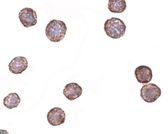
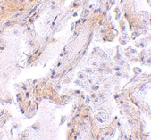
Left: Western blot analysis of PIG-Y in human spleen tissue lysate with PIG-Y antibody at (A) 1 and (B) 2 µg/ml.
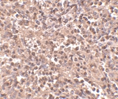
Below: Immunohistochemistry of PIG-Y in human spleen tissue with PIG-Y antibody at 2.5 µg/ml.
Other Product Images
 Source : PIG-Y antibody was raised against a 16 amino acid peptide near the center of human PIG-Y.
Purification : Affinity chromatography purified via peptide column
Clonality and Clone : This is a polyclonal antibody.
Host : PIG-Y antibody was raised in rabbit. Please use anti-rabbit secondary antibodies.
Application : PIG-Y antibody can be used for detection of PIG-Y by Western blot at 1 – 2 µg/ml.
Tested Application(s) : E, WB, IHC
Buffer : Antibody is supplied in PBS containing 0.02% sodium azide.
Blocking Peptide :Cat.No. 4955P - PIG-Y Peptide
Long-Term Storage : PIG-Y antibody can be stored at 4ºC, stable for one year. As with all antibodies care should be taken to avoid repeated freeze thaw cycles. Antibodies should not be exposed to prolonged high temperatures.
Positive Control :
1. Cat. No. 1306 - Human Spleen Tissue Lysate
Species Reactivity : H, M
GI Number : 14249680
Accession Number : NP_116295
Short Description : (IN) Phosphatidylinositol glycan anchor biosynthesis class Y
References
1. Eisenhaber B, Maurer-Stroh S, Novatchkova M, et al. Enzymes and auxiliary factors for GPI lipid anchor biosynthesis and post-translational transfer to proteins. Bioessays 2003; 25:367-85.
2. Watanabe R, Murakami Y, Marmor MD, et al. Initial enzyme for glycosylphosphatidylinositol biosynthesis requires PIG-P and is regulated by DPM2. EMBO J. 2000; 19:4402-11.
3. Murakami Y, Siripanyaphinoyo U, Hong Y, et al. The initial enzyme for glycosylphosphatidylinositol biosynthesis requires PIG-Y, a seventh component. Mol. Biol. Cell 2005; 16:5236-46.
PCDH12 Antibody
Catalog# :5071
Protocadherins comprise the largest group within the cadherin family of calcium-dependent cell-cell adhesion molecules. Protocadherin 12 (PCDH12) was initially identified through PCR screening of mouse heart microvascular endothelial cell RNA; further experiments revealed its mRNA to be strongly expressed in highly vascularized organs such as lung and kidney, in addition to glycogen-rich trophoblasts in the placenta. PCDH12-null mice are viable and fertile, but show reduced placental and embryonic sizes when compared to wild-type mice. Further studies showed significant expression changes in 2,289 genes, including those involved in tissue morphogenesis, angiogenesis, cell-matrix adhesion and migration, immune response and chromatin remodeling. This antibody is predicted to not cross-react with PCDH18.
Additional Names : PCDH12, Protocadherin 12, vascular endothelial cadherin 2, VE-cadherin 2, VECAD2
 Description
Left: Western blot analysis of PCDH12 in K562 cell lysate with PCDH12 antibody at 2 µg/ml.
Below: Immunohistochemistry of PCDH12 in rat lung tissue with PCDH12 antibody at 5 µg/ml.
Other Product Images
Source :
PCDH12 antibody was raised against a 17 amino acid peptide near the amino terminus of human PCDH12.
Purification : Affinity chromatography purified via peptide column
Clonality and Clone : This is a polyclonal antibody.
Host : PCDH12 antibody was raised in rabbit. Please use anti-rabbit secondary antibodies.
Application : PCDH12 antibody can be used for detection of PCDH12 by Western blot at 1 – 2 µg/ml.
Tested Application(s) : E, WB, IHC
Buffer : Antibody is supplied in PBS containing 0.02% sodium azide.
Blocking Peptide :Cat.No. 5071P - PCDH12 Peptide
Long-Term Storage : PCDH12 antibody can be stored at 4ºC, stable for one year. As with all antibodies care should be taken to avoid repeated freeze thaw cycles. Antibodies should not be exposed to prolonged high temperatures.
Positive Control :
1. Cat. No. 1204 - K562 Cell Lysate
Species Reactivity : H, M, R
GI Number : 7706113
Accession Number : NP_057664
Short Description : Protocadherin 12
References
1. Frank M and Kemler R. Protocadherins. Curr. Opin. Cell Biol. 2002; 14:557-62.
2. Telo P, Breviaro F, Huber P, et al. Identification of a novel cadherin (vascular endothelial cadherin-2) located at intercellular junctions in endothelial cells. J. Biol. Chem. 1998; 28:17565-72.
3. Rampon C, Prandini MH, Bouillot S, et al. Protocadherin 12 (VE-cadherin 2) is expressed in endothelial, trophoblast, and mesangial cells. Exp. Cell Res. 2005; 302:48-60.
4. Rampon C, Bouillot S, Climescu-Haulica A, et al. Protocadherin 12 deficiency alters morphogenesis and transcriptional profile of the placenta. Physiol. Genomics 2008; 34:193-204.
JMJD8 Antibody
Catalog# : 5387
The jumonji domain-containing protein (JMJD) family is defined by the presence of the JmjC domain that is observed in several diverse species. While several JMJD proteins have been identified as being involved in chromatin regulation, histone demethylation and development, the function of JMJD8 has not been identified.
Additional Names : JMJD8, Jumonji domain-containing protein 8, JmjC domain-containing protein 8
 Description
Left: Western blot analysis of JMJD8 in rat kidney tissue lysate with JMJD8 antibody at 1µg/ml.
Source : JMJD8 antibody was raised against an 18 amino acid peptide from near the carboxy terminus of human JMJD8.
Purification : Affinity chromatography purified via peptide column
Clonality and Clone : Polyclonal
Host : JMJD8 antibody was raised in rabbit. Please use anti-rabbit secondary antibodies.
Application : JMJD8 antibody can be used for detection of JMJD8 by Western blot at 1 - 2 µg/ml.
Tested Application(s) : E, WB
Buffer : Antibody is supplied in PBS containing 0.02% sodium azide.
Blocking Peptide : Cat.No. 5387P - JMJD8 Peptide
Long-Term Storage : JMJD8 antibody can be stored at 4ºC, stable for one year. As with all antibodies care should be taken to avoid repeated freeze thaw cycles. Antibodies should not be exposed to prolonged high temperatures.
Positive Control
1. Cat. No. 1465 - Rat Kidney Tissue Lysate
Species Reactivity :H, M, R
GI Number : 74717267
Accession Number : Q96S16
Short Description : Jumonji domain-containing protein 8
References
1. Takeuchi T, Watanabe Y, Takano-Shimizu T, et al. Roles of jumonji and jumonji family genes in chromatin regulation and development. Dev. Dyn. 2006; 235:2449-59.
GALNT10 Antibody
Catalog# : 5281
Protein glycosylation is an important biological process that is carried out by a large family of glycosyltransferases that catalyze the synthesis of oligosaccharides and glycoconjugates. Polypeptide GalNAc transferases initiate the synthesis of mucin-type oligosaccharides by transferring GalNAc from UDP-GalNAc to the hydroxyl group of either a serine or threonine residue on the polypeptide acceptor. Polypeptide galactoaminyltransferase 10 (GALNT10) belongs to the polypeptide N-acetylgalactosaminyl-transferase (pp-GalNAc-T) protein family. Following expression in insect cells, recombinant GALNT10 showed significant GalNAcT activity toward mucin-derived peptides, and it utilized both non-glycosylated and glycosylated peptide substrates. GALNT10 mRNA is highly expressed in several distinct hypothalamic, thalamic, and amygdaloid nuclei in mouse brain. At least four isoforms of GALNT10 are known to exist.
Additional Names : GALNT10 (NT),Polypeptide galactoaminyltransferase 10, GalNAc transferase 10
 Description
Left: Western blot analysis of GALNT10 in rat brain tissue lysate with GALNT10 antibody at (A) 1 and (B) 2 µg/ml.
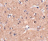
Below: Immunohistochemistry of GALNT10 in human brain tissue with GALNT10 antibody at 2.5 μg/ml.
Other Product Images
Source : GALNT10 antibody was raised against a 16 amino acid peptide near the amino terminus of human GALNT10.
Purification : Affinity chromatography purified via peptide column
Clonality and Clone : Polyclonal
Host : GALNT10 antibody was raised in rabbit. Please use anti-rabbit secondary antibodies.
Application : GALNT10 antibody can be used for detection of GALNT10 by Western blot at 1 - 2 µg/ml.
Tested Application(s) : E, WB
Buffer : Antibody is supplied in PBS containing 0.02% sodium azide.
Blocking Peptide : Cat.No. 5281P - GALNT10 Peptide
Long-Term Storage : GALNT10 antibody can be stored at 4ºC, stable for one year. As with all antibodies care should be taken to avoid repeated freeze thaw cycles. Antibodies should not be exposed to prolonged high temperatures.
Positive Control
1. Cat. No. 1463 - Rat Brain Tissue Lysate
Species Reactivity :H, M, R
GI Number : 38195091
Accession Number : NP_938080
Short Description : (NT) Polypeptide galactoaminyltransferase 10
References
1. Amado M, Almeida R, Schwientek T, et al. Identification and characterization of large galactosyltransferase gene families: galactosyltransferases for all functions. Biochim. Biophys. Acta 1999; 1473:35-53.
2. Nelson PA, Sutcliffe JG, and Thomas EA. A new UDP-GalNAc:polypeptide N-acetylgalactosaminyltransferase mRNA exhibits predominant expression in the hypothalamus, thalamus and amygdala for mouse forebrain. Gene Express. Patterns 2002; 1:95-9.
EVER1 Antibody
Catalog# : 4547
Epidermodysplasia verruciformis (EV) is an autosomal recessive dermatosis characterized by abnormal susceptibility to human papillomaviruses (HPVs) and a high rate of progression to squamous cell carcinoma on sun-exposed skin. EV is caused by mutations in either of two adjacent genes, EVER1 and EVER2, located on chromosome 17q25.3. Both of these genes encode integral membrane proteins that localize to the endoplasmic reticulum and are predicted to form transmembrane channels. Both EVER1 and EVER2 are members of the transmembrane channel-like (TMC) protein family. EVER1 possesses eight trans-membrane domains and two leucine zipper motifs. EVER1 and EVER2 form a complex and interact with the zinc transporter 1 (ZnT-1), suggesting that EVER1 and EVER2 act to regulate cellular zinc balance. At least four isoforms of EVER1 are known to exist. This EVER1 antibody does not cross-react with EVER2.
Additional Names : EVER1 (NT), EV1, EVIN1, Transmembrane protein channel-like protein 6, TMC6
 Description
Left: Western blot analysis of EVER1 in A-20 cell lysate with EVER1 antibody at (A) 1 and (B) 2 µg/ml.
Below: Immunohistochemistry of EVER1 in human spleen with EVER1 antibody at 2.5 µg/ml.
Other Product Images
Source : EVER1 antibody was raised against a 14 amino acid peptide from near the amino terminus of human EVER1.
Purification : Affinity chromatography purified via peptide column
Clonality and Clone : This is a polyclonal antibody.
Host : EVER1 antibody was raised in rabbit. Please use anti-rabbit secondary antibodies.
Application : EVER1 antibody can be used for the detection of EVER1 by Western blot at 1 – 2 µg/ml.
Tested Application(s) : E, WB, IHC
Buffer : Antibody is supplied in PBS containing 0.02% sodium azide.
Blocking Peptide : Cat.No. 4547P - EVER1 Peptide
Long-Term Storage : EVER1 antibody can be stored at 4ºC, stable for one year. As with all antibodies care should be taken to avoid repeated freeze thaw cycles. Antibodies should not be exposed to prolonged high temperatures.
Positive Control
1. Cat. No. 1288 - A20 Cell Lysate
Species Reactivity :H, M, R
GI Number : 25527208
Accession Number : AAM44452
Short Description : (NT) Transmembrane protein channel-like protein 6
References
1. Majewski S, Jablonska J and Orth G. Epidermodysplasia verruciformis. Immunological and nonimmunological surveillance mechanisms: role in tumor progression. Clin. Dermatol. 1997; 15:321-34.
2. Ramoz N, Rueda L-A, Bouadjar B, et al. Mutations in two adjacent novel genes are associated with epidermodysplasia verruciformis. Nat. Genetics 2002; 32:579-81.
3. Keresztes G, Mutai H and Heller S. TMC and EVER genes belong to a larger novel family, the TMC gene family encoding transmembrane proteins. BMC Genomics 2003; 4:24-34.
4. Lazarczyk L, Pons C, Mendoza JA, et al. Regulation of cellular zinc balance as a potential mechanism of EVER-mediated protection against pathogenesis by cutaneous oncogenic human papillomaviruses. J. Exp. Med. 2008; 205:35-42.
Beta-actin Antibody
Catalog# : 3777
Actins are highly conserved proteins that are involved in cell motility, structure and integrity, processes that are crucial for tissue development and the development of organism (reviewed in 1). The actin cytoskeleton is one of the principal drivers of cell motility and is capable of responding to complex signaling cascades. Recent evidence suggests that it may play key roles in regulating apoptosis and aging (2). beta actin is one of six different actin isoforms which have been identified. Like GAPDH, Β-actin is constitutively expressed at high levels in almost all tissues and cell lines making it ideal for use as a loading control marker in immunoblots.
Additional Names : Beta-actin (CT), Beta actin
 Description
Left: Western blot analysis of beta-actin in HeLa cell lysate with beta-actin antibody at (A) 0.5, (B) 1 and (C) 2 µg/ml.
Below: Immunocytochemistry of beta-actin in HeLa cells with beta-actin antibody at 10µg/ml.
Other Product Images
 Source : b-actin antibody was raised against a 16 amino acid peptide from near the carboxy-terminus of human b-actin.
Purification : Affinity chromatography purified via peptide column
Clonality and Clone : This is a polyclonal antibody.
Host : Beta-actin antibody was raised in rabbit. Please use anti-rabbit secondary antibodies.
Immunogen : Human b-actin / Beta actin (C-Terminus) Peptide (Cat. No. 3777P)
Application : b-actin antibody can be used for the detection of b-actin by Western blot at 1 – 2 µg/ml.
Tested Application(s) : E, WB, ICC
Buffer : Antibody is supplied in PBS containing 0.02% sodium azide.
Blocking Peptide : Cat. No. 3777P - B-actin Peptide
Long-Term Storage : Beta-actin antibody can be stored at 4ºC, stable for one year. As with all antibodies care should be taken to avoid repeated freeze thaw cycles. Antibodies should not be exposed to prolonged high temperatures.
Positive Control
1. Cat. No. 1201 - HeLa Cell Lysate
Species Reactivity :H, M, R
GI Number : 12803203
Accession Number : AAH02409
Short Description : (CT) a major cytoskeletal protein, C Terminus
References
1. Lambrechts A, Van Troys, M and Ampe C. The actin cytoskeleton in normal and pathological cell motility. Int. J. Biochem. Cell Biol. 2004; 36:1890-909.
2. Gourlay CW and Ayscough KR. The actin cytoskeleton: a key regulator of apoptosis and ageing. Nat. Rev. 2005; 6:583-9.
Albumin Antibody
Catalog# : 5159
Albumin is a soluble, monomeric protein which comprises about one-half of the blood serum protein. It functions primarily as a carrier protein for steroids, fatty acids, and thyroid hormones and plays a role in stabilizing extracellular fluid volume. Albumin is synthesized in the liver as preproalbumin which has an N-terminal peptide that is removed before the nascent protein is released from the rough endoplasmic reticulum. The resulting product, proalbumin, is in turn cleaved in the Golgi vesicles to produce the secreted albumin. At least two isoforms of Albumin are known to exist.
Additional Names : Albumin,Alb, serum albumin
 Description
Left: Western blot analysis of Albumin in mouse liver tissue lysate with Albumin antibody at (A) 1 and (B) 2 µg/ml.
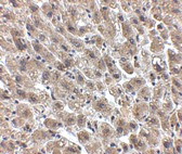
Below: Immunohistochemistry of Albumin in human liver tissue with Albumin antibody at 2.5 μg/ml.
Other Product Images
 Source : Albumin antibody was raised against a 13 amino acid peptide near the center of human Albumin.
Purification : Affinity chromatography purified via peptide column
Clonality and Clone : This is a polyclonal antibody.
Host : Albumin antibody was raised in chicken. Please use anti-chicken secondary antibodies.
Application : Albumin antibody can be used for detection of Albumin by Western blot at 1 – 2 µg/ml.
Tested Application(s) : E, WB
Buffer : Antibody is supplied in PBS containing 0.02% sodium azide.
Blocking Peptide : Cat.No. 5159P - Albumin Peptide
Long-Term Storage : Albumin antibody can be stored at 4ºC, stable for one year. As with all antibodies care should be taken to avoid repeated freeze thaw cycles. Antibodies should not be exposed to prolonged high temperatures.
Positive Control
1. Cat. No. 1404 - Mouse Liver Tissue Lysate
Species Reactivity :H, M, R
GI Number : 113576
Accession Number : P02768
Short Description : Serum albumin
References
1. Rothschild MA, Oratz M, and Schreiber SS. Serum Albumin. Hepatology
Bio-Synthesis has been producing synthetic peptides for over 25 years. Our expertise in custom synthetic polypeptide manufacturing allows us to produce the high-quality, large-scale, and GMP peptides with the highest success rate with long standing records. We have been delivered more than 100,000 peptides to customers worldwide, including very hydrophobic polypeptide, peptide with multiple disulfide bonds, multi-phosph0rylated peptides and extremely long peptides.
GMP Peptides of Biologically Active Products
Our large scale non-GMP ever delivered 5 kilograms of peptides on a single order and has the capacity of 10,000 peptides per month. Our capacity of GMP peptide is 10 kilograms. In the last few years, after the completion of the human genome project, there have been more targets that are being worked on and the numbers of GMP facilities and the cost for GMP peptides have been continuously improving. Our forecast is that synthetic peptide chemistry will be an important source of many medically/clinically relevant peptides and proteins in the years to come.
Custom Large-Scale Peptide Synthesis
In the human body, most if not all biological/ physiological processes are regulated by various forms of molecular recognition. Most of these processes involve initiation or inhibition trough protein-protein interaction. As we know peptides and proteins due to the vast number of conformational possibilities are ideal to carry out such complex control functions. The last 40 years have seen an enormous growth in the methodologies available to obtain peptide and protein molecules. Through recombinant methods, most labs can now assemble genes, subcloned them into expression vectors and obtain a wide range of endogenous proteins; likewise the pioneering work of Bruce Merrifield, makes it possible to obtain multikilo amounts of a number of biologically active peptides.
Synthetic Peptides for Clinical Applications
However, the number of peptides that have entered the pharmaceutical market is relatively low; perhaps due to a number of reasons, among them: antigenicity, immunogenicity , bioavailability and stability of the product upon administration into the patient. To some degree the relative scarcity of large scale peptide production plants that can make the peptide products in large amounts (perhaps at the ton levels) at more affordable costs, have also been a factor.
gp120 Antibody
Catalog# : 4777
Human immunodeficiency virus type 1 (HIV-1) entry into target cells is directed by the envelope (Env) glycoproteins, which are present on the surface of HIV-1 virion or infected cells in the form of trimers consisting of gp120/gp41 complexes. The surface subunit, gp120, initiates the entry process by interacting sequentially with the CD4 receptor and a co-receptor CCR5 or CXCR4, thereby inducing a conformational change that allows the transmembrane (TM) gp41 subunit to mediate fusion between viral and target cell membranes. Cleavage of Env into its gp120 and gp41 components is necessary for activation of its fusogenic activity.
Additional Names : gp120 (IN), HIV-1 glycoprotein 120
 Description
Left: Western blot analysis of (A) 25 and (B) 100 ng of gp120 with gp120 antibody at 1 µg/ml.
Source : gp120 antibody was raised against 15 amino acids from near the center of gp120.
Purification : Affinity chromatography purified via peptide column
Clonality and Clone : This is a polyclonal antibody.
Host : gp120 antibody was raised in goat. Please use anti-goat secondary antibodies.
Application : Gp120 antibody can be used for detection of gp120 by Western blot at 0.5 – 1 µg/ml.
Tested Application(s) : E, WB
Buffer : Antibody is supplied in PBS containing 0.02% sodium azide.
Blocking Peptide : Cat.No. 4777P - gp120 Peptide
Long-Term Storage : gp120 antibody can be stored at 4ºC, stable for one year. As with all antibodies care should be taken to avoid repeated freeze thaw cycles. Antibodies should not be exposed to prolonged high temperatures.
Species Reactivity :V
GI Number : 1465781
Accession Number : AAB05604
Short Description : (IN) HIV-1 glycoprotein 120
References
1. Pinter A. Roles of HIV-1 Env variable regions in viral neutralization and vaccine development. Curr. HIV Res. 2007; 5:542-53.
2. Alkhatib G and Berger EA. HIV coreceptors: from discovery and designation to new paradigms and promise. Eur. J. Med. Res. 2007; 12:375-84.
DC-SIGN Monoclonal Antibody
Catalog# : PM-2347
Dendritic cells (DCs) that control immune responses were recently found to capture and transport HIV from the mucosal area to remote lymph nodes (1), where DCs hand over HIV to CD4+ T lymphocytes. DCs also amplify the amount of virus and extend the duration of viral infectivity. Multiple strains of HIV-1, HIV-2 and SIV bind to DCs via DC-SIGN (2). ICAM-3 is the natural ligand for DC-SIGN (3). A DC-SIGN homologue (termed DC-SIGNR, L-SIGN, and DC-SIGN2) was identified recently (4-8). DC-SIGN forms a novel gene family with DC-SIGNR and many alternatively spliced isoforms of DC-SIGN and DC-SIGNR (8). The expression of DC-SIGN was found in mucosal tissues including placenta, small intestine, and rectum.
Additional Names : DC-SIGN (5D7), Dendritic cell-specific ICAM-3-grabbing nonintegrin 1
 Description
Left: Western blot detection of DC-SIGN fusion protein in human placenta at (A) 1 and (B) 2 µg/ml.
Below: Immunohistochemistry of DC-SIGN in lymph node tissue with DC-SIGN antibody at 5 µg/ml.
Other Product Images
 Source : Mouse monoclonal DC-SIGN antibody was raised against a recombinant His-tagged protein fragment corresponding to the extracellular region of human DC-SIGN (1).
Purification : Immunoaffinity chromotography purified IgG
Clonality and Clone : This is a monoclonal antibody. (Clone 5D7)
Host : DC-SIGN monoclonal antibody was raised in mouse. Please use anti-mouse secondary antibodies.
Immunogen : A peptide corresponding to amino acids near the carboxy terminus of human DC-SIGN.
Application : DC-SIGN antibody can be used for detection of DC-SIGN in immunoblots at 1 – 2 mg/ml and in immunohistochemistry at 5 – 10 mg/ml.
Tested Application(s) : E, WB, IHC
Buffer : Antibody is supplied in PBS containing 0.02% sodium azide.
Blocking Peptide :
Long-Term Storage : DC-SIGN monoclonal antibody can be stored at 4ºC, stable for one year.
Positive Control
1. Cat. No. 1384 - Human Uterus Tissue Lysate
2. Cat. No. 1369 - Human Lymph Node Tissue Lysate
Species Reactivity :H
Short Description : HIV binding receptor
References
1. Geijtenbeek TB, Kwon DS, Torensma R, et al. DC-SIGN, a dendritic cell-specific HIV-1-binding protein that enhances trans-infection of T cells. Cell 2000; 100:587-97.
2. Pohlmann S, Baribaud F, Lee B, et al. DC-SIGN interactions with human immunodeficiency virus type 1 and 2 and simian immunodeficiency virus. J. Virol. 2001;75:4664-72.
3. Geijtenbeek TB, Torensma R, van Vliet SJ, et al. Identification of DC-SIGN, a novel dendritic cell-specific ICAM-3 receptor that supports primary immune responses. Cell 2000; 100:575-85.
4. Soilleux EJ, Barten R, Trowsdale J. DC-SIGN; a related gene, DC-SIGNR; and CD23 form a cluster on 19p13. J. Immunol. 2000; 165:2937-42.
CXCR4-Lo Antibody
Catalog# : 4443
Human immunodeficiency virus (HIV) and related viruses require coreceptors, in addition to CD4, to infect target cells. Some G protein-coupled receptors including CCR5, CXCR4, CCR3, CCR2b and CCR8 in the chemokine receptor family, and four new human molecules GPR15, STRL33, GPR1 and V28 were recently identified as HIV coreceptors. Among them, CXCR4 is a principal coreceptor for T-cell tropic strains of HIV-1 fusion and entry of human white blood cells. CXCR4 is also required for the infection by dual-tropic strains of HIV-1 and mediates CD-4 independent infection by HIV-2. The a-chemokine SDF-1 is the ligand for CXCR4 and prevents infection by T-tropic HIV-1. CXCR4 associates with the surface CD4-gp120 complex before HIV enters target cells. CXCR4 messenger RNA levels correlated with HIV-1 permissiveness in diverse human cell types. Antibodies to CXCR4 block HIV-1 and HIV-2 fusion and infection of human target cells. The amino-terminal domain and the second extracellular loop of CXCR4 serve as HIV binding sites. This antibody is specific for the longer isoform of CXCR4.
Additional Names : CXCR4-Lo, Fusin, LESTR, HUMSTR
 Description
Left: Western blot analysis of CXCR4 in (A) human spleen and (B) human thymus tissue lysate with CXCR4-Lo antibody at 10 µg/ml.
Below: Immunohistochemistry of CXCR4Lo in HeLa cells with CXCR4Lo antibody at 2 µg/ml.
Other Product Images
Source : CXCR4-Lo antibody was raised against a peptide corresponding to nine amino acids near the amino terminus of human CXCR4 isoform a.
Purification : Purified IgG
Clonality and Clone : This is a polyclonal antibody.
Host : CXCR4-Lo antibody was raised in rabbit. Please use anti-rabbit secondary antibodies.
Application : CXCR4-Lo antibody can be used for Western blot at 5 – 1 µg/ml.
Tested Application(s) : E, WB, ICC
Buffer : Antibody is supplied in PBS containing 0.02% sodium azide.
Blocking Peptide : Cat.No. 4443P - CXCR4-Lo Peptide
Long-Term Storage : CXCR4-Lo antibody can be stored at 4ºC, stable for one year. As with all antibodies care should be taken to avoid repeated freeze thaw cycles. Antibodies should not be exposed to prolonged high temperatures.
Positive Control
1. Cat. No. 1306 - Human Spleen Tissue Lysate
Species Reactivity :H
GI Number : 56790927
Accession Number : NP_001008540
Short Description : Isoform a of HIV & chemokine receptor
References
1. Dimitrov DS. Cell 1997; 91:721-30.
2. Feng Y et al. Science 1996; 272:872-7.
3. Berson JF et al. J. Virol. 1996; 70:6288-95.
4. Doranz BJ et al. Cell 1996; 85:1149-58.
CAPN6 Antibody
Catalog# : 4759
Calpains make up a ubiquitously expressed, well-conserved family of calcium-dependent cysteine proteases. The calpain proteins are heterodimers consisting of an invariant small subunit and variable large subunits. This large subunit possesses a cysteine protease domain, and both subunits possess calcium-binding domains. Calpains have been implicated in neurodegenerative processes as their activation can be triggered by calcium influx and oxidative stress. Calpain 6 (CAPN6) is most similar to Calpain 5; the C-terminal region of CAPN6 lacks homology to the calmodulin-like domain of other vertebrate calpains. CAPN6 is thought to be involved in the regulation of microtubule dynamics and cytoskeletal organization. CAPN6 has also been recently identified as an HIV dependency factor (HDF), suggesting that CAPN6 may be an important drug target in HIV treatment.
Additional Names : CAPN6, Calpain 6, CANPX, CalpM
 Description
Left: Western blot analysis of CAPN6 in rat lung tissue lysate with CAPN6 antibody at (A) 0.5 and (B) 1 µg/ml.
Below: Immunohistochemistry of CAPN6 in human lung tissue with CAPN6 antibody at 2.5 μg/ml.
Other Product Images
 Source : CAPN6 antibody was raised against a 18 amino acid peptide from near the carboxy terminus of human CAPN6.
Purification : Affinity chromatography purified via peptide column
Clonality and Clone : This is a polyclonal antibody.
Host : CAPN6 antibody was raised in rabbit. Please use anti-rabbit secondary antibodies.
Application : CAPN6 antibody can be used for the detection of StrepII by Western blot at 0.5 – 1 µg/ml.
Tested Application(s) : E, WB
Buffer : Antibody is supplied in PBS containing 0.02% sodium azide.
Blocking Peptide : Cat.No. 4759P - CAPN6 Peptide
Long-Term Storage : CAPN6 antibody can be stored at 4ºC, stable for one year. As with all antibodies care should be taken to avoid repeated freeze thaw cycles. Antibodies should not be exposed to prolonged high temperatures.
Positive Control
1. Cat. No. 1462 - Rat Lung Tissue Lysate
Species Reactivity :H, M, R
GI Number : 13186316
Accession Number : NP_055104
Short Description : Calpain 6
References
1. Croall DE and Ersfeld K. The calpains: modular designs and functional diversity. Genome Biol. 2007; 8:216.
2. Dear N, Matena K, Vingron M, et al. A new subfamily of vertebrate calpains lacking a calmodulin-like domain: implications for calpain regulation and evolution. Genomics 1997; 45:175-84.
3. Tonami K, Kurihara Y, Aburatani J, et al. Calpain 6 is involved in microtubule stabilization and cytoskeletal organization. Mol. Cell. Biol. 2007; 27:2548-61.
4. Brass AL, Dykxhoorn DM, Benita Y, et al. Identification of host proteins required for HIV infection through a functional genomic screen. Science 2008; 319:921-6.
IL-21 Receptor Antibody
Catalog# : 2469
A novel cytokine related to IL-2 and IL-15 was recently identified and designated IL-21 . The receptor for IL-21 (IL-21R, also termed NILR for novel Interleukin receptor) is a new member of the class I cytokine receptor family (1,2). IL-21R forms a complex with the common cytokine receptor g chain, gc, and mediates IL-21 signaling (3,4). Both IL-21R and the gc are necessary for the IL-21 function. IL-21 and its receptor activate JAK-STAT signaling pathway. IL-21R is expressed in spleen, thymus, natural killer (NK), T and B cell lines. IL-21 plays a role in the proliferation and maturation of NK, B and T cell populations.
Additional Names : IL-21 Receptor (ED), IL21R
 Description
Left: Western blot analysis of IL-21 receptor expression in human Raji cell lysate with IL-21 Receptor antibody at 1 µg /ml.
Below: Immunohistochemistry of IL-21 receptor in rat lung with IL-21 receptor antibody at 10 µg/ml.
Other Product Images
 Source : IL-21 receptor antibody was raised against a synthetic peptide corresponding to amino acids 97 to 111 of human IL-21 receptor precursor .
Purification : Affinity chromatography purified via peptide column
Clonality and Clone : This is a polyclonal antibody.
Host : IL-21 Receptor antibody was raised in rabbit. Please use anti-rabbit secondary antibodies.
Immunogen : Human IL-21 Receptor (Extracellular Domain) Peptide (Cat. No. 2469P)
Application : IL-21 receptor antibody can be used for detection of IL-21 receptor by Western blot at 0.5 to 1 µg/ml.An approximately 60 kDa band can be detected.
Tested Application(s) : E, WB, IHC
Buffer : Antibody is supplied in PBS containing 0.02% sodium azide.
Blocking Peptide : Cat. No. 2469P - IL-21 Receptor Peptide
Long-Term Storage : IL-21 Receptor antibody can be stored at 4ºC, stable for one year. As with all antibodies care should be taken to avoid repeated freeze thaw cycles. Antibodies should not be exposed to prolonged high temperatures.
Positive Control
1. Cat. No. 1207 - Raji Cell Lysate
2. Cat. No. 1462 - Rat Lung Tissue Lysate
Species Reactivity :H, M, R
GI Number : 11141869
Accession Number : NP_068570
Short Description : (ED) Interleukin 21 receptor
References
1. Parrish-Novak J, Dillon SR, Nelson A, et al. Interleukin 21 and its receptor are involved in NK cell expansion and regulation of lymphocyte function. Nature. 2000;408(6808):57-63.
2. Ozaki K, Kikly K, Michalovich D, et al. Cloning of a type I cytokine receptor most related to the IL-2 receptor beta chain. Proc Natl Acad Sci USA . 2000;97(21):11439-44.
3. Asao H, Okuyama C, Kumaki S, et al. the common gamma-chain is an indispensable subunit of the IL-21 receptor complex. J Immunol. 2001;167(1):1-5.
4. Vosshenrich CA, Di Santo JP. Cytokines: IL-21 joins the gamma(c)-dependent network.Curr Biol. 2001;11(5):R175-7.
IRGM Antibody
Catalog# : 4543
Autophagy, the process of bulk degradation of cellular proteins through an autophagosomic-lysosomal pathway is important for normal growth control and may be defective in tumor cells. It is involved in the preservation of cellular nutrients under starvation conditions as well as the normal turnover of cytosolic components. Two of the strongest hits implicate genes IRGM and ATG16L1, which encode proteins thought to be critical to the autophagy pathway and being significantly associated with Crohn’s disease. In mouse, IRGM belongs to a family of gamma-interferon-induced GTP-binding proteins of approximately 48 kDa. Murine IRGM induces autophagy and generates large autolysosomal organelles as a mechanism for the elimination of intracellular Mycobacterium tuberculosis. Human IRGM is also involved in autophagy and plays a role in the control of intracellular pathogens and in the reduction of intracellular bacillary load.
Additional Names : IRGM (NT), Immunity related GTPase family, IFL1, IRGM1, LRG47, LRG-47
 Description
Left: Western blot analysis of IRGM in Rat brain lysate with IRGM antibody at (A) 1 and (B) 2 µg/ml.
Source : IRGM antibody was raised against a 17 amino acid peptide near the amino terminus of the human IRGM.
Purification : Affinity chromatography purified via peptide column
Clonality and Clone : This is a polyclonal antibody.
Host : IRGM antibody was raised in rabbit. Please use anti-rabbit secondary antibodies.
Application : IRGM antibody can be used for detection of IRGM by Western blot at 1 – 2 µg/ml.
Tested Application(s) : E, WB
Buffer : Antibody is supplied in PBS containing 0.02% sodium azide.
Blocking Peptide : Cat.No. 4543P - IRGM Peptide
Long-Term Storage : IRGM antibody can be stored at 4ºC, stable for one year. As with all antibodies care should be taken to avoid repeated freeze thaw cycles. Antibodies should not be exposed to prolonged high temperatures.
Positive Control
1. Cat. No. 1463 - Rat Brain Tissue Lysate
Species Reactivity :H, M, R
GI Number : 118764009
Accession Number : AAI28169
Short Description : (NT) gamma-interferon-induced GTP-binding protein
References
1. Gozuacik D and Kimchi A. Autophagy as a cell death and tumor suppressor mechanism. Oncogene 2004; 23:2891-906.
2. Massey DC and Parkes M. Genome-wide association scanning highlights two autophagy genes, ATG16L1 and IRGM, as being significantly associated with Crohn's disease. Autophagy 2007; 3:649-51.
3. Fisher SA, Tremelling M, Anderson CA, et al. Genetic determinants of ulcerative colitis include the ECM1 locus and five loci implicated in Crohn's disease. Nat. Genet. 2008; 40:710-2.
4. Singh SB, Davis AS, Taylor GA, et al. Human IRGM induces autophagy to eliminate intracellular mycobacteria. Science 2006; 313:1438-41.
ATG5 Antibody
Catalog# : 4441
Autophagy, the process of bulk degradation of cellular proteins through an autophagosomic-lysosomal pathway is important for normal growth control and may be defective in tumor cells. It is involved in the preservation of cellular nutrients under starvation conditions as well as the normal turnover of cytosolic components. This process is negatively regulated by TOR (Target of rapamycin) through phosphorylation of autophagy protein APG1. ATG5, another member of the autophagy protein family, forms a conjugate with ATG12; this conjugate has a ubiquitin-protein ligase (E3)-like activity for protein lipidation in autophagy. This conjugate also associates with innate immune response proteins such as RIG-I and VISA (also known as IPS-1), inhibiting type I interferon production and permitting viral replication in host cells. Three isoforms of ATG5 are known to exist; this ATG5 antibody will only detect the longest isoform.
Additional Names : ATG5, Autophagy protein 5, Autophagy related protein 5, ATG5L, ASP
 Description
Left: Western blot analysis of ATG5 in rat spleen tissue lysate with ATG5 antibody at (A) 1 and (B) 2 µg/ml.
Below: Immunohistochemistry of ATG5 in human spleen tissue with ATG5 antibody at 2.5 µg/ml.
Other Product Images
 Source : ATG5 antibody was raised against a 16 amino acid peptide from near the amino terminus of human ATG5.
Purification : Affinity chromatography purified via peptide column
Clonality and Clone : This is a polyclonal antibody.
Host : ATG5 antibody was raised in rabbit. Please use anti-rabbit secondary antibodies.
Application : ATG5 antibody can be used for the detection of ATG5 by Western blot at 1 – 2 µg/ml.
Tested Application(s) : E, WB, IHC
Buffer : Antibody is supplied in PBS containing 0.02% sodium azide.
Blocking Peptide : Cat.No. 4441P - ATG5 Peptide
Long-Term Storage : ATG5 antibody can be stored at 4ºC, stable for one year. As with all antibodies care should be taken to avoid repeated freeze thaw cycles. Antibodies should not be exposed to prolonged high temperatures.
Positive Control
1. Cat. No. 1466 - Rat Spleen Tissue Lysate
Species Reactivity :H, M, R
GI Number : 119568800
Accession Number : EAW48415
Short Description : Autophagy related protein 5
References
1. Gozuacik D and Kimchi A. Autophagy as a cell death and tumor suppressor mechanism. Oncogene 2004; 23:2891-906.
2. Kisen GO, Tessitore L, Costelli P, et al. Reduced autophagic activity in primary rat hepatocellular carcinoma and ascites hepatoma cells. Carcinogenesis 1993; 14:2501-5.
3. Kamada Y, Funakoshi T, Shintani T, et al. Tor-mediated induction of autophagy via Apg1 protein kinase complex. J. Cell. Biol. 2000; 150:1507-13.
4. Hanada T, Noda NN, Satomi Y, et al. The Atg12-Atg5 conjugate has a novel E3-like activity for protein lipidation in autophagy. J. Biol. Chem. 2007; 282:37298-302.
ATG12 Antibody
Catalog# : 4423
Autophagy, the process of bulk degradation of cellular proteins through an autophagosomic-lysosomal pathway is important for normal growth control and may be defective in tumor cells. It is involved in the preservation of cellular nutrients under starvation conditions as well as the normal turnover of cytosolic components. This process is negatively regulated by TOR (Target of rapamycin) through phosphorylation of autophagy protein APG1. ATG12, another member of the autophagy protein family, forms a conjugate with ATG5; this conjugate has a ubiquitin-protein ligase (E3)-like activity for protein lipidation in autophagy. This conjugate also associates with innate immune response proteins such as RIG-I and VISA (also known as IPS-1), inhibiting type I interferon production and permitting viral replication in host cells. ATG12 has also been shown to interact with ATG10 in human embryonic kidney cells in the presence of ATG7. At least two isoforms of ATG12 are known to exist.
Additional Names : ATG12 (IN), Autophagy protein 12, Autophagy related protein 12, APG12, APG12L, HAPG12
 Description
Left: Western blot analysis of ATG12 in mouse heart tissue lysate with ATG12 antibody at 1 µg/ml in (A) the absence and (B) the presence of blocking peptide.
Below: Immunohistochemistry of ATG12 in human brain tissue with ATG12 antibody at 2.5 µg/ml.
Other Product Images
 Source : ATG12 antibody was raised against a 15 amino acid peptide from near the center of human ATG12.
Purification : Affinity chromatography purified via peptide column
Clonality and Clone : This is a polyclonal antibody.
Host : ATG12 antibody was raised in rabbit. Please use anti-rabbit secondary antibodies.
Application : ATG12 antibody can be used for the detection of ATG10 by Western blot at 0.5 – 1 µg/ml.
Tested Application(s) : E, WB, IHC
Buffer : Antibody is supplied in PBS containing 0.02% sodium azide.
Blocking Peptide : Cat.No. 4423P - ATG12 Peptide
Long-Term Storage : ATG12 antibody can be stored at 4ºC, stable for one year. As with all antibodies care should be taken to avoid repeated freeze thaw cycles. Antibodies should not be exposed to prolonged high temperatures.
Positive Control
1. Cat. No. 1401 - Mouse Heart Tissue Lysate
Species Reactivity :H, M, R
GI Number : 119569340
Accession Number : EAW48955
Short Description : (IN) Autophagy protein 12
References
1. Gozuacik D and Kimchi A. Autophagy as a cell death and tumor suppressor mechanism. Oncogene 2004; 23:2891-906.
2. Kisen GO, Tessitore L, Costelli P, et al. Reduced autophagic activity in primary rat hepatocellular carcinoma and ascites hepatoma cells. Carcinogenesis 1993; 14:2501-5.
3. Kamada Y, Funakoshi T, Shintani T, et al. Tor-mediated induction of autophagy via Apg1 protein kinase complex. J. Cell. Biol. 2000; 150:1507-13.
4. Hanada T, Noda NN, Satomi Y, et al. The Atg12-Atg5 conjugate has a novel E3-like activity for protein lipidation in autophagy. J. Biol. Chem. 2007; 282:37298-302.
ATG12 Antibody
Catalog# : 4421
Autophagy, the process of bulk degradation of cellular proteins through an autophagosomic-lysosomal pathway is important for normal growth control and may be defective in tumor cells. It is involved in the preservation of cellular nutrients under starvation conditions as well as the normal turnover of cytosolic components. This process is negatively regulated by TOR (Target of rapamycin) through phosphorylation of autophagy protein APG1. ATG12, another member of the autophagy protein family, forms a conjugate with ATG5; this conjugate has a ubiquitin-protein ligase (E3)-like activity for protein lipidation in autophagy. This conjugate also associates with innate immune response proteins such as RIG-I and VISA (also known as IPS-1), inhibiting type I interferon production and permitting viral replication in host cells. ATG12 has also been shown to interact with ATG10 in human embryonic kidney cells in the presence of ATG7. At least two isoforms of ATG12 are known to exist.
Additional Names : ATG12 (NT), Autophagy protein 12, Autophagy related protein 12, APG12, APG12L, HAPG12
 Description
Left: Western blot analysis of ATG12 in human brain tissue lysate with ATG12 antibody at (A) 0.5, and (B) 1 µg/ml.
Below: Immunohistochemistry of ATG12 in human brain tissue with ATG12 antibody at 2.5 µg/ml.
Other Product Images
 Source : ATG12 antibody was raised against a 16 amino acid peptide from near the amino terminus of human ATG12.
Purification : Affinity chromatography purified via peptide column
Clonality and Clone : This is a polyclonal antibody.
Host : ATG12 antibody was raised in rabbit.Please use anti-rabbit secondary antibodies.
Application : ATG12 antibody can be used for the detection of ATG10 by Western blot at 0.5 – 1 µg/ml.
Tested Application(s) : E, WB, IHC
Buffer : Antibody is supplied in PBS containing 0.02% sodium azide.
Blocking Peptide : Cat.No. 4421P - ATG12 Peptide
Long-Term Storage : ATG12 antibody can be stored at 4ºC, stable for one year. As with all antibodies care should be taken to avoid repeated freeze thaw cycles. Antibodies should not be exposed to prolonged high temperatures.
Positive Control
1. Cat. No. 1303 - Human Brain Tissue Lysate
Species Reactivity :H, M, R
GI Number : 119569340
Accession Number : EAW48955
Short Description : (NT) Autophagy protein 12
References
1. Gozuacik D and Kimchi A. Autophagy as a cell death and tumor suppressor mechanism. Oncogene 2004; 23:2891-906.
2. Kisen GO, Tessitore L, Costelli P, et al. Reduced autophagic activity in primary rat hepatocellular carcinoma and ascites hepatoma cells. Carcinogenesis 1993; 14:2501-5.
3. Kamada Y, Funakoshi T, Shintani T, et al. Tor-mediated induction of autophagy via Apg1 protein kinase complex. J. Cell. Biol. 2000; 150:1507-13.
4. Hanada T, Noda NN, Satomi Y, et al. The Atg12-Atg5 conjugate has a novel E3-like activity for protein lipidation in autophagy. J. Biol. Chem. 2007; 282:37298-302.
ATG10 Antibody
Catalog# : 4399
Autophagy, the process of bulk degradation of cellular proteins through an autophagosomic-lysosomal pathway is important for normal growth control and may be defective in tumor cells. It is involved in the preservation of cellular nutrients under starvation conditions as well as the normal turnover of cytosolic components. This process is negatively regulated by TOR (Target of rapamycin) through phosphorylation of autophagy protein APG1. Another member of the autophagy protein family is ATG10, an E2-like enzyme involved in two ubiquitin-like modifications essential for autophagosome formation: ATG12-ATG5 conjugation and the modification of a soluble form of MAP-LC3, a homolog of yeast Apg8, to a membrane-bound form. ATG10 has also been shown to interact with ATG12 in human embryonic kidney cells in the presence of ATG7. Multiple isoforms of ATG10 are known to exist.
Additional Names : ATG10, Autophagy protein 10, Autophagy related protein 10, APG10L
 Description
Left: Western blot analysis of ATG10 in SK-N-SH cell lysate with ATG10 antibody at (A) 0.5, (B) 1 and (C) 2 µg/ml.
Source : ATG10 antibody was raised against a 15 amino acid peptide from near the carboxy terminus of human ATG10.
Purification : Affinity chromatography purified via peptide column
Clonality and Clone : This is a polyclonal antibody.
Host : ATG10 antibody was raised in rabbit.Please use anti-rabbit secondary antibodies.
Application : ATG10 antibody can be used for the detection of ATG10 by Western blot at 0.5 – 1 µg/ml.
Tested Application(s) : E, WB
Buffer : Antibody is supplied in PBS containing 0.02% sodium azide.
Blocking Peptide : Cat.No. 4399P - ATG10 Peptide
Long-Term Storage : ATG10 antibody can be stored at 4ºC, stable for one year. As with all antibodies care should be taken to avoid repeated freeze thaw cycles. Antibodies should not be exposed to prolonged high temperatures.
Positive Control
1. Cat. No. 1220 - SK-N-SH Cell Lysate
Species Reactivity :H, M, R
GI Number : 119616290
Accession Number : EAW95884
Short Description : Autophagy protein 10
References
1. Gozuacik D and Kimchi A. Autophagy as a cell death and tumor suppressor mechanism. Oncogene 2004; 23:2891-906.
2. Kisen GO, Tessitore L, Costelli P, et al. Reduced autophagic activity in primary rat hepatocellular carcinoma and ascites hepatoma cells. Carcinogenesis 1993; 14:2501-5.
3. Kamada Y, Funakoshi T, Shintani T, et al. Tor-mediated induction of autophagy via Apg1 protein kinase complex. J. Cell. Biol. 2000; 150:1507-13.
4. Nemoto T, Tanida I, Tanida–Miyake E, et al. The mouse APG10 homologue, an E2-like enzyme for APG12p conjugation, facilitates MAP-LC3 modification. J. Biol. Chem. 2003; 278:39517-26.
Ambra1 Antibody
Catalog# : 4557
Autophagy, the process of bulk degradation of cellular proteins through an autophagosomic-lysosomal pathway is important for normal growth control and may be defective in tumor cells. It is involved in the preservation of cellular nutrients under starvation conditions as well as the normal turnover of cytosolic components. Beclin-1, a principal regulator of autophagosome formation, is in turn regulated by Ambra1. Ambra1 associates with Beclin-1 through a region near its center as determined by yeast two-hybrid assay. Null mutations in this gene in mice resulted in embryonic lethality with severe neural tube defects associated with autophagy impairment, accumulation of ubiquitinated proteins, unbalanced cell proliferation and excessive apoptotic death . Furthermore, down-regulation of Ambra1 in cultured cells though RNA interference decreased the level of rapamycin- and nutrient starvation-induced autophagy. Multiple isoforms of Ambra1 are known to exist.
Additional Names : Ambra1 (NT), Activating molecule in beclin-1-regulated autophagy, WDR94
 Description
Left: Western blot analysis of Ambra1 in rat brain tissue lysate with Ambra1 antibody at 2 µg/ml.
Below: Immunohistochemistry of Ambra1 in human brain with Ambra1 antibody at 5 µg/ml.
Other Product Images
 Source : Ambra1 antibody was raised against a 18 amino acid peptide from near the amino terminus of human Ambra1.
Purification : Affinity chromatography purified via peptide column
Clonality and Clone : This is a polyclonal antibody.
Host : Ambra1 antibody was raised in rabbit. Please use anti-rabbit secondary antibodies.
Application : Ambra1 antibody can be used for the detection of Ambra1 by Western blot at 2 – 4 µg/ml.
Tested Application(s) : E, WB, IHC
Buffer : Antibody is supplied in PBS containing 0.02% sodium azide.
Blocking Peptide : Cat.No. 4557P - Ambra1 Peptide
Long-Term Storage : Ambra1 antibody can be stored at 4ºC, stable for one year. As with all antibodies care should be taken to avoid repeated freeze thaw cycles. Antibodies should not be exposed to prolonged high temperatures.
Positive Control
1. Cat. No. 1463 - Rat Brain Tissue Lysate
Species Reactivity :H, M
GI Number : 166215833
Accession Number : Q9C0C7
Short Description : (NT) autophagy regulating protein
References
1. Gozuacik D and Kimchi A. Autophagy as a cell death and tumor suppressor mechanism. Oncogene 2004; 23:2891-906.
2. Kisen GO, Tessitore L, Costelli P, et al. Reduced autophagic activity in primary rat hepatocellular carcinoma and ascites hepatoma cells. Carcinogenesis 1993; 14:2501-5.
3. Liang XH, Jackson S, Seaman M, et al. Induction of autophagy and inhibition of tumorigenesis by beclin 1. Nature 1999; 402:672-6.
4. Fimia GM, Stoykova A, Romagnoli A, et al. Ambra1 regulates autophagy and development of the nervous system. Nature 2007; 447:1121-5.