Bio-Synthesis offer custom construction of polymer bioconjugation by covalently attached synthetic PEG (polyethylene glycol) polymer to nucleic acids, peptides, enzymes, carbohydrates. These polymer bioconjugates, sometimes called "bio-hybrid" or "macromolecular chimeras" have many benefits. Benefits range beyond biomedical field and includes issues as diverse as bio-sensors, artificial enzymes, biometrics, and much more.
PEG (Polyethylene glycol) Polymer
PEG (polyethylene glyco), the most often used polymer. This chemical compound composed of repeating ethylene glycol units. It also known as poly ethylene oxide (PEO) and polyoxyethylene (POE) with molecular weights from 300g/mol to 10,000,000 g/mol.
PEG Advantage
The attachment of a PEG polymer to a therapeutic molecule called PEGylation. This technology is now widely used for the modification of proteins, peptides, antibody fragments, oligonucleotides and the like for use as drugs. There are advantages like: Increase bioavailability, Reduce toxicity an immunogenicity, Hydrophilic (aqueous-soluble), Highly flexible etc.
Type of PEG for Conjugation
There are various type of PEG like: Linear Activated PEGs, Multi-arm PEG conjugation, Branched PEG conjugation, Heterofuntional PEGs conjugation, Comb-Shaped Copolymers, PEG containing Biotin.
Showing posts with label Peptides. Show all posts
Showing posts with label Peptides. Show all posts
Monday, October 4, 2010
Wednesday, September 8, 2010
IRGM Antibody
IRGM Antibody
Catalog# : 4543
Autophagy, the process of bulk degradation of cellular proteins through an autophagosomic-lysosomal pathway is important for normal growth control and may be defective in tumor cells. It is involved in the preservation of cellular nutrients under starvation conditions as well as the normal turnover of cytosolic components. Two of the strongest hits implicate genes IRGM and ATG16L1, which encode proteins thought to be critical to the autophagy pathway and being significantly associated with Crohn’s disease. In mouse, IRGM belongs to a family of gamma-interferon-induced GTP-binding proteins of approximately 48 kDa. Murine IRGM induces autophagy and generates large autolysosomal organelles as a mechanism for the elimination of intracellular Mycobacterium tuberculosis. Human IRGM is also involved in autophagy and plays a role in the control of intracellular pathogens and in the reduction of intracellular bacillary load.
Additional Names : IRGM (NT), Immunity related GTPase family, IFL1, IRGM1, LRG47, LRG-47
 Description
Left: Western blot analysis of IRGM in Rat brain lysate with IRGM antibody at (A) 1 and (B) 2 µg/ml.
Source : IRGM antibody was raised against a 17 amino acid peptide near the amino terminus of the human IRGM.
Purification : Affinity chromatography purified via peptide column
Clonality and Clone : This is a polyclonal antibody.
Host : IRGM antibody was raised in rabbit. Please use anti-rabbit secondary antibodies.
Application : IRGM antibody can be used for detection of IRGM by Western blot at 1 – 2 µg/ml.
Tested Application(s) : E, WB
Buffer : Antibody is supplied in PBS containing 0.02% sodium azide.
Blocking Peptide : Cat.No. 4543P - IRGM Peptide
Long-Term Storage : IRGM antibody can be stored at 4ºC, stable for one year. As with all antibodies care should be taken to avoid repeated freeze thaw cycles. Antibodies should not be exposed to prolonged high temperatures.
Positive Control
1. Cat. No. 1463 - Rat Brain Tissue Lysate
Species Reactivity :H, M, R
GI Number : 118764009
Accession Number : AAI28169
Short Description : (NT) gamma-interferon-induced GTP-binding protein
References
1. Gozuacik D and Kimchi A. Autophagy as a cell death and tumor suppressor mechanism. Oncogene 2004; 23:2891-906.
2. Massey DC and Parkes M. Genome-wide association scanning highlights two autophagy genes, ATG16L1 and IRGM, as being significantly associated with Crohn's disease. Autophagy 2007; 3:649-51.
3. Fisher SA, Tremelling M, Anderson CA, et al. Genetic determinants of ulcerative colitis include the ECM1 locus and five loci implicated in Crohn's disease. Nat. Genet. 2008; 40:710-2.
4. Singh SB, Davis AS, Taylor GA, et al. Human IRGM induces autophagy to eliminate intracellular mycobacteria. Science 2006; 313:1438-41.
Catalog# : 4543
Autophagy, the process of bulk degradation of cellular proteins through an autophagosomic-lysosomal pathway is important for normal growth control and may be defective in tumor cells. It is involved in the preservation of cellular nutrients under starvation conditions as well as the normal turnover of cytosolic components. Two of the strongest hits implicate genes IRGM and ATG16L1, which encode proteins thought to be critical to the autophagy pathway and being significantly associated with Crohn’s disease. In mouse, IRGM belongs to a family of gamma-interferon-induced GTP-binding proteins of approximately 48 kDa. Murine IRGM induces autophagy and generates large autolysosomal organelles as a mechanism for the elimination of intracellular Mycobacterium tuberculosis. Human IRGM is also involved in autophagy and plays a role in the control of intracellular pathogens and in the reduction of intracellular bacillary load.
Additional Names : IRGM (NT), Immunity related GTPase family, IFL1, IRGM1, LRG47, LRG-47
DescriptionLeft: Western blot analysis of IRGM in Rat brain lysate with IRGM antibody at (A) 1 and (B) 2 µg/ml.
Source : IRGM antibody was raised against a 17 amino acid peptide near the amino terminus of the human IRGM.
Purification : Affinity chromatography purified via peptide column
Clonality and Clone : This is a polyclonal antibody.
Host : IRGM antibody was raised in rabbit. Please use anti-rabbit secondary antibodies.
Application : IRGM antibody can be used for detection of IRGM by Western blot at 1 – 2 µg/ml.
Tested Application(s) : E, WB
Buffer : Antibody is supplied in PBS containing 0.02% sodium azide.
Blocking Peptide : Cat.No. 4543P - IRGM Peptide
Long-Term Storage : IRGM antibody can be stored at 4ºC, stable for one year. As with all antibodies care should be taken to avoid repeated freeze thaw cycles. Antibodies should not be exposed to prolonged high temperatures.
Positive Control
1. Cat. No. 1463 - Rat Brain Tissue Lysate
Species Reactivity :H, M, R
GI Number : 118764009
Accession Number : AAI28169
Short Description : (NT) gamma-interferon-induced GTP-binding protein
References
1. Gozuacik D and Kimchi A. Autophagy as a cell death and tumor suppressor mechanism. Oncogene 2004; 23:2891-906.
2. Massey DC and Parkes M. Genome-wide association scanning highlights two autophagy genes, ATG16L1 and IRGM, as being significantly associated with Crohn's disease. Autophagy 2007; 3:649-51.
3. Fisher SA, Tremelling M, Anderson CA, et al. Genetic determinants of ulcerative colitis include the ECM1 locus and five loci implicated in Crohn's disease. Nat. Genet. 2008; 40:710-2.
4. Singh SB, Davis AS, Taylor GA, et al. Human IRGM induces autophagy to eliminate intracellular mycobacteria. Science 2006; 313:1438-41.
ATG5 Antibody
ATG5 Antibody
Catalog# : 4441
Autophagy, the process of bulk degradation of cellular proteins through an autophagosomic-lysosomal pathway is important for normal growth control and may be defective in tumor cells. It is involved in the preservation of cellular nutrients under starvation conditions as well as the normal turnover of cytosolic components. This process is negatively regulated by TOR (Target of rapamycin) through phosphorylation of autophagy protein APG1. ATG5, another member of the autophagy protein family, forms a conjugate with ATG12; this conjugate has a ubiquitin-protein ligase (E3)-like activity for protein lipidation in autophagy. This conjugate also associates with innate immune response proteins such as RIG-I and VISA (also known as IPS-1), inhibiting type I interferon production and permitting viral replication in host cells. Three isoforms of ATG5 are known to exist; this ATG5 antibody will only detect the longest isoform.
Additional Names : ATG5, Autophagy protein 5, Autophagy related protein 5, ATG5L, ASP
 Description
Left: Western blot analysis of ATG5 in rat spleen tissue lysate with ATG5 antibody at (A) 1 and (B) 2 µg/ml.
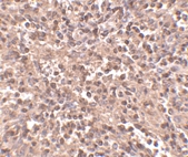
Below: Immunohistochemistry of ATG5 in human spleen tissue with ATG5 antibody at 2.5 µg/ml.
Other Product Images
 Source : ATG5 antibody was raised against a 16 amino acid peptide from near the amino terminus of human ATG5.
Purification : Affinity chromatography purified via peptide column
Clonality and Clone : This is a polyclonal antibody.
Host : ATG5 antibody was raised in rabbit. Please use anti-rabbit secondary antibodies.
Application : ATG5 antibody can be used for the detection of ATG5 by Western blot at 1 – 2 µg/ml.
Tested Application(s) : E, WB, IHC
Buffer : Antibody is supplied in PBS containing 0.02% sodium azide.
Blocking Peptide : Cat.No. 4441P - ATG5 Peptide
Long-Term Storage : ATG5 antibody can be stored at 4ºC, stable for one year. As with all antibodies care should be taken to avoid repeated freeze thaw cycles. Antibodies should not be exposed to prolonged high temperatures.
Positive Control
1. Cat. No. 1466 - Rat Spleen Tissue Lysate
Species Reactivity :H, M, R
GI Number : 119568800
Accession Number : EAW48415
Short Description : Autophagy related protein 5
References
1. Gozuacik D and Kimchi A. Autophagy as a cell death and tumor suppressor mechanism. Oncogene 2004; 23:2891-906.
2. Kisen GO, Tessitore L, Costelli P, et al. Reduced autophagic activity in primary rat hepatocellular carcinoma and ascites hepatoma cells. Carcinogenesis 1993; 14:2501-5.
3. Kamada Y, Funakoshi T, Shintani T, et al. Tor-mediated induction of autophagy via Apg1 protein kinase complex. J. Cell. Biol. 2000; 150:1507-13.
4. Hanada T, Noda NN, Satomi Y, et al. The Atg12-Atg5 conjugate has a novel E3-like activity for protein lipidation in autophagy. J. Biol. Chem. 2007; 282:37298-302.
Catalog# : 4441
Autophagy, the process of bulk degradation of cellular proteins through an autophagosomic-lysosomal pathway is important for normal growth control and may be defective in tumor cells. It is involved in the preservation of cellular nutrients under starvation conditions as well as the normal turnover of cytosolic components. This process is negatively regulated by TOR (Target of rapamycin) through phosphorylation of autophagy protein APG1. ATG5, another member of the autophagy protein family, forms a conjugate with ATG12; this conjugate has a ubiquitin-protein ligase (E3)-like activity for protein lipidation in autophagy. This conjugate also associates with innate immune response proteins such as RIG-I and VISA (also known as IPS-1), inhibiting type I interferon production and permitting viral replication in host cells. Three isoforms of ATG5 are known to exist; this ATG5 antibody will only detect the longest isoform.
Additional Names : ATG5, Autophagy protein 5, Autophagy related protein 5, ATG5L, ASP
DescriptionLeft: Western blot analysis of ATG5 in rat spleen tissue lysate with ATG5 antibody at (A) 1 and (B) 2 µg/ml.
Below: Immunohistochemistry of ATG5 in human spleen tissue with ATG5 antibody at 2.5 µg/ml.
Other Product Images
Source : ATG5 antibody was raised against a 16 amino acid peptide from near the amino terminus of human ATG5.Purification : Affinity chromatography purified via peptide column
Clonality and Clone : This is a polyclonal antibody.
Host : ATG5 antibody was raised in rabbit. Please use anti-rabbit secondary antibodies.
Application : ATG5 antibody can be used for the detection of ATG5 by Western blot at 1 – 2 µg/ml.
Tested Application(s) : E, WB, IHC
Buffer : Antibody is supplied in PBS containing 0.02% sodium azide.
Blocking Peptide : Cat.No. 4441P - ATG5 Peptide
Long-Term Storage : ATG5 antibody can be stored at 4ºC, stable for one year. As with all antibodies care should be taken to avoid repeated freeze thaw cycles. Antibodies should not be exposed to prolonged high temperatures.
Positive Control
1. Cat. No. 1466 - Rat Spleen Tissue Lysate
Species Reactivity :H, M, R
GI Number : 119568800
Accession Number : EAW48415
Short Description : Autophagy related protein 5
References
1. Gozuacik D and Kimchi A. Autophagy as a cell death and tumor suppressor mechanism. Oncogene 2004; 23:2891-906.
2. Kisen GO, Tessitore L, Costelli P, et al. Reduced autophagic activity in primary rat hepatocellular carcinoma and ascites hepatoma cells. Carcinogenesis 1993; 14:2501-5.
3. Kamada Y, Funakoshi T, Shintani T, et al. Tor-mediated induction of autophagy via Apg1 protein kinase complex. J. Cell. Biol. 2000; 150:1507-13.
4. Hanada T, Noda NN, Satomi Y, et al. The Atg12-Atg5 conjugate has a novel E3-like activity for protein lipidation in autophagy. J. Biol. Chem. 2007; 282:37298-302.
Tuesday, September 7, 2010
ATG12 Antibody
ATG12 Antibody
Catalog# : 4423
Autophagy, the process of bulk degradation of cellular proteins through an autophagosomic-lysosomal pathway is important for normal growth control and may be defective in tumor cells. It is involved in the preservation of cellular nutrients under starvation conditions as well as the normal turnover of cytosolic components. This process is negatively regulated by TOR (Target of rapamycin) through phosphorylation of autophagy protein APG1. ATG12, another member of the autophagy protein family, forms a conjugate with ATG5; this conjugate has a ubiquitin-protein ligase (E3)-like activity for protein lipidation in autophagy. This conjugate also associates with innate immune response proteins such as RIG-I and VISA (also known as IPS-1), inhibiting type I interferon production and permitting viral replication in host cells. ATG12 has also been shown to interact with ATG10 in human embryonic kidney cells in the presence of ATG7. At least two isoforms of ATG12 are known to exist.
Additional Names : ATG12 (IN), Autophagy protein 12, Autophagy related protein 12, APG12, APG12L, HAPG12
 Description
Left: Western blot analysis of ATG12 in mouse heart tissue lysate with ATG12 antibody at 1 µg/ml in (A) the absence and (B) the presence of blocking peptide.
Below: Immunohistochemistry of ATG12 in human brain tissue with ATG12 antibody at 2.5 µg/ml.
Other Product Images
 Source : ATG12 antibody was raised against a 15 amino acid peptide from near the center of human ATG12.
Purification : Affinity chromatography purified via peptide column
Clonality and Clone : This is a polyclonal antibody.
Host : ATG12 antibody was raised in rabbit. Please use anti-rabbit secondary antibodies.
Application : ATG12 antibody can be used for the detection of ATG10 by Western blot at 0.5 – 1 µg/ml.
Tested Application(s) : E, WB, IHC
Buffer : Antibody is supplied in PBS containing 0.02% sodium azide.
Blocking Peptide : Cat.No. 4423P - ATG12 Peptide
Long-Term Storage : ATG12 antibody can be stored at 4ºC, stable for one year. As with all antibodies care should be taken to avoid repeated freeze thaw cycles. Antibodies should not be exposed to prolonged high temperatures.
Positive Control
1. Cat. No. 1401 - Mouse Heart Tissue Lysate
Species Reactivity :H, M, R
GI Number : 119569340
Accession Number : EAW48955
Short Description : (IN) Autophagy protein 12
References
1. Gozuacik D and Kimchi A. Autophagy as a cell death and tumor suppressor mechanism. Oncogene 2004; 23:2891-906.
2. Kisen GO, Tessitore L, Costelli P, et al. Reduced autophagic activity in primary rat hepatocellular carcinoma and ascites hepatoma cells. Carcinogenesis 1993; 14:2501-5.
3. Kamada Y, Funakoshi T, Shintani T, et al. Tor-mediated induction of autophagy via Apg1 protein kinase complex. J. Cell. Biol. 2000; 150:1507-13.
4. Hanada T, Noda NN, Satomi Y, et al. The Atg12-Atg5 conjugate has a novel E3-like activity for protein lipidation in autophagy. J. Biol. Chem. 2007; 282:37298-302.
Catalog# : 4423
Autophagy, the process of bulk degradation of cellular proteins through an autophagosomic-lysosomal pathway is important for normal growth control and may be defective in tumor cells. It is involved in the preservation of cellular nutrients under starvation conditions as well as the normal turnover of cytosolic components. This process is negatively regulated by TOR (Target of rapamycin) through phosphorylation of autophagy protein APG1. ATG12, another member of the autophagy protein family, forms a conjugate with ATG5; this conjugate has a ubiquitin-protein ligase (E3)-like activity for protein lipidation in autophagy. This conjugate also associates with innate immune response proteins such as RIG-I and VISA (also known as IPS-1), inhibiting type I interferon production and permitting viral replication in host cells. ATG12 has also been shown to interact with ATG10 in human embryonic kidney cells in the presence of ATG7. At least two isoforms of ATG12 are known to exist.
Additional Names : ATG12 (IN), Autophagy protein 12, Autophagy related protein 12, APG12, APG12L, HAPG12
DescriptionLeft: Western blot analysis of ATG12 in mouse heart tissue lysate with ATG12 antibody at 1 µg/ml in (A) the absence and (B) the presence of blocking peptide.
Below: Immunohistochemistry of ATG12 in human brain tissue with ATG12 antibody at 2.5 µg/ml.
Other Product Images
Source : ATG12 antibody was raised against a 15 amino acid peptide from near the center of human ATG12.Purification : Affinity chromatography purified via peptide column
Clonality and Clone : This is a polyclonal antibody.
Host : ATG12 antibody was raised in rabbit. Please use anti-rabbit secondary antibodies.
Application : ATG12 antibody can be used for the detection of ATG10 by Western blot at 0.5 – 1 µg/ml.
Tested Application(s) : E, WB, IHC
Buffer : Antibody is supplied in PBS containing 0.02% sodium azide.
Blocking Peptide : Cat.No. 4423P - ATG12 Peptide
Long-Term Storage : ATG12 antibody can be stored at 4ºC, stable for one year. As with all antibodies care should be taken to avoid repeated freeze thaw cycles. Antibodies should not be exposed to prolonged high temperatures.
Positive Control
1. Cat. No. 1401 - Mouse Heart Tissue Lysate
Species Reactivity :H, M, R
GI Number : 119569340
Accession Number : EAW48955
Short Description : (IN) Autophagy protein 12
References
1. Gozuacik D and Kimchi A. Autophagy as a cell death and tumor suppressor mechanism. Oncogene 2004; 23:2891-906.
2. Kisen GO, Tessitore L, Costelli P, et al. Reduced autophagic activity in primary rat hepatocellular carcinoma and ascites hepatoma cells. Carcinogenesis 1993; 14:2501-5.
3. Kamada Y, Funakoshi T, Shintani T, et al. Tor-mediated induction of autophagy via Apg1 protein kinase complex. J. Cell. Biol. 2000; 150:1507-13.
4. Hanada T, Noda NN, Satomi Y, et al. The Atg12-Atg5 conjugate has a novel E3-like activity for protein lipidation in autophagy. J. Biol. Chem. 2007; 282:37298-302.
ATG12 Antibody
ATG12 Antibody
Catalog# : 4421
Autophagy, the process of bulk degradation of cellular proteins through an autophagosomic-lysosomal pathway is important for normal growth control and may be defective in tumor cells. It is involved in the preservation of cellular nutrients under starvation conditions as well as the normal turnover of cytosolic components. This process is negatively regulated by TOR (Target of rapamycin) through phosphorylation of autophagy protein APG1. ATG12, another member of the autophagy protein family, forms a conjugate with ATG5; this conjugate has a ubiquitin-protein ligase (E3)-like activity for protein lipidation in autophagy. This conjugate also associates with innate immune response proteins such as RIG-I and VISA (also known as IPS-1), inhibiting type I interferon production and permitting viral replication in host cells. ATG12 has also been shown to interact with ATG10 in human embryonic kidney cells in the presence of ATG7. At least two isoforms of ATG12 are known to exist.
Additional Names : ATG12 (NT), Autophagy protein 12, Autophagy related protein 12, APG12, APG12L, HAPG12
 Description
Left: Western blot analysis of ATG12 in human brain tissue lysate with ATG12 antibody at (A) 0.5, and (B) 1 µg/ml.
Below: Immunohistochemistry of ATG12 in human brain tissue with ATG12 antibody at 2.5 µg/ml.
Other Product Images
 Source : ATG12 antibody was raised against a 16 amino acid peptide from near the amino terminus of human ATG12.
Purification : Affinity chromatography purified via peptide column
Clonality and Clone : This is a polyclonal antibody.
Host : ATG12 antibody was raised in rabbit.Please use anti-rabbit secondary antibodies.
Application : ATG12 antibody can be used for the detection of ATG10 by Western blot at 0.5 – 1 µg/ml.
Tested Application(s) : E, WB, IHC
Buffer : Antibody is supplied in PBS containing 0.02% sodium azide.
Blocking Peptide : Cat.No. 4421P - ATG12 Peptide
Long-Term Storage : ATG12 antibody can be stored at 4ºC, stable for one year. As with all antibodies care should be taken to avoid repeated freeze thaw cycles. Antibodies should not be exposed to prolonged high temperatures.
Positive Control
1. Cat. No. 1303 - Human Brain Tissue Lysate
Species Reactivity :H, M, R
GI Number : 119569340
Accession Number : EAW48955
Short Description : (NT) Autophagy protein 12
References
1. Gozuacik D and Kimchi A. Autophagy as a cell death and tumor suppressor mechanism. Oncogene 2004; 23:2891-906.
2. Kisen GO, Tessitore L, Costelli P, et al. Reduced autophagic activity in primary rat hepatocellular carcinoma and ascites hepatoma cells. Carcinogenesis 1993; 14:2501-5.
3. Kamada Y, Funakoshi T, Shintani T, et al. Tor-mediated induction of autophagy via Apg1 protein kinase complex. J. Cell. Biol. 2000; 150:1507-13.
4. Hanada T, Noda NN, Satomi Y, et al. The Atg12-Atg5 conjugate has a novel E3-like activity for protein lipidation in autophagy. J. Biol. Chem. 2007; 282:37298-302.
Catalog# : 4421
Autophagy, the process of bulk degradation of cellular proteins through an autophagosomic-lysosomal pathway is important for normal growth control and may be defective in tumor cells. It is involved in the preservation of cellular nutrients under starvation conditions as well as the normal turnover of cytosolic components. This process is negatively regulated by TOR (Target of rapamycin) through phosphorylation of autophagy protein APG1. ATG12, another member of the autophagy protein family, forms a conjugate with ATG5; this conjugate has a ubiquitin-protein ligase (E3)-like activity for protein lipidation in autophagy. This conjugate also associates with innate immune response proteins such as RIG-I and VISA (also known as IPS-1), inhibiting type I interferon production and permitting viral replication in host cells. ATG12 has also been shown to interact with ATG10 in human embryonic kidney cells in the presence of ATG7. At least two isoforms of ATG12 are known to exist.
Additional Names : ATG12 (NT), Autophagy protein 12, Autophagy related protein 12, APG12, APG12L, HAPG12
DescriptionLeft: Western blot analysis of ATG12 in human brain tissue lysate with ATG12 antibody at (A) 0.5, and (B) 1 µg/ml.
Below: Immunohistochemistry of ATG12 in human brain tissue with ATG12 antibody at 2.5 µg/ml.
Other Product Images
Source : ATG12 antibody was raised against a 16 amino acid peptide from near the amino terminus of human ATG12.Purification : Affinity chromatography purified via peptide column
Clonality and Clone : This is a polyclonal antibody.
Host : ATG12 antibody was raised in rabbit.Please use anti-rabbit secondary antibodies.
Application : ATG12 antibody can be used for the detection of ATG10 by Western blot at 0.5 – 1 µg/ml.
Tested Application(s) : E, WB, IHC
Buffer : Antibody is supplied in PBS containing 0.02% sodium azide.
Blocking Peptide : Cat.No. 4421P - ATG12 Peptide
Long-Term Storage : ATG12 antibody can be stored at 4ºC, stable for one year. As with all antibodies care should be taken to avoid repeated freeze thaw cycles. Antibodies should not be exposed to prolonged high temperatures.
Positive Control
1. Cat. No. 1303 - Human Brain Tissue Lysate
Species Reactivity :H, M, R
GI Number : 119569340
Accession Number : EAW48955
Short Description : (NT) Autophagy protein 12
References
1. Gozuacik D and Kimchi A. Autophagy as a cell death and tumor suppressor mechanism. Oncogene 2004; 23:2891-906.
2. Kisen GO, Tessitore L, Costelli P, et al. Reduced autophagic activity in primary rat hepatocellular carcinoma and ascites hepatoma cells. Carcinogenesis 1993; 14:2501-5.
3. Kamada Y, Funakoshi T, Shintani T, et al. Tor-mediated induction of autophagy via Apg1 protein kinase complex. J. Cell. Biol. 2000; 150:1507-13.
4. Hanada T, Noda NN, Satomi Y, et al. The Atg12-Atg5 conjugate has a novel E3-like activity for protein lipidation in autophagy. J. Biol. Chem. 2007; 282:37298-302.
ATG10 Antibody
ATG10 Antibody
Catalog# : 4399
Autophagy, the process of bulk degradation of cellular proteins through an autophagosomic-lysosomal pathway is important for normal growth control and may be defective in tumor cells. It is involved in the preservation of cellular nutrients under starvation conditions as well as the normal turnover of cytosolic components. This process is negatively regulated by TOR (Target of rapamycin) through phosphorylation of autophagy protein APG1. Another member of the autophagy protein family is ATG10, an E2-like enzyme involved in two ubiquitin-like modifications essential for autophagosome formation: ATG12-ATG5 conjugation and the modification of a soluble form of MAP-LC3, a homolog of yeast Apg8, to a membrane-bound form. ATG10 has also been shown to interact with ATG12 in human embryonic kidney cells in the presence of ATG7. Multiple isoforms of ATG10 are known to exist.
Additional Names : ATG10, Autophagy protein 10, Autophagy related protein 10, APG10L
 Description
Left: Western blot analysis of ATG10 in SK-N-SH cell lysate with ATG10 antibody at (A) 0.5, (B) 1 and (C) 2 µg/ml.
Source : ATG10 antibody was raised against a 15 amino acid peptide from near the carboxy terminus of human ATG10.
Purification : Affinity chromatography purified via peptide column
Clonality and Clone : This is a polyclonal antibody.
Host : ATG10 antibody was raised in rabbit.Please use anti-rabbit secondary antibodies.
Application : ATG10 antibody can be used for the detection of ATG10 by Western blot at 0.5 – 1 µg/ml.
Tested Application(s) : E, WB
Buffer : Antibody is supplied in PBS containing 0.02% sodium azide.
Blocking Peptide : Cat.No. 4399P - ATG10 Peptide
Long-Term Storage : ATG10 antibody can be stored at 4ºC, stable for one year. As with all antibodies care should be taken to avoid repeated freeze thaw cycles. Antibodies should not be exposed to prolonged high temperatures.
Positive Control
1. Cat. No. 1220 - SK-N-SH Cell Lysate
Species Reactivity :H, M, R
GI Number : 119616290
Accession Number : EAW95884
Short Description : Autophagy protein 10
References
1. Gozuacik D and Kimchi A. Autophagy as a cell death and tumor suppressor mechanism. Oncogene 2004; 23:2891-906.
2. Kisen GO, Tessitore L, Costelli P, et al. Reduced autophagic activity in primary rat hepatocellular carcinoma and ascites hepatoma cells. Carcinogenesis 1993; 14:2501-5.
3. Kamada Y, Funakoshi T, Shintani T, et al. Tor-mediated induction of autophagy via Apg1 protein kinase complex. J. Cell. Biol. 2000; 150:1507-13.
4. Nemoto T, Tanida I, Tanida–Miyake E, et al. The mouse APG10 homologue, an E2-like enzyme for APG12p conjugation, facilitates MAP-LC3 modification. J. Biol. Chem. 2003; 278:39517-26.
Catalog# : 4399
Autophagy, the process of bulk degradation of cellular proteins through an autophagosomic-lysosomal pathway is important for normal growth control and may be defective in tumor cells. It is involved in the preservation of cellular nutrients under starvation conditions as well as the normal turnover of cytosolic components. This process is negatively regulated by TOR (Target of rapamycin) through phosphorylation of autophagy protein APG1. Another member of the autophagy protein family is ATG10, an E2-like enzyme involved in two ubiquitin-like modifications essential for autophagosome formation: ATG12-ATG5 conjugation and the modification of a soluble form of MAP-LC3, a homolog of yeast Apg8, to a membrane-bound form. ATG10 has also been shown to interact with ATG12 in human embryonic kidney cells in the presence of ATG7. Multiple isoforms of ATG10 are known to exist.
Additional Names : ATG10, Autophagy protein 10, Autophagy related protein 10, APG10L
DescriptionLeft: Western blot analysis of ATG10 in SK-N-SH cell lysate with ATG10 antibody at (A) 0.5, (B) 1 and (C) 2 µg/ml.
Source : ATG10 antibody was raised against a 15 amino acid peptide from near the carboxy terminus of human ATG10.
Purification : Affinity chromatography purified via peptide column
Clonality and Clone : This is a polyclonal antibody.
Host : ATG10 antibody was raised in rabbit.Please use anti-rabbit secondary antibodies.
Application : ATG10 antibody can be used for the detection of ATG10 by Western blot at 0.5 – 1 µg/ml.
Tested Application(s) : E, WB
Buffer : Antibody is supplied in PBS containing 0.02% sodium azide.
Blocking Peptide : Cat.No. 4399P - ATG10 Peptide
Long-Term Storage : ATG10 antibody can be stored at 4ºC, stable for one year. As with all antibodies care should be taken to avoid repeated freeze thaw cycles. Antibodies should not be exposed to prolonged high temperatures.
Positive Control
1. Cat. No. 1220 - SK-N-SH Cell Lysate
Species Reactivity :H, M, R
GI Number : 119616290
Accession Number : EAW95884
Short Description : Autophagy protein 10
References
1. Gozuacik D and Kimchi A. Autophagy as a cell death and tumor suppressor mechanism. Oncogene 2004; 23:2891-906.
2. Kisen GO, Tessitore L, Costelli P, et al. Reduced autophagic activity in primary rat hepatocellular carcinoma and ascites hepatoma cells. Carcinogenesis 1993; 14:2501-5.
3. Kamada Y, Funakoshi T, Shintani T, et al. Tor-mediated induction of autophagy via Apg1 protein kinase complex. J. Cell. Biol. 2000; 150:1507-13.
4. Nemoto T, Tanida I, Tanida–Miyake E, et al. The mouse APG10 homologue, an E2-like enzyme for APG12p conjugation, facilitates MAP-LC3 modification. J. Biol. Chem. 2003; 278:39517-26.
Ambra1 Antibody
Ambra1 Antibody
Catalog# : 4557
Autophagy, the process of bulk degradation of cellular proteins through an autophagosomic-lysosomal pathway is important for normal growth control and may be defective in tumor cells. It is involved in the preservation of cellular nutrients under starvation conditions as well as the normal turnover of cytosolic components. Beclin-1, a principal regulator of autophagosome formation, is in turn regulated by Ambra1. Ambra1 associates with Beclin-1 through a region near its center as determined by yeast two-hybrid assay. Null mutations in this gene in mice resulted in embryonic lethality with severe neural tube defects associated with autophagy impairment, accumulation of ubiquitinated proteins, unbalanced cell proliferation and excessive apoptotic death . Furthermore, down-regulation of Ambra1 in cultured cells though RNA interference decreased the level of rapamycin- and nutrient starvation-induced autophagy. Multiple isoforms of Ambra1 are known to exist.
Additional Names : Ambra1 (NT), Activating molecule in beclin-1-regulated autophagy, WDR94
 Description
Left: Western blot analysis of Ambra1 in rat brain tissue lysate with Ambra1 antibody at 2 µg/ml.
Below: Immunohistochemistry of Ambra1 in human brain with Ambra1 antibody at 5 µg/ml.
Other Product Images
 Source : Ambra1 antibody was raised against a 18 amino acid peptide from near the amino terminus of human Ambra1.
Purification : Affinity chromatography purified via peptide column
Clonality and Clone : This is a polyclonal antibody.
Host : Ambra1 antibody was raised in rabbit. Please use anti-rabbit secondary antibodies.
Application : Ambra1 antibody can be used for the detection of Ambra1 by Western blot at 2 – 4 µg/ml.
Tested Application(s) : E, WB, IHC
Buffer : Antibody is supplied in PBS containing 0.02% sodium azide.
Blocking Peptide : Cat.No. 4557P - Ambra1 Peptide
Long-Term Storage : Ambra1 antibody can be stored at 4ºC, stable for one year. As with all antibodies care should be taken to avoid repeated freeze thaw cycles. Antibodies should not be exposed to prolonged high temperatures.
Positive Control
1. Cat. No. 1463 - Rat Brain Tissue Lysate
Species Reactivity :H, M
GI Number : 166215833
Accession Number : Q9C0C7
Short Description : (NT) autophagy regulating protein
References
1. Gozuacik D and Kimchi A. Autophagy as a cell death and tumor suppressor mechanism. Oncogene 2004; 23:2891-906.
2. Kisen GO, Tessitore L, Costelli P, et al. Reduced autophagic activity in primary rat hepatocellular carcinoma and ascites hepatoma cells. Carcinogenesis 1993; 14:2501-5.
3. Liang XH, Jackson S, Seaman M, et al. Induction of autophagy and inhibition of tumorigenesis by beclin 1. Nature 1999; 402:672-6.
4. Fimia GM, Stoykova A, Romagnoli A, et al. Ambra1 regulates autophagy and development of the nervous system. Nature 2007; 447:1121-5.
Catalog# : 4557
Autophagy, the process of bulk degradation of cellular proteins through an autophagosomic-lysosomal pathway is important for normal growth control and may be defective in tumor cells. It is involved in the preservation of cellular nutrients under starvation conditions as well as the normal turnover of cytosolic components. Beclin-1, a principal regulator of autophagosome formation, is in turn regulated by Ambra1. Ambra1 associates with Beclin-1 through a region near its center as determined by yeast two-hybrid assay. Null mutations in this gene in mice resulted in embryonic lethality with severe neural tube defects associated with autophagy impairment, accumulation of ubiquitinated proteins, unbalanced cell proliferation and excessive apoptotic death . Furthermore, down-regulation of Ambra1 in cultured cells though RNA interference decreased the level of rapamycin- and nutrient starvation-induced autophagy. Multiple isoforms of Ambra1 are known to exist.
Additional Names : Ambra1 (NT), Activating molecule in beclin-1-regulated autophagy, WDR94
DescriptionLeft: Western blot analysis of Ambra1 in rat brain tissue lysate with Ambra1 antibody at 2 µg/ml.
Below: Immunohistochemistry of Ambra1 in human brain with Ambra1 antibody at 5 µg/ml.
Other Product Images
Source : Ambra1 antibody was raised against a 18 amino acid peptide from near the amino terminus of human Ambra1.Purification : Affinity chromatography purified via peptide column
Clonality and Clone : This is a polyclonal antibody.
Host : Ambra1 antibody was raised in rabbit. Please use anti-rabbit secondary antibodies.
Application : Ambra1 antibody can be used for the detection of Ambra1 by Western blot at 2 – 4 µg/ml.
Tested Application(s) : E, WB, IHC
Buffer : Antibody is supplied in PBS containing 0.02% sodium azide.
Blocking Peptide : Cat.No. 4557P - Ambra1 Peptide
Long-Term Storage : Ambra1 antibody can be stored at 4ºC, stable for one year. As with all antibodies care should be taken to avoid repeated freeze thaw cycles. Antibodies should not be exposed to prolonged high temperatures.
Positive Control
1. Cat. No. 1463 - Rat Brain Tissue Lysate
Species Reactivity :H, M
GI Number : 166215833
Accession Number : Q9C0C7
Short Description : (NT) autophagy regulating protein
References
1. Gozuacik D and Kimchi A. Autophagy as a cell death and tumor suppressor mechanism. Oncogene 2004; 23:2891-906.
2. Kisen GO, Tessitore L, Costelli P, et al. Reduced autophagic activity in primary rat hepatocellular carcinoma and ascites hepatoma cells. Carcinogenesis 1993; 14:2501-5.
3. Liang XH, Jackson S, Seaman M, et al. Induction of autophagy and inhibition of tumorigenesis by beclin 1. Nature 1999; 402:672-6.
4. Fimia GM, Stoykova A, Romagnoli A, et al. Ambra1 regulates autophagy and development of the nervous system. Nature 2007; 447:1121-5.
Ambra1 Antibody
Ambra1 Antibody
Catalog# : 4555
Autophagy, the process of bulk degradation of cellular proteins through an autophagosomic-lysosomal pathway is important for normal growth control and may be defective in tumor cells. It is involved in the preservation of cellular nutrients under starvation conditions as well as the normal turnover of cytosolic components. Beclin-1, a principal regulator of autophagosome formation, is in turn regulated by Ambra1. Ambra1 associates with Beclin-1 through a region near its center as determined by yeast two-hybrid assay. Null mutations in this gene in mice resulted in embryonic lethality with severe neural tube defects associated with autophagy impairment, accumulation of ubiquitinated proteins, unbalanced cell proliferation and excessive apoptotic death . Furthermore, down-regulation of Ambra1 in cultured cells though RNA interference decreased the level of rapamycin- and nutrient starvation-induced autophagy. Multiple isoforms of Ambra1 are known to exist.
Additional Names : Ambra1 (CT), Activating molecule in beclin-1-regulated autophagy, WDR94
 Description
Left: Western blot analysis of Ambra1 in 3T3 cell lysate with Ambra1 antibody at 1 µg/ml.
Below: Immunohistochemistry of Ambra1 in human brain with Ambra1 antibody at 5 µg/ml.
Other Product Images
 Source : Ambra1 antibody was raised against a 15 amino acid peptide from near the carboxy terminus of human Ambra1.
Purification : Affinity chromatography purified via peptide column
Clonality and Clone : This is a polyclonal antibody.
Host : Ambra1 antibody was raised in rabbit. Please use anti-rabbit secondary antibodies.
Application : Ambra1 antibody can be used for the detection of Ambra1 by Western blot at 1 – 2 µg/ml.
Tested Application(s) : E, WB, IHC
Buffer : Antibody is supplied in PBS containing 0.02% sodium azide.
Blocking Peptide : Cat.No. 4555P - Ambra1 Peptide
Long-Term Storage : Ambra1 antibody can be stored at 4ºC, stable for one year. As with all antibodies care should be taken to avoid repeated freeze thaw cycles. Antibodies should not be exposed to prolonged high temperatures.
Positive Control
1. Cat. No. 1282 - 3T3 (NIH) Cell Lysate
Species Reactivity :H, M, R
GI Number : 166215833
Accession Number : Q9C0C7
Short Description : (CT) autophagy regulating protein
References
1. Gozuacik D and Kimchi A. Autophagy as a cell death and tumor suppressor mechanism. Oncogene 2004; 23:2891-906.
2. Kisen GO, Tessitore L, Costelli P, et al. Reduced autophagic activity in primary rat hepatocellular carcinoma and ascites hepatoma cells. Carcinogenesis 1993; 14:2501-5.
3. Liang XH, Jackson S, Seaman M, et al. Induction of autophagy and inhibition of tumorigenesis by beclin 1. Nature 1999; 402:672-6.
4. Fimia GM, Stoykova A, Romagnoli A, et al. Ambra1 regulates autophagy and development of the nervous system. Nature 2007; 447:1121-5.
Catalog# : 4555
Autophagy, the process of bulk degradation of cellular proteins through an autophagosomic-lysosomal pathway is important for normal growth control and may be defective in tumor cells. It is involved in the preservation of cellular nutrients under starvation conditions as well as the normal turnover of cytosolic components. Beclin-1, a principal regulator of autophagosome formation, is in turn regulated by Ambra1. Ambra1 associates with Beclin-1 through a region near its center as determined by yeast two-hybrid assay. Null mutations in this gene in mice resulted in embryonic lethality with severe neural tube defects associated with autophagy impairment, accumulation of ubiquitinated proteins, unbalanced cell proliferation and excessive apoptotic death . Furthermore, down-regulation of Ambra1 in cultured cells though RNA interference decreased the level of rapamycin- and nutrient starvation-induced autophagy. Multiple isoforms of Ambra1 are known to exist.
Additional Names : Ambra1 (CT), Activating molecule in beclin-1-regulated autophagy, WDR94
DescriptionLeft: Western blot analysis of Ambra1 in 3T3 cell lysate with Ambra1 antibody at 1 µg/ml.
Below: Immunohistochemistry of Ambra1 in human brain with Ambra1 antibody at 5 µg/ml.
Other Product Images
Source : Ambra1 antibody was raised against a 15 amino acid peptide from near the carboxy terminus of human Ambra1.Purification : Affinity chromatography purified via peptide column
Clonality and Clone : This is a polyclonal antibody.
Host : Ambra1 antibody was raised in rabbit. Please use anti-rabbit secondary antibodies.
Application : Ambra1 antibody can be used for the detection of Ambra1 by Western blot at 1 – 2 µg/ml.
Tested Application(s) : E, WB, IHC
Buffer : Antibody is supplied in PBS containing 0.02% sodium azide.
Blocking Peptide : Cat.No. 4555P - Ambra1 Peptide
Long-Term Storage : Ambra1 antibody can be stored at 4ºC, stable for one year. As with all antibodies care should be taken to avoid repeated freeze thaw cycles. Antibodies should not be exposed to prolonged high temperatures.
Positive Control
1. Cat. No. 1282 - 3T3 (NIH) Cell Lysate
Species Reactivity :H, M, R
GI Number : 166215833
Accession Number : Q9C0C7
Short Description : (CT) autophagy regulating protein
References
1. Gozuacik D and Kimchi A. Autophagy as a cell death and tumor suppressor mechanism. Oncogene 2004; 23:2891-906.
2. Kisen GO, Tessitore L, Costelli P, et al. Reduced autophagic activity in primary rat hepatocellular carcinoma and ascites hepatoma cells. Carcinogenesis 1993; 14:2501-5.
3. Liang XH, Jackson S, Seaman M, et al. Induction of autophagy and inhibition of tumorigenesis by beclin 1. Nature 1999; 402:672-6.
4. Fimia GM, Stoykova A, Romagnoli A, et al. Ambra1 regulates autophagy and development of the nervous system. Nature 2007; 447:1121-5.
Enzyme Bioconjugation
Various enzymes, such as HRP and AP, can be attached to antibodies and proteins to act as signal-generating molecules. Enzymes may be modified to contain homobifunctional or heterobifunctional reactive groups that can covalently couple to some chemical target on the enzyme and result in a terminally reactive group that can crosslink with another molecule.
Methods of Covalently Coupled Enzymes with Antiboby
Enzymes can be covalently coupled to our clients’ specific antibody using various methods like SMCC-Activated Enzymes, Periodate oxidation methods, Glutaraldehyde methods, Hydrazide-activated enzymes, SPDP heterobifunctional cross linking chemistry. As each conjugation is unique, our support team will work closely with each client to identify their objective and determine proper coupling chemistry. As we are the experts in enzyme conjugation techniques, BSI can provide a properly conjugated product quickly and efficiently.
Type of Enzyme
There are various types of enzymes like Horseradish Peroxidase (HRP), Alkaline Phosphatase (AP), ß-Galactosidease (also called lactase), Glucose Oxidase.
Services
Bio-Synthesis offer various services like: Oligonucleotide-enzyme conjugation, Enzyme-antibody conjugation, Enzyme-peptide conjugation and more.
Monday, September 6, 2010
ZIPK Antibody
ZIPK Antibody
Catalog# : 2067
Apoptosis is mediated by death domain containing adapter molecules and a caspase family of proteases. Certain serine/threonine protein kinases, such as ASK-1 and RIP, are mediators of apoptosis. A novel serine/threonine kinase that mediates apoptosis was recently identified and designated ZIP kinase . ZIP kinase contains an N-terminal kinase domain and a C-terminal leucine zipper structure and binds to ATF4 that is a member of ATF/CREB family. ZIP kinase has high sequence homology to DAP kinase (death-associated protein kinase), which is a mediator of apoptosis induced by gamma interferon. Overexpression of ZIP kinase induces apoptosis. ZIP and DAP kinases represent a novel kinase family, which mediates apoptosis through their catalytic activities. The messenger RNA was ubiquitously expressed in various tissues .
Additional Names : ZIPK (IN), ZIPK
 Description
Left: Western blot analysis of ZIP kinase in HeLa (H) and Jurkat (J) whole cell lysates with ZIP kinase antibody at 1 µg/ml.
Below: Immunocytochemistry of ZIP kinase in Jurkat cells with ZIP kinase antibody at 10 µg/ml.
Other Product Images
Source : ZIP kinase antibody was raised against a peptide corresponding to amino acids near the center of human ZIP kinase.
Purification : Affinity chromatography purified via peptide column
Clonality and Clone : This is a polyclonal antibody.
Host : ZIPK antibody was raised in rabbit. Please use anti-rabbit secondary antibodies.
Immunogen : Human ZIPK (Intermediate Domain) Peptide (Cat. No. 2067P)
Application : ZIPK antibody can be used for detection of DNase II expression by Western blot at 1:500 to 1:1000 dilution. An approximate 40 kDa band can be detected, which represents the pro-enzyme of DNase II.
Tested Application(s) : E, WB, ICC
Buffer : Antibody is supplied in PBS containing 0.02% sodium azide.
Blocking Peptide : Cat. No. 2067P - ZIPK Peptide
Long-Term Storage : ZIPK antibody can be stored at 4ºC, stable for one year. As with all antibodies care should be taken to avoid repeated freeze thaw cycles. Antibodies should not be exposed to prolonged high temperatures.
Positive Control
1. Cat. No. 1306 - Human Spleen Tissue Lysate
2. Cat. No. 1208 - THP-1 Whole Cell Lysate
Species Reactivity :H, M, R
GI Number : 5162884
Accession Number : BAA81746
Short Description : (IN) Apoptosis inducing kinase
References
1. Kawai T, Matsumoto M, Takeda K, Sanjo H, Akira S. ZIP kinase, a novel serine/threonine kinase which mediates apoptosis. Mol Cell Biol 1998;18:1642-51 (RD1299)
Catalog# : 2067
Apoptosis is mediated by death domain containing adapter molecules and a caspase family of proteases. Certain serine/threonine protein kinases, such as ASK-1 and RIP, are mediators of apoptosis. A novel serine/threonine kinase that mediates apoptosis was recently identified and designated ZIP kinase . ZIP kinase contains an N-terminal kinase domain and a C-terminal leucine zipper structure and binds to ATF4 that is a member of ATF/CREB family. ZIP kinase has high sequence homology to DAP kinase (death-associated protein kinase), which is a mediator of apoptosis induced by gamma interferon. Overexpression of ZIP kinase induces apoptosis. ZIP and DAP kinases represent a novel kinase family, which mediates apoptosis through their catalytic activities. The messenger RNA was ubiquitously expressed in various tissues .
Additional Names : ZIPK (IN), ZIPK
DescriptionLeft: Western blot analysis of ZIP kinase in HeLa (H) and Jurkat (J) whole cell lysates with ZIP kinase antibody at 1 µg/ml.
Below: Immunocytochemistry of ZIP kinase in Jurkat cells with ZIP kinase antibody at 10 µg/ml.
Other Product Images
Source : ZIP kinase antibody was raised against a peptide corresponding to amino acids near the center of human ZIP kinase.
Purification : Affinity chromatography purified via peptide column
Clonality and Clone : This is a polyclonal antibody.
Host : ZIPK antibody was raised in rabbit. Please use anti-rabbit secondary antibodies.
Immunogen : Human ZIPK (Intermediate Domain) Peptide (Cat. No. 2067P)
Application : ZIPK antibody can be used for detection of DNase II expression by Western blot at 1:500 to 1:1000 dilution. An approximate 40 kDa band can be detected, which represents the pro-enzyme of DNase II.
Tested Application(s) : E, WB, ICC
Buffer : Antibody is supplied in PBS containing 0.02% sodium azide.
Blocking Peptide : Cat. No. 2067P - ZIPK Peptide
Long-Term Storage : ZIPK antibody can be stored at 4ºC, stable for one year. As with all antibodies care should be taken to avoid repeated freeze thaw cycles. Antibodies should not be exposed to prolonged high temperatures.
Positive Control
1. Cat. No. 1306 - Human Spleen Tissue Lysate
2. Cat. No. 1208 - THP-1 Whole Cell Lysate
Species Reactivity :H, M, R
GI Number : 5162884
Accession Number : BAA81746
Short Description : (IN) Apoptosis inducing kinase
References
1. Kawai T, Matsumoto M, Takeda K, Sanjo H, Akira S. ZIP kinase, a novel serine/threonine kinase which mediates apoptosis. Mol Cell Biol 1998;18:1642-51 (RD1299)
XIAP Antibody
XIAP Antibody
Catalog# : 3331
Apoptosis, or programmed cell death, is related to many diseases, such as cancer. Apoptosis is triggered by a variety of stimuli including members in the TNF family and can be prevented by the inhibitor of apoptosis (IAP) proteins. IAP proteins form a conserved gene family that binds to and inhibits cell death proteases (1 for review). The X-chromosome linked inhibitor of apoptosis (XIAP) contains 3 baculoviral IAP repeat (BIR) motifs that are essential and sufficient for the binding and inhibition of caspases–3, –7, and –9 (2,3). Upregulation of XIAP expression can protect cells from apoptosis induced by low level radiation; conversely, decreased XIAP expression by antisense targeting resulted in increased cell death following low level radiation (4). Two negative regulators, termed XAF-1 (5) and Smac (6), can bind and inhibit XIAP activity.
Additional Names : XIAP (CT), XIAP
 Description
Left: Western blot analysis of XIAP in human kidney lysate with XIAP antibody at 0.5 (lane A), 1 (lane B), and 2 (lane C) µg/ml, respectively.
Below: Immunohistochemical staining of human kidney tissue using XIAP antibody at 2 µg/ml.
Other Product Images
 Source : XIAP antibody was raised against a synthetic peptide corresponding to 13 amino acids at the C-terminus of human XIAP.
Purification : Affinity chromatography purified via peptide column
Clonality and Clone : This is a polyclonal antibody.
Host : XIAP antibody was raised in rabbit. Please use anti-rabbit secondary antibodies.
Immunogen : Human XIAP (C-Terminus) Peptide (Cat. No. 3331P)
Application : XIAP antibody can be used for the detection of XIAP by Western blot at 0.5 to 2 µg/ml.Human kidney cell lysate can be used as positive control. Anti-XIAP is human and mouse reactive.
Tested Application(s) : E, WB, IHC
Buffer : Antibody is supplied in PBS containing 0.02% sodium azide.
Blocking Peptide : Cat. No. 3331P - XIAP Peptide
Long-Term Storage : XIAP antibody can be stored at 4ºC, stable for one year. As with all antibodies care should be taken to avoid repeated freeze thaw cycles. Antibodies should not be exposed to prolonged high temperatures.
Positive Control
1. Cat. No. 1324 - Human Kidney Tissue Lysate
Species Reactivity :H, M
GI Number : 32528299
Accession Number : NP_001158
Short Description : (CT) X-linked inhibitor of apoptosis protein
References
1. Schimmer AD. Inhibitor of apoptosis proteins: translating basic knowledge into clinical practice. Cancer Res. 2004; 64:7183-90.
2. Deveraux QL, Takahashi R, Savesan GS, et al. X-linked IAP is a direct inhibitor of cell-death proteases. Nature 1997; 388:300-4.
3. Deveraux QL, Leo E, Stennicke HR, et al. Cleavage of human inhibitor of apoptosis protein XIAP results in fragments with distinct specificities for caspases. EMBO J. 1999; 18:5242-51.
4. Holcik M, Yeh C, Korneluk RG, et al. Translational upregulation of X-linked inhibitor of apoptosis (XIAP) increases resistance to radiation induced cell death. Oncogene 2000; 19:4174-7.
Catalog# : 3331
Apoptosis, or programmed cell death, is related to many diseases, such as cancer. Apoptosis is triggered by a variety of stimuli including members in the TNF family and can be prevented by the inhibitor of apoptosis (IAP) proteins. IAP proteins form a conserved gene family that binds to and inhibits cell death proteases (1 for review). The X-chromosome linked inhibitor of apoptosis (XIAP) contains 3 baculoviral IAP repeat (BIR) motifs that are essential and sufficient for the binding and inhibition of caspases–3, –7, and –9 (2,3). Upregulation of XIAP expression can protect cells from apoptosis induced by low level radiation; conversely, decreased XIAP expression by antisense targeting resulted in increased cell death following low level radiation (4). Two negative regulators, termed XAF-1 (5) and Smac (6), can bind and inhibit XIAP activity.
Additional Names : XIAP (CT), XIAP
DescriptionLeft: Western blot analysis of XIAP in human kidney lysate with XIAP antibody at 0.5 (lane A), 1 (lane B), and 2 (lane C) µg/ml, respectively.
Below: Immunohistochemical staining of human kidney tissue using XIAP antibody at 2 µg/ml.
Other Product Images
Source : XIAP antibody was raised against a synthetic peptide corresponding to 13 amino acids at the C-terminus of human XIAP.Purification : Affinity chromatography purified via peptide column
Clonality and Clone : This is a polyclonal antibody.
Host : XIAP antibody was raised in rabbit. Please use anti-rabbit secondary antibodies.
Immunogen : Human XIAP (C-Terminus) Peptide (Cat. No. 3331P)
Application : XIAP antibody can be used for the detection of XIAP by Western blot at 0.5 to 2 µg/ml.Human kidney cell lysate can be used as positive control. Anti-XIAP is human and mouse reactive.
Tested Application(s) : E, WB, IHC
Buffer : Antibody is supplied in PBS containing 0.02% sodium azide.
Blocking Peptide : Cat. No. 3331P - XIAP Peptide
Long-Term Storage : XIAP antibody can be stored at 4ºC, stable for one year. As with all antibodies care should be taken to avoid repeated freeze thaw cycles. Antibodies should not be exposed to prolonged high temperatures.
Positive Control
1. Cat. No. 1324 - Human Kidney Tissue Lysate
Species Reactivity :H, M
GI Number : 32528299
Accession Number : NP_001158
Short Description : (CT) X-linked inhibitor of apoptosis protein
References
1. Schimmer AD. Inhibitor of apoptosis proteins: translating basic knowledge into clinical practice. Cancer Res. 2004; 64:7183-90.
2. Deveraux QL, Takahashi R, Savesan GS, et al. X-linked IAP is a direct inhibitor of cell-death proteases. Nature 1997; 388:300-4.
3. Deveraux QL, Leo E, Stennicke HR, et al. Cleavage of human inhibitor of apoptosis protein XIAP results in fragments with distinct specificities for caspases. EMBO J. 1999; 18:5242-51.
4. Holcik M, Yeh C, Korneluk RG, et al. Translational upregulation of X-linked inhibitor of apoptosis (XIAP) increases resistance to radiation induced cell death. Oncogene 2000; 19:4174-7.
XAF-1 Antibody
XAF-1 Antibody
Catalog# : 3207
XAF-1 binds to XIAP, an inhibitor of caspases-3, -7, and -9, and triggers its relocation from the cytosol to the nucleus (1,2). Overexpression of XAF-1 results in the neutralization of XIAP's ability to inhibit cell death (1). XAF-1 is normally expressed in all adult and fetal tissues but was found to be present in very low levels in a variety of cancer cell lines (3). In contrast, XIAP levels have been shown to be high in a majority of cell lines. Low XAF-1 and high basal expression of XIAP may therefore play a critical role in maintaining survival of cancer cell lines (2,3). Both IFN-alpha2 and IFN-beta can induce XAF-1 mRNA in all cells examined but induction of XAF-1 protein (as observed by immunoblot analysis) was seen only in cell lines sensitive to the apoptotic effects of IFNs (4).
Additional Names : XAF-1
 Description
Left: Western blot analysis of XAF-1 in human spleen lysate with XAF-1 antibody at 0.5 (lane A), 1 (lane B), and 2 (lane C) µg/ml, respectively.
Below: Immunohistochemical staining of human spleen tissue using XAF-1 antibody at 2 µg/ml.
Other Product Images
 Source : XAF-1 antibody was raised against a synthetic peptide corresponding to amino acids at the C-terminus of human XAF-1.
Purification : Affinity chromatography purified via peptide column
Clonality and Clone : This is a polyclonal antibody.
Host : XAF-1 antibody was raised in rabbit. Please use anti-rabbit secondary antibodies.
Immunogen : Human XAF-1 (C-Terminus) Peptide (Cat. No. 3207P)
Application : XAF-1 antibody can be used for the detection of XAF-1 by Western blot at 0.5 to 2 µg/ml.A 32 kDa band can be detected.
Tested Application(s) : E, WB, IHC
Buffer : Antibody is supplied in PBS containing 0.02% sodium azide.
Blocking Peptide : Cat. No. 3207P - XAF-1 Peptide
Long-Term Storage : XAF-1 antibody can be stored at 4ºC, stable for one year. As with all antibodies care should be taken to avoid repeated freeze thaw cycles. Antibodies should not be exposed to prolonged high temperatures.
Positive Control
1. Cat. No. 1306 - Human Spleen Tissue Lysate
Species Reactivity :H, M
GI Number : 1869901
Accession Number : CAA68030
Short Description : Inhibitor of XIAP
References
1. Liston P, Fong W, Kelly NL, et al. Identification of XAF1 as an antagonist of XIAP anticaspase activity. Nature Cell Biol. 2001;3:128-33.
2. Deveraux QL, Takahashi R, Savesan GS, and Reed JC. X-linked IAP is a direct inhibitor of cell-death proteases. Nature 1997;388:300-4.
3. Fong WG, Liston P, Rajcan-Separovic E, et al. Expression and genetic analysis of XIAP-associated factor 1 (XAF1) in cancer cell lines. Genomics 2000;70:113-122.
4. Leaman DW, Chawla-Sarkar M, Vyas K, et al. Identification of X-linked inhibitor of apoptosis associated factor-1 as an interferon-stimulated gene that augments TRAIL/Apo2L-induced apoptosis. J. Biol. Chem. 2002;277:28504-11.
Catalog# : 3207
XAF-1 binds to XIAP, an inhibitor of caspases-3, -7, and -9, and triggers its relocation from the cytosol to the nucleus (1,2). Overexpression of XAF-1 results in the neutralization of XIAP's ability to inhibit cell death (1). XAF-1 is normally expressed in all adult and fetal tissues but was found to be present in very low levels in a variety of cancer cell lines (3). In contrast, XIAP levels have been shown to be high in a majority of cell lines. Low XAF-1 and high basal expression of XIAP may therefore play a critical role in maintaining survival of cancer cell lines (2,3). Both IFN-alpha2 and IFN-beta can induce XAF-1 mRNA in all cells examined but induction of XAF-1 protein (as observed by immunoblot analysis) was seen only in cell lines sensitive to the apoptotic effects of IFNs (4).
Additional Names : XAF-1
DescriptionLeft: Western blot analysis of XAF-1 in human spleen lysate with XAF-1 antibody at 0.5 (lane A), 1 (lane B), and 2 (lane C) µg/ml, respectively.
Below: Immunohistochemical staining of human spleen tissue using XAF-1 antibody at 2 µg/ml.
Other Product Images
Source : XAF-1 antibody was raised against a synthetic peptide corresponding to amino acids at the C-terminus of human XAF-1.Purification : Affinity chromatography purified via peptide column
Clonality and Clone : This is a polyclonal antibody.
Host : XAF-1 antibody was raised in rabbit. Please use anti-rabbit secondary antibodies.
Immunogen : Human XAF-1 (C-Terminus) Peptide (Cat. No. 3207P)
Application : XAF-1 antibody can be used for the detection of XAF-1 by Western blot at 0.5 to 2 µg/ml.A 32 kDa band can be detected.
Tested Application(s) : E, WB, IHC
Buffer : Antibody is supplied in PBS containing 0.02% sodium azide.
Blocking Peptide : Cat. No. 3207P - XAF-1 Peptide
Long-Term Storage : XAF-1 antibody can be stored at 4ºC, stable for one year. As with all antibodies care should be taken to avoid repeated freeze thaw cycles. Antibodies should not be exposed to prolonged high temperatures.
Positive Control
1. Cat. No. 1306 - Human Spleen Tissue Lysate
Species Reactivity :H, M
GI Number : 1869901
Accession Number : CAA68030
Short Description : Inhibitor of XIAP
References
1. Liston P, Fong W, Kelly NL, et al. Identification of XAF1 as an antagonist of XIAP anticaspase activity. Nature Cell Biol. 2001;3:128-33.
2. Deveraux QL, Takahashi R, Savesan GS, and Reed JC. X-linked IAP is a direct inhibitor of cell-death proteases. Nature 1997;388:300-4.
3. Fong WG, Liston P, Rajcan-Separovic E, et al. Expression and genetic analysis of XIAP-associated factor 1 (XAF1) in cancer cell lines. Genomics 2000;70:113-122.
4. Leaman DW, Chawla-Sarkar M, Vyas K, et al. Identification of X-linked inhibitor of apoptosis associated factor-1 as an interferon-stimulated gene that augments TRAIL/Apo2L-induced apoptosis. J. Biol. Chem. 2002;277:28504-11.
Sunday, September 5, 2010
Liposome Bioconjugation
Bio Synthesis offer custom conjugation of liposome by covalently attached peptides, oligonucleotides, antibodies to the surface of liposomes or lipid emulsions. Lipsome bioconjugations are used as delivery devices to encapsulate cosmetics, drugs, fluorescent detection reagents and as vehicles to transport nucleic acids peptides, and proteins to cellular site in vivo. Antibody can be attached to liposomal surfaces and used to create large antigen- specific complexes for generation of specific antibodies or as vaccine carrier to elicit protective immunity.
End-Products of Liposome Technology
The various end products are Phosphatidyl Choline (PC), Lyso-phosphatidylcholine, Phosphatidyl Glycerol (PG), Phosphatidic acid, Phosphatidyl Inositol (PI), Phosphatidyl Ethanolamine (PE), Phosphatidyl Serine (PS), PEG-phospholipids, Polyglycerine phospholipids and other derivatives, Diacylglycerol-PEG, Cholesterol-PEG derivatives, Fluorescened phospholipids, Myristic acid, Palmitic acid, Stearic acid.
Use of End-Products of Liposome Technology
The end-products of liposome technology are widely used in retail markets, for the diagnosis of disease, as therapeutic agents, as vaccines, and assay designed to detect and quantify certain analytes.
Trail Antibody
Trail Antibody
Catalog# : 1113
Apoptosis, or programmed cell death, occurs during normal cellular differentiation and development of multicellular organisms. Apoptosis is induced by certain cytokines including TNF and Fas ligand in the TNF family through their death domain containing receptors, TNFR1 and Fas. A novel member in the TNF family was recently identified and designated TRAIL (for TNF-related apoptosis-inducing ligand) and Apo-2L (for Apo-2 ligand)1,TRAIL is a type II membrane protein and expressed in a variety of human tissues. Two novel death domain containing receptors DR4 and DR5 have been identified as the receptor for TRAIL3-6. Like TNF and Fas ligand, TRAIL induces apoptosis and NF-kappaB activation in many tissues and cells.
Additional Names : Trail (CT), Apo-2L
 Description
Left: Western blot analysis of TRAIL in HeLa cell lysate containing 10, 2.5, or 1 ng of recombinant protein containing extracellular domain of TRAIL with TRAIL antibody at 1 µg/ml.
Below: Immunohistochemistry of TRAIL in human brain tissue with TRAIL antibody at 20 µg/ml.
Other Product Images
 Source : TRAIL antibody was raised against a peptide corresponding to amino acids near the carboxy terminus of human TRAIL.
Purification : Ion exchange chromatography purified
Clonality and Clone : This is a polyclonal antibody.
Host : Trail antibody was raised in rabbit. Please use anti-rabbit secondary antibodies.
Immunogen : Human TRAIL (C-Terminus) Peptide (Cat. No. 1113P)
Application : TRAIL antibody can be used for detection of TRAIL by Western blot at 1 µg/ml dilution.
Tested Application(s) : E, WB, IHC
Buffer : Antibody is supplied in PBS containing 0.02% sodium azide.
Blocking Peptide : Cat. No. 1113P - Trail Peptide
Long-Term Storage : Trail antibody can be stored at 4ºC, stable for one year. As with all antibodies care should be taken to avoid repeated freeze thaw cycles. Antibodies should not be exposed to prolonged high temperatures.
Positive Control
1. Cat. No. 1201 - HeLa Cell Lysate
2. Cat. No. 1303 - Human Brain Tissue Lysate
Species Reactivity :H
GI Number : 4507593
Accession Number : NP_003801
Short Description : (CT) Ligand for DR4 and DR5
References
1. Wiley SR, Schooley K, Smolak PJ, Din WS, Huang CP, Nicholl JK, Sutherland GR, Smith TD, Rauch C, Smith CA, et al. Identification and characterization of a new member of the TNF family that induces apoptosis. Immunity 1995;3:673-682
2. Pitti RM; Marsters SA; Ruppert S; Donahue CJ; Moore A; Ashkenazi A. Induction of apoptosis by Apo-2 ligand, a new member of the tumor necrosis factor cytokine family. J. Biol. Chem. 1996;271:12687-90
3. Pan G; O'Rourke K; Chinnaiyan AM; O'Rourke K; Gentz R; Ebner R; Ni J; Dixit VM. The receptor for the cytotoxic ligand TRAIL. Science; 1997;276:111-113
4. Schneider P, Thome M, Burns K, Bodmer JL, Hofmann K, Kataoka T, Holler N, Tschopp J. TRAIL receptors 1 (DR4) and 2 (DR5) signal FADD-dependent apoptosis and activate NF-kappaB. Immunity 1997;7:831-836
Catalog# : 1113
Apoptosis, or programmed cell death, occurs during normal cellular differentiation and development of multicellular organisms. Apoptosis is induced by certain cytokines including TNF and Fas ligand in the TNF family through their death domain containing receptors, TNFR1 and Fas. A novel member in the TNF family was recently identified and designated TRAIL (for TNF-related apoptosis-inducing ligand) and Apo-2L (for Apo-2 ligand)1,TRAIL is a type II membrane protein and expressed in a variety of human tissues. Two novel death domain containing receptors DR4 and DR5 have been identified as the receptor for TRAIL3-6. Like TNF and Fas ligand, TRAIL induces apoptosis and NF-kappaB activation in many tissues and cells.
Additional Names : Trail (CT), Apo-2L
DescriptionLeft: Western blot analysis of TRAIL in HeLa cell lysate containing 10, 2.5, or 1 ng of recombinant protein containing extracellular domain of TRAIL with TRAIL antibody at 1 µg/ml.
Below: Immunohistochemistry of TRAIL in human brain tissue with TRAIL antibody at 20 µg/ml.
Other Product Images
Source : TRAIL antibody was raised against a peptide corresponding to amino acids near the carboxy terminus of human TRAIL.Purification : Ion exchange chromatography purified
Clonality and Clone : This is a polyclonal antibody.
Host : Trail antibody was raised in rabbit. Please use anti-rabbit secondary antibodies.
Immunogen : Human TRAIL (C-Terminus) Peptide (Cat. No. 1113P)
Application : TRAIL antibody can be used for detection of TRAIL by Western blot at 1 µg/ml dilution.
Tested Application(s) : E, WB, IHC
Buffer : Antibody is supplied in PBS containing 0.02% sodium azide.
Blocking Peptide : Cat. No. 1113P - Trail Peptide
Long-Term Storage : Trail antibody can be stored at 4ºC, stable for one year. As with all antibodies care should be taken to avoid repeated freeze thaw cycles. Antibodies should not be exposed to prolonged high temperatures.
Positive Control
1. Cat. No. 1201 - HeLa Cell Lysate
2. Cat. No. 1303 - Human Brain Tissue Lysate
Species Reactivity :H
GI Number : 4507593
Accession Number : NP_003801
Short Description : (CT) Ligand for DR4 and DR5
References
1. Wiley SR, Schooley K, Smolak PJ, Din WS, Huang CP, Nicholl JK, Sutherland GR, Smith TD, Rauch C, Smith CA, et al. Identification and characterization of a new member of the TNF family that induces apoptosis. Immunity 1995;3:673-682
2. Pitti RM; Marsters SA; Ruppert S; Donahue CJ; Moore A; Ashkenazi A. Induction of apoptosis by Apo-2 ligand, a new member of the tumor necrosis factor cytokine family. J. Biol. Chem. 1996;271:12687-90
3. Pan G; O'Rourke K; Chinnaiyan AM; O'Rourke K; Gentz R; Ebner R; Ni J; Dixit VM. The receptor for the cytotoxic ligand TRAIL. Science; 1997;276:111-113
4. Schneider P, Thome M, Burns K, Bodmer JL, Hofmann K, Kataoka T, Holler N, Tschopp J. TRAIL receptors 1 (DR4) and 2 (DR5) signal FADD-dependent apoptosis and activate NF-kappaB. Immunity 1997;7:831-836
Toso Antibody
Toso Antibody
Catalog# : 2273
Apoptosis is an important process by which normal tissue homeostasis and function are maintained. One of the major signals that regulate this process is mediated by the activation of the Fas receptor by its ligand. This leads to the formation of a Fas-associated death domain (FADD)- containing death-inducing signaling complex and the activation of caspase-8, which in turn activates downstream effector caspases, such as caspase-3 and -7. Recent experiments have shown that overexpression of Toso, a novel regulator of Fas-induced apoptosis in lymphoid cells, in Jurkat cells as well as transgenic mice render these cells resistant to Fas-induced apoptosis but not to TRAIL-induced apoptosis. Furthermore, Toso was found to associate with FADD, suggesting that Toso functions by disrupting the formation of the death-inducing signaling complex. Despite its predicted molecular weight, Toso often migrates at 60 kDa in SDS-PAGE.
Additional Names : Toso, Regulator of Fas-induced apoptosis, Fas apoptotic inhibitory molecule 3, FAIM3
 Description
Left: Western blot analysis of Toso in human lung tissue lysate with Toso antibody at 1 µg/ml in either the (A) presence, or (B) absence of blocking peptide.
Source : Toso antibody was raised against a 13 amino acid peptide from near the carboxy terminus of human Toso.
Purification : Affinity chromatography purified via peptide column
Clonality and Clone : This is a polyclonal antibody.
Host : Toso antibody was raised in rabbit. Please use anti-rabbit secondary antibodies.
Application : Toso antibody can be used for detection of Toso by Western blot at 1 µg/ml.
Tested Application(s) : E, WB
Buffer : Antibody is supplied in PBS containing 0.02% sodium azide.
Blocking Peptide : Cat.No. 2273P - Toso Peptide
Long-Term Storage : Toso antibody can be stored at 4ºC, stable for one year. As with all antibodies care should be taken to avoid repeated freeze thaw cycles. Antibodies should not be exposed to prolonged high temperatures.
Positive Control
1. Cat. No. 1302 - Human Lung Tissue Lysate
Species Reactivity :H
GI Number : 4885641
Accession Number : NP_005440
Short Description : a regulator of the death-inducing signaling complex
References
1. Curtin JF and Cotter TG. Live and let die: regulatory mechanisms in Fas-mediated apoptosis. Cell Signal. 2003; 15:983-92.
2. Hitoshi Y, Lorens J, Kitada S-I, et al. Toso, a cell surface, specific regulator of Fas-induced apoptosis in T cells. Immunity 1998; 8:461-71.
3. Song Y and Jacob CO. The mouse cell surface protein Toso regulates Fas/Fas ligand-induced apoptosis through its binding to Fas-associated death domain. J. Biol. Chem. 2005; 280:9618-26.
Catalog# : 2273
Apoptosis is an important process by which normal tissue homeostasis and function are maintained. One of the major signals that regulate this process is mediated by the activation of the Fas receptor by its ligand. This leads to the formation of a Fas-associated death domain (FADD)- containing death-inducing signaling complex and the activation of caspase-8, which in turn activates downstream effector caspases, such as caspase-3 and -7. Recent experiments have shown that overexpression of Toso, a novel regulator of Fas-induced apoptosis in lymphoid cells, in Jurkat cells as well as transgenic mice render these cells resistant to Fas-induced apoptosis but not to TRAIL-induced apoptosis. Furthermore, Toso was found to associate with FADD, suggesting that Toso functions by disrupting the formation of the death-inducing signaling complex. Despite its predicted molecular weight, Toso often migrates at 60 kDa in SDS-PAGE.
Additional Names : Toso, Regulator of Fas-induced apoptosis, Fas apoptotic inhibitory molecule 3, FAIM3
DescriptionLeft: Western blot analysis of Toso in human lung tissue lysate with Toso antibody at 1 µg/ml in either the (A) presence, or (B) absence of blocking peptide.
Source : Toso antibody was raised against a 13 amino acid peptide from near the carboxy terminus of human Toso.
Purification : Affinity chromatography purified via peptide column
Clonality and Clone : This is a polyclonal antibody.
Host : Toso antibody was raised in rabbit. Please use anti-rabbit secondary antibodies.
Application : Toso antibody can be used for detection of Toso by Western blot at 1 µg/ml.
Tested Application(s) : E, WB
Buffer : Antibody is supplied in PBS containing 0.02% sodium azide.
Blocking Peptide : Cat.No. 2273P - Toso Peptide
Long-Term Storage : Toso antibody can be stored at 4ºC, stable for one year. As with all antibodies care should be taken to avoid repeated freeze thaw cycles. Antibodies should not be exposed to prolonged high temperatures.
Positive Control
1. Cat. No. 1302 - Human Lung Tissue Lysate
Species Reactivity :H
GI Number : 4885641
Accession Number : NP_005440
Short Description : a regulator of the death-inducing signaling complex
References
1. Curtin JF and Cotter TG. Live and let die: regulatory mechanisms in Fas-mediated apoptosis. Cell Signal. 2003; 15:983-92.
2. Hitoshi Y, Lorens J, Kitada S-I, et al. Toso, a cell surface, specific regulator of Fas-induced apoptosis in T cells. Immunity 1998; 8:461-71.
3. Song Y and Jacob CO. The mouse cell surface protein Toso regulates Fas/Fas ligand-induced apoptosis through its binding to Fas-associated death domain. J. Biol. Chem. 2005; 280:9618-26.
TL1A Antibody
TL1A Antibody
Catalog# : 3789
Members in the TNF and its receptor superfamilies regulate immune responses and induce apoptosis. DR3 (also termed Wsl-1, Apo-3, TRAMP, and LARD) (1-5) is preferentially expressed by T lymphocytes and upregulated during T cell activation. The ligand for DR3 was recently identified and designated TL1A (6). TL1A also binds decoy receptor DcR3/TR6 (7,8), which is expressed in several lung and colon carcinomas and in some normal tissues. TL1A induces apoptosis and NF-kappaB activation in DR3 expressing cells, which is antagonized by DcR3. TL1A is upregulated by proinflammatory cytokines TNF and IL-1. TL1A is a longer variant of TL1 (also called VEGI).
Additional Names : TL1A
 Description
Left: Western blot analysis of TL1A in mouse lung cell lysates with TL1A antibody at (A) 0.5, (B) 1, and (C) 2 µg/ml.
Source : TL1A antibody was raised against recombinant human TL1A.
Purification : Antibody is DEAE purified
Clonality and Clone : This is a polyclonal antibody.
Host : TL1A antibody was raised in rabbit. Please use anti-rabbit secondary antibodies.
Immunogen : Recombinant human TL1A (GenBank accession no. AAM77366).
Application : TL1A antibody can be used for detection of TL1A by Western blot at 0.5 to 1 µg/ml.TL1A will not cross-react with TL1.
Tested Application(s) : E, WB
Buffer : Antibody is supplied in PBS containing 0.02% sodium azide.
Blocking Peptide : Cat.No. 3789P - TL1A Peptide
Long-Term Storage : TL1A antibody can be stored at 4ºC, stable for one year. As with all antibodies care should be taken to avoid repeated freeze thaw cycles. Antibodies should not be exposed to prolonged high temperatures.
Positive Control
1. Cat. No. 1402 - Mouse Lung Tissue Lysate
Species Reactivity :H, M
GI Number : 21745392
Accession Number : AAM77366
Short Description : DR3 ligand
References
1. Chinnayan AM, O’Rourke K, Yu GL, et al. Signal transduction by DR3, a death domain-containing receptor related to TNFR-1 and CD95. Science 1996; 274:990-2.
2. Kitson J, Raven T, Jiang YP, et al. A death-domain-containing receptor that mediates apoptosis. Nature 1996; 384:372-5.
3. Screaton GR, Xu XN, Olsen AL, et al. LARD: a new lymphoid-specific death domain containing receptor regulated by pre-mRNA splicing. Proc. Natl. Acad. Sci. USA 1997; 94:4615-9.
4. Bodmer JL, Burns K, Schneider P. TRAMP, a novel apoptosis-mediating receptor with sequence homology to tumor necrosis factor receptor 1 and Fas (Apo-1/CD95). Immunity 1997; 6:79-88.
Catalog# : 3789
Members in the TNF and its receptor superfamilies regulate immune responses and induce apoptosis. DR3 (also termed Wsl-1, Apo-3, TRAMP, and LARD) (1-5) is preferentially expressed by T lymphocytes and upregulated during T cell activation. The ligand for DR3 was recently identified and designated TL1A (6). TL1A also binds decoy receptor DcR3/TR6 (7,8), which is expressed in several lung and colon carcinomas and in some normal tissues. TL1A induces apoptosis and NF-kappaB activation in DR3 expressing cells, which is antagonized by DcR3. TL1A is upregulated by proinflammatory cytokines TNF and IL-1. TL1A is a longer variant of TL1 (also called VEGI).
Additional Names : TL1A
DescriptionLeft: Western blot analysis of TL1A in mouse lung cell lysates with TL1A antibody at (A) 0.5, (B) 1, and (C) 2 µg/ml.
Source : TL1A antibody was raised against recombinant human TL1A.
Purification : Antibody is DEAE purified
Clonality and Clone : This is a polyclonal antibody.
Host : TL1A antibody was raised in rabbit. Please use anti-rabbit secondary antibodies.
Immunogen : Recombinant human TL1A (GenBank accession no. AAM77366).
Application : TL1A antibody can be used for detection of TL1A by Western blot at 0.5 to 1 µg/ml.TL1A will not cross-react with TL1.
Tested Application(s) : E, WB
Buffer : Antibody is supplied in PBS containing 0.02% sodium azide.
Blocking Peptide : Cat.No. 3789P - TL1A Peptide
Long-Term Storage : TL1A antibody can be stored at 4ºC, stable for one year. As with all antibodies care should be taken to avoid repeated freeze thaw cycles. Antibodies should not be exposed to prolonged high temperatures.
Positive Control
1. Cat. No. 1402 - Mouse Lung Tissue Lysate
Species Reactivity :H, M
GI Number : 21745392
Accession Number : AAM77366
Short Description : DR3 ligand
References
1. Chinnayan AM, O’Rourke K, Yu GL, et al. Signal transduction by DR3, a death domain-containing receptor related to TNFR-1 and CD95. Science 1996; 274:990-2.
2. Kitson J, Raven T, Jiang YP, et al. A death-domain-containing receptor that mediates apoptosis. Nature 1996; 384:372-5.
3. Screaton GR, Xu XN, Olsen AL, et al. LARD: a new lymphoid-specific death domain containing receptor regulated by pre-mRNA splicing. Proc. Natl. Acad. Sci. USA 1997; 94:4615-9.
4. Bodmer JL, Burns K, Schneider P. TRAMP, a novel apoptosis-mediating receptor with sequence homology to tumor necrosis factor receptor 1 and Fas (Apo-1/CD95). Immunity 1997; 6:79-88.
Thursday, September 2, 2010
TIGAR Antibody
TIGAR Antibody
Catalog# : 4049
The p53 tumor-suppressor gene integrates numerous signals that control cell life and death; loss of its functions contributes to the development of most cancers. Recent studies have demonstrated the ability of p53 to regulate the expression of several proteins involved in glycolysis and oxidative phosphorylation, such as TIGAR, SCO2, and phosphoglycerate mutase. TIGAR is a recently discovered protein that functions to regulate glycolysis and protect cells against oxidative stress. TIGAR is similar in structure to proteins in the phosphoglycerate mutase family, most notably 6-phosphofructo-2-kinase, suggesting TIGAR may function as a fructose bisphosphatase. Expression of TIGAR in transfected cells correlated with an inhibition of glycolysis and decreased levels of reactive oxygen species and p53-induced apoptosis, indicating that TIGAR may act to modulate the apoptotic response to p53, thereby allowing cells to survive mild or transient stresses.
Additional Names : TIGAR (IN1), Tp53-induced glycolysis and apoptosis regulator
 Description
Left: Western blot analysis of TIGAR in EL4 cell lysate with TIGAR antibody at (A) 0.5, (B) 1 and (C) 2 µg/ml.
Below: Immunohistochemistry of TIGAR in human brain tissue with TIGAR antibody at 2.5 µg/ml.
Other Product Images
 Source : TIGAR antibody was raised against a 18 amino acid peptide from near the center of human TIGAR.
Purification : Affinity chromatography purified via peptide column
Clonality and Clone : This is a polyclonal antibody.
Host : TIGAR antibody was raised in rabbit. Please use anti-rabbit secondary antibodies.
Application : TIGAR antibody can be used for detection of TIGAR by Western blot at 0.5 – 1 µg/ml.
Tested Application(s) : E, WB, IHC
Buffer : Antibody is supplied in PBS containing 0.02% sodium azide.
Blocking Peptide : Cat.No. 4049P - TIGAR Peptide
Long-Term Storage : TIGAR antibody can be stored at 4ºC, stable for one year. As with all antibodies care should be taken to avoid repeated freeze thaw cycles. Antibodies should not be exposed to prolonged high temperatures.
Positive Control
1. Cat. No. 1287 - EL4 Cell Lysate
2. Cat. No. 1303 - Human Brain Tissue Lysate
Species Reactivity :H, M, R
GI Number : 9966849
Accession Number : NP_065108
Short Description : (IN1) p53-induced glycolysis and apoptosis regulator
References
1. Guimaraes DP and Hainaut P. TP53: a key gene in human cancer. Biochimie 2002; 84:83-93.
2. Corcoran CA, Huang Y, and Sheikh MS. The regulation of energy generating pathways by p53. Cancer Biol. Ther. 2006; 5:1610-3.
3. Bensaad K, Tsuruta A, Selak MA, et al. TIGAR, a p53-inducible regulator of glycolysis and apoptosis. Cell 2006; 126:107-20.
Catalog# : 4049
The p53 tumor-suppressor gene integrates numerous signals that control cell life and death; loss of its functions contributes to the development of most cancers. Recent studies have demonstrated the ability of p53 to regulate the expression of several proteins involved in glycolysis and oxidative phosphorylation, such as TIGAR, SCO2, and phosphoglycerate mutase. TIGAR is a recently discovered protein that functions to regulate glycolysis and protect cells against oxidative stress. TIGAR is similar in structure to proteins in the phosphoglycerate mutase family, most notably 6-phosphofructo-2-kinase, suggesting TIGAR may function as a fructose bisphosphatase. Expression of TIGAR in transfected cells correlated with an inhibition of glycolysis and decreased levels of reactive oxygen species and p53-induced apoptosis, indicating that TIGAR may act to modulate the apoptotic response to p53, thereby allowing cells to survive mild or transient stresses.
Additional Names : TIGAR (IN1), Tp53-induced glycolysis and apoptosis regulator
DescriptionLeft: Western blot analysis of TIGAR in EL4 cell lysate with TIGAR antibody at (A) 0.5, (B) 1 and (C) 2 µg/ml.
Below: Immunohistochemistry of TIGAR in human brain tissue with TIGAR antibody at 2.5 µg/ml.
Other Product Images
Source : TIGAR antibody was raised against a 18 amino acid peptide from near the center of human TIGAR.Purification : Affinity chromatography purified via peptide column
Clonality and Clone : This is a polyclonal antibody.
Host : TIGAR antibody was raised in rabbit. Please use anti-rabbit secondary antibodies.
Application : TIGAR antibody can be used for detection of TIGAR by Western blot at 0.5 – 1 µg/ml.
Tested Application(s) : E, WB, IHC
Buffer : Antibody is supplied in PBS containing 0.02% sodium azide.
Blocking Peptide : Cat.No. 4049P - TIGAR Peptide
Long-Term Storage : TIGAR antibody can be stored at 4ºC, stable for one year. As with all antibodies care should be taken to avoid repeated freeze thaw cycles. Antibodies should not be exposed to prolonged high temperatures.
Positive Control
1. Cat. No. 1287 - EL4 Cell Lysate
2. Cat. No. 1303 - Human Brain Tissue Lysate
Species Reactivity :H, M, R
GI Number : 9966849
Accession Number : NP_065108
Short Description : (IN1) p53-induced glycolysis and apoptosis regulator
References
1. Guimaraes DP and Hainaut P. TP53: a key gene in human cancer. Biochimie 2002; 84:83-93.
2. Corcoran CA, Huang Y, and Sheikh MS. The regulation of energy generating pathways by p53. Cancer Biol. Ther. 2006; 5:1610-3.
3. Bensaad K, Tsuruta A, Selak MA, et al. TIGAR, a p53-inducible regulator of glycolysis and apoptosis. Cell 2006; 126:107-20.
THEM4 Antibody
THEM4 Antibody
Catalog# : 5227
THEM4, also known as CTMP, binds specifically to the carboxy-terminal regulatory domain of PKB/Akt at the plasma membrane and acts as a negative regulator, reversing the phenotype of v-Akt-transformed cells. Hypermethylation of the THEM4 promoter and transcriptional downregulation of the gene has been reported in multiple glioblastomas, suggesting that epigenetic regulation of THEM4 may play a role in the progression of this cancer. Bioinformatic analysis, confirmed by in vitro testing, indicates that THEM4 is a broad-range, high activity acyl-CoA thioesterase. Recent reports have also indicated that TMEM4 is a mitochondrial protein whose overexpression is associated with an increase in mitochondrial membrane depolarization and caspase-3 and PARP cleavage, suggesting that THEM4 is involved in the apoptotic program. The additional higher molecular weight bands seen in the immunoblot may represent post-translationally modified TMEM4.
Additional Names : THEM4, Thioesterase superfamily member 4, Carboxy-terminal modulator protein, CTMP
 Description
Left: Western blot analysis of THEM4 in human liver tissue lysate with THEM4 antibody at (A) 1 and (B) 2 µg/ml.
Below: Immunohistochemistry of THEM4 in human liver tissue with THEM4 antibody at 2.5 μg/ml.
Other Product Images
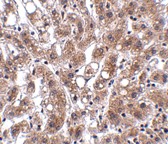 Source : THEM4 antibody was raised against an 18 amino acid peptide near the center of human THEM4.
Purification : Affinity chromatography purified via peptide column
Clonality and Clone : This is a polyclonal antibody.
Host : THEM4 antibody was raised in rabbit. Please use anti-rabbit secondary antibodies.
Application : THEM4 antibody can be used for detection of THEM4 by Western blot at 1 - 2 µg/ml.
Tested Application(s) : E, WB
Buffer : Antibody is supplied in PBS containing 0.02% sodium azide.
Blocking Peptide : Cat.No. 5227P - THEM4 Peptide
Long-Term Storage : THEM4 antibody can be stored at 4ºC, stable for one year. As with all antibodies care should be taken to avoid repeated freeze thaw cycles. Antibodies should not be exposed to prolonged high temperatures.
Positive Control
1. Cat. No. 1304 - Human Liver Tissue Lysate
Species Reactivity :H, M, R
GI Number : 76159293
Accession Number : NP_444283
Short Description : Thioesterase superfamily member 4
References
1. Maira SM, Galetic I, Brazil DP, et al. Carboxyl-terminal modulator protein (CTMP), a negative regulator of PKB/Akt and v-Akt at the plasma membrane. Science 2001; 294:374-80.
2. Knobbe CB, Reifberger J, Blaschke B, et al. Hypermethylation and transcriptional downregulation of the carboxyl-terminal modulator protein gene in glioblastomas. J. Natl. Cancer Inst. 2004; 96:483-6.
3. Zhao H, Martin BM, Bisoffi M, et al. The Akt C-terminal modulator protein is an acyl-CoA thioesterase of the hotdog-fold family. Biochemistry 2009; 48:5507-9.
4. Parcellier A, Tintignac LA, Zhuravleva E, et al. Carboxy-terminal modulator protein (CTMP) is a mitochondrial protein that sensitizes cells to apoptosis. Cell Signal. 2009; 21:639-50.
Catalog# : 5227
THEM4, also known as CTMP, binds specifically to the carboxy-terminal regulatory domain of PKB/Akt at the plasma membrane and acts as a negative regulator, reversing the phenotype of v-Akt-transformed cells. Hypermethylation of the THEM4 promoter and transcriptional downregulation of the gene has been reported in multiple glioblastomas, suggesting that epigenetic regulation of THEM4 may play a role in the progression of this cancer. Bioinformatic analysis, confirmed by in vitro testing, indicates that THEM4 is a broad-range, high activity acyl-CoA thioesterase. Recent reports have also indicated that TMEM4 is a mitochondrial protein whose overexpression is associated with an increase in mitochondrial membrane depolarization and caspase-3 and PARP cleavage, suggesting that THEM4 is involved in the apoptotic program. The additional higher molecular weight bands seen in the immunoblot may represent post-translationally modified TMEM4.
Additional Names : THEM4, Thioesterase superfamily member 4, Carboxy-terminal modulator protein, CTMP
DescriptionLeft: Western blot analysis of THEM4 in human liver tissue lysate with THEM4 antibody at (A) 1 and (B) 2 µg/ml.
Below: Immunohistochemistry of THEM4 in human liver tissue with THEM4 antibody at 2.5 μg/ml.
Other Product Images
Source : THEM4 antibody was raised against an 18 amino acid peptide near the center of human THEM4.Purification : Affinity chromatography purified via peptide column
Clonality and Clone : This is a polyclonal antibody.
Host : THEM4 antibody was raised in rabbit. Please use anti-rabbit secondary antibodies.
Application : THEM4 antibody can be used for detection of THEM4 by Western blot at 1 - 2 µg/ml.
Tested Application(s) : E, WB
Buffer : Antibody is supplied in PBS containing 0.02% sodium azide.
Blocking Peptide : Cat.No. 5227P - THEM4 Peptide
Long-Term Storage : THEM4 antibody can be stored at 4ºC, stable for one year. As with all antibodies care should be taken to avoid repeated freeze thaw cycles. Antibodies should not be exposed to prolonged high temperatures.
Positive Control
1. Cat. No. 1304 - Human Liver Tissue Lysate
Species Reactivity :H, M, R
GI Number : 76159293
Accession Number : NP_444283
Short Description : Thioesterase superfamily member 4
References
1. Maira SM, Galetic I, Brazil DP, et al. Carboxyl-terminal modulator protein (CTMP), a negative regulator of PKB/Akt and v-Akt at the plasma membrane. Science 2001; 294:374-80.
2. Knobbe CB, Reifberger J, Blaschke B, et al. Hypermethylation and transcriptional downregulation of the carboxyl-terminal modulator protein gene in glioblastomas. J. Natl. Cancer Inst. 2004; 96:483-6.
3. Zhao H, Martin BM, Bisoffi M, et al. The Akt C-terminal modulator protein is an acyl-CoA thioesterase of the hotdog-fold family. Biochemistry 2009; 48:5507-9.
4. Parcellier A, Tintignac LA, Zhuravleva E, et al. Carboxy-terminal modulator protein (CTMP) is a mitochondrial protein that sensitizes cells to apoptosis. Cell Signal. 2009; 21:639-50.
TCCR Antibody
TCCR Antibody
Catalog# : 2483
Upon antigen challenge, T-helper cells differentiate into two functional distinct subsets, Th1 and Th2. Th1 cells produce IL-2, IFN-gamma and lymphotoxin-beta that augment cell mediated immune response while Th2 cells secrete IL-4, IL-5, and IL-10 that enhance humoral immunity. The function of T-helper cells is regulated by cytokines. A novel cytokine receptor was recently identified and cloned (1,2). It is a new member in the type I cytokine receptor family and designated TCCR for T-cell cytokine receptor and WSX-1 (1,2). TCCR deficient mice had impaired Th1 responses to protein antigen challenge, including decreased levels of IFN-gamma and Th1-dependent antibody IgG2a . TCCR is predominately expressed in thymus, spleen, lymph notes and peripheral blood leukocytes.
Additional Names : TCCR (CT), Type I T-cell cytokine receptor, IL-27 receptor alpha, IL-27Ra
 Description
Left: Western blot analysis of TCCR expression in human spleen tissue lysate with TCCR antibody at 1 µg /ml.
Below: Immunohistochemistry of TCCR in human spleen tissue with TCCR antibody at 5 µg/ml.
Other Product Images
 Source : TCCR antibody was raised against a synthetic peptide corresponding to amino acids near the carboxy terminus of human TCCR precursor This sequence is identical to that of mouse TCCR.
Purification : Affinity chromatography purified via peptide column
Clonality and Clone : This is a polyclonal antibody.
Host : TCCR antibody was raised in rabbit. Please use anti-rabbit secondary antibodies.
Immunogen : Human TCCR (C-Terminus) Peptide (Cat. No. 2483P)
Application : TCCR antibody can be used for detection of TCCR by Western blot at 0.5 to 1 µg/ml.An approximately 70 kDa band can be detected. It is human, mouse, and rat reactive.
Tested Application(s) : E, WB, IHC
Buffer : Antibody is supplied in PBS containing 0.02% sodium azide.
Blocking Peptide : Cat. No. 2483P - TCCR Peptide
Long-Term Storage : TCCR antibody can be stored at 4ºC, stable for one year. As with all antibodies care should be taken to avoid repeated freeze thaw cycles. Antibodies should not be exposed to prolonged high temperatures.
Positive Control
1. Cat. No. 1306 - Human Spleen Tissue Lysate
Species Reactivity :H
GI Number : 4759328
Accession Number : NP_004834
Short Description : (CT) A new T cell cytokine receptor
References
1. Chen Q, Ghilardi N, Wang H, Baker T, Xie MH, Gurney A, Grewal IS and de Sauvage FJ. Development of Th1-type immune responses requires the type I cytokine receptor TCCR Nature 2000;407(6806):916-920
2. Sprecher,C.A., Grant,F.J., Baumgartner,J.W., Presnell,S.R., Schrader,S.K., Yamagiwa,T., Whitmore,T.E., O'Hara,P.J. and Foster,D.F. Cloning and characterization of a novel class I cytokine receptor Biochem. Biophys. Res. Commun. 1998;246(1):82-90
Catalog# : 2483
Upon antigen challenge, T-helper cells differentiate into two functional distinct subsets, Th1 and Th2. Th1 cells produce IL-2, IFN-gamma and lymphotoxin-beta that augment cell mediated immune response while Th2 cells secrete IL-4, IL-5, and IL-10 that enhance humoral immunity. The function of T-helper cells is regulated by cytokines. A novel cytokine receptor was recently identified and cloned (1,2). It is a new member in the type I cytokine receptor family and designated TCCR for T-cell cytokine receptor and WSX-1 (1,2). TCCR deficient mice had impaired Th1 responses to protein antigen challenge, including decreased levels of IFN-gamma and Th1-dependent antibody IgG2a . TCCR is predominately expressed in thymus, spleen, lymph notes and peripheral blood leukocytes.
Additional Names : TCCR (CT), Type I T-cell cytokine receptor, IL-27 receptor alpha, IL-27Ra
DescriptionLeft: Western blot analysis of TCCR expression in human spleen tissue lysate with TCCR antibody at 1 µg /ml.
Below: Immunohistochemistry of TCCR in human spleen tissue with TCCR antibody at 5 µg/ml.
Other Product Images
Source : TCCR antibody was raised against a synthetic peptide corresponding to amino acids near the carboxy terminus of human TCCR precursor This sequence is identical to that of mouse TCCR.Purification : Affinity chromatography purified via peptide column
Clonality and Clone : This is a polyclonal antibody.
Host : TCCR antibody was raised in rabbit. Please use anti-rabbit secondary antibodies.
Immunogen : Human TCCR (C-Terminus) Peptide (Cat. No. 2483P)
Application : TCCR antibody can be used for detection of TCCR by Western blot at 0.5 to 1 µg/ml.An approximately 70 kDa band can be detected. It is human, mouse, and rat reactive.
Tested Application(s) : E, WB, IHC
Buffer : Antibody is supplied in PBS containing 0.02% sodium azide.
Blocking Peptide : Cat. No. 2483P - TCCR Peptide
Long-Term Storage : TCCR antibody can be stored at 4ºC, stable for one year. As with all antibodies care should be taken to avoid repeated freeze thaw cycles. Antibodies should not be exposed to prolonged high temperatures.
Positive Control
1. Cat. No. 1306 - Human Spleen Tissue Lysate
Species Reactivity :H
GI Number : 4759328
Accession Number : NP_004834
Short Description : (CT) A new T cell cytokine receptor
References
1. Chen Q, Ghilardi N, Wang H, Baker T, Xie MH, Gurney A, Grewal IS and de Sauvage FJ. Development of Th1-type immune responses requires the type I cytokine receptor TCCR Nature 2000;407(6806):916-920
2. Sprecher,C.A., Grant,F.J., Baumgartner,J.W., Presnell,S.R., Schrader,S.K., Yamagiwa,T., Whitmore,T.E., O'Hara,P.J. and Foster,D.F. Cloning and characterization of a novel class I cytokine receptor Biochem. Biophys. Res. Commun. 1998;246(1):82-90
TCCR Antibody
TCCR Antibody
Catalog# : 2481
Upon antigen challenge, T-helper cells differentiate into two functional distinct subsets, Th1 and Th2. Th1 cells produce IL-2, IFN-gamma and lymphotoxin-beta that augment cell mediated immune response while Th2 cells secrete IL-4, IL-5, and IL-10 that enhance humoral immunity. The function of T-helper cells is regulated by cytokines. A novel cytokine receptor was recently identified and cloned (1,2). It is a new member in the type I cytokine receptor family and designated TCCR for T-cell cytokine receptor and WSX-1 (1,2). TCCR deficient mice had impaired Th1 responses to protein antigen challenge, including decreased levels of IFN-gamma and Th1-dependent antibody IgG2a . TCCR is predominately expressed in thymus, spleen, lymph notes and peripheral blood leukocytes.
Additional Names : TCCR (NT), Type I T-cell cytokine receptor, IL-27 receptor alpha, IL-27Ra
 Description
Left: Western blot analysis of TCCR expression in human spleen tissue lysates with TCCR antibody at 1 µg /ml.
Below: Immunohistochemistry of TCCR in human spleen tissue with TCCR antibody at 10 µg/ml.
Other Product Images
 Source : TCCR antibody was raised against a synthetic peptide corresponding to amino acids near the amino terminus of human TCCR precursor.
Purification : Affinity chromatography purified via peptide column
Clonality and Clone : This is a polyclonal antibody.
Host : TCCR antibody was raised in rabbit. Please use anti-rabbit secondary antibodies.
Immunogen : Human TCCR (N-Terminus) Peptide (Cat. No. 2481P)
Application : TCCR antibody can be used for detection of TCCR by Western blot at 0.5 to 1 µg/ml.An approximately 70 kDa band can be detected.
Tested Application(s) : E, WB, IHC
Buffer : Antibody is supplied in PBS containing 0.02% sodium azide.
Blocking Peptide : Cat. No. 2481P - TCCR Peptide
Long-Term Storage : TCCR antibody can be stored at 4ºC, stable for one year. As with all antibodies care should be taken to avoid repeated freeze thaw cycles. Antibodies should not be exposed to prolonged high temperatures.
Positive Control
1. Cat. No. 1306 - Human Spleen Tissue Lysate
Species Reactivity :H
GI Number : 4759328
Accession Number : NP_004834
Short Description : (NT) A new T cell cytokine receptor
References
1. Chen Q, Ghilardi N, Wang H, Baker T, Xie MH, Gurney A, Grewal IS and de Sauvage FJ. Development of Th1-type immune responses requires the type I cytokine receptor TCCR Nature 2000;407(6806):916-920
2. Sprecher,C.A., Grant,F.J., Baumgartner,J.W., Presnell,S.R., Schrader,S.K., Yamagiwa,T., Whitmore,T.E., O'Hara,P.J. and Foster,D.F. Cloning and characterization of a novel class I cytokine receptor Biochem. Biophys. Res. Commun. 1998;246(1):82-90
Catalog# : 2481
Upon antigen challenge, T-helper cells differentiate into two functional distinct subsets, Th1 and Th2. Th1 cells produce IL-2, IFN-gamma and lymphotoxin-beta that augment cell mediated immune response while Th2 cells secrete IL-4, IL-5, and IL-10 that enhance humoral immunity. The function of T-helper cells is regulated by cytokines. A novel cytokine receptor was recently identified and cloned (1,2). It is a new member in the type I cytokine receptor family and designated TCCR for T-cell cytokine receptor and WSX-1 (1,2). TCCR deficient mice had impaired Th1 responses to protein antigen challenge, including decreased levels of IFN-gamma and Th1-dependent antibody IgG2a . TCCR is predominately expressed in thymus, spleen, lymph notes and peripheral blood leukocytes.
Additional Names : TCCR (NT), Type I T-cell cytokine receptor, IL-27 receptor alpha, IL-27Ra
DescriptionLeft: Western blot analysis of TCCR expression in human spleen tissue lysates with TCCR antibody at 1 µg /ml.
Below: Immunohistochemistry of TCCR in human spleen tissue with TCCR antibody at 10 µg/ml.
Other Product Images
Source : TCCR antibody was raised against a synthetic peptide corresponding to amino acids near the amino terminus of human TCCR precursor.Purification : Affinity chromatography purified via peptide column
Clonality and Clone : This is a polyclonal antibody.
Host : TCCR antibody was raised in rabbit. Please use anti-rabbit secondary antibodies.
Immunogen : Human TCCR (N-Terminus) Peptide (Cat. No. 2481P)
Application : TCCR antibody can be used for detection of TCCR by Western blot at 0.5 to 1 µg/ml.An approximately 70 kDa band can be detected.
Tested Application(s) : E, WB, IHC
Buffer : Antibody is supplied in PBS containing 0.02% sodium azide.
Blocking Peptide : Cat. No. 2481P - TCCR Peptide
Long-Term Storage : TCCR antibody can be stored at 4ºC, stable for one year. As with all antibodies care should be taken to avoid repeated freeze thaw cycles. Antibodies should not be exposed to prolonged high temperatures.
Positive Control
1. Cat. No. 1306 - Human Spleen Tissue Lysate
Species Reactivity :H
GI Number : 4759328
Accession Number : NP_004834
Short Description : (NT) A new T cell cytokine receptor
References
1. Chen Q, Ghilardi N, Wang H, Baker T, Xie MH, Gurney A, Grewal IS and de Sauvage FJ. Development of Th1-type immune responses requires the type I cytokine receptor TCCR Nature 2000;407(6806):916-920
2. Sprecher,C.A., Grant,F.J., Baumgartner,J.W., Presnell,S.R., Schrader,S.K., Yamagiwa,T., Whitmore,T.E., O'Hara,P.J. and Foster,D.F. Cloning and characterization of a novel class I cytokine receptor Biochem. Biophys. Res. Commun. 1998;246(1):82-90
Subscribe to:
Comments (Atom)