Wednesday, May 5, 2010

Caspase-12 Antibody (Large)

Caspase-12 Antibody (Large)

Catalog# : 3195

Three distinct signaling pathways lead to programmed cell death (apoptosis). The death receptor and mitochondrion pathways are the main, in which the key apoptotic proteases capase-8 and caspase-9, respectively, are involved. The endoplasmic reticulum (ER) stress is the third apoptotic pathway and caspase-12 is involved (1,2). Caspase-12 is localized to the ER but not to cytoplasm or mitochondrion. Caspase-12 is activated by ER stress, including disruption of ER calcium homeostasis, and mediates ER stress-induced apoptosis. Caspase-12 is co-localized to the ER with several proteins that are involved in Alzheimer's disease including ?-secretase presenilin and beta-amyloid precursor protein (APP). Caspase-12 mediates cytotoxicity induced by amyloid-beta. Caspase-12 is ubiquitously expressed in mouse tissues (1).

Additional Names : Caspase-12 (large), Casp-12 (lrg)
Description
Left: Western blot analysis of caspase-12 in human heart lysate with caspase-12 antibody (large) at 0.5 (lane A), 1 (lane B), and 2 µg/ml (lane C), respectively.

Below: Immunohistochemical staining of human heart tissue using caspase-12 antibody (large) at 2 µg/ml.

Source : Caspase-12 antibody was raised against a synthetic peptide corresponding to amino acids within the large cleavage product of murine caspase-12.
Purification : Affinity chromatography purified via peptide column
Clonality and Clone : This is a polyclonal antibody.
Host : Caspase-12 antibody (Large) was raised in rabbit. Please use anti-rabbit secondary antibodies.
Immunogen : Mouse Caspase-12 (Large) Peptide (Cat. No. 3195P)
Application : Caspase-12 (large) antibody can be used for the detection of both the propeptide and cleaved forms of caspase-12 by Western blot at 0.5 to 2 µg/ml.
Tested Application(s) : E, WB, IHC
Buffer : Antibody is supplied in PBS containing 0.02% sodium azide.
Blocking Peptide : Cat. No. 3195P - Caspase-12 (Large) Peptide
Long-Term Storage : Caspase-12 antibody (Large) can be stored at 4ºC, stable for one year. As with all antibodies care should be taken to avoid repeated freeze thaw cycles. Antibodies should not be exposed to prolonged high temperatures.
Positive Control

1. Cat. No. 1301 - Human Heart Tissue Lysate
Species Reactivity :H, M, R
GI Number : 2094806
Accession Number : CAA73532
Short Description : A new caspase
References


1. Nakagawa T, Zhu H, Morishima N, Li E, Xu J, Yankner BA, Yuan J. Caspase-12 mediates endoplasmic-reticulum-specific apoptosis and cytotoxicity by amyloid-beta. Nature 2000;403:98-103.
2. Mehmet H. Caspases find a new place to hide. Nature 2000;403:29-30
3. Van de Craen M, Vandenabeele P, Declercq W, Van den Brande I, Van Loo G, Molemans F, Schotte P, Van Criekinge W, Beyaert R, Fiers W. Characterization of seven murine caspase family members. FEBS Lett 1997;403:61-9

For more detail regarding Peptide please visit here : Custom Peptide Synthesis

Caspase-12 Antibody

Caspase-12 Antibody

Catalog# : 2327

Three distinct signaling pathways lead to programmed cell death (apoptosis). The death receptor and mitochondrion pathways are the mains, in which the key apoptotic proteases capase-8 and caspase-9, respectively, are involved. The endoplasmic reticulum (ER) stress is the third apoptotic pathway and caspase-12 is involved (1,2). Caspase-12 is localized to the ER but not to cytoplasm or mitochondrion. Caspase-12 is activated by ER stress, including disruption of ER calcium homeostasis, and mediates ER stress-induced apoptosis. Caspase-12 is co-localized to the ER with several proteins that are involved in Alzheimer's disease including ?-secretase presenilin and ?-amyloid precursor protein (APP). Caspase-12 mediates cytotoxicity induced by amyloid-beta. Caspase-12 is ubiquitously expressed in mouse tissues (1).

Additional Names : Caspase-12 (IN), Casp-12

Description
Left: Western blot analysis of caspase-12 in mouse brain tissue lysate in the absence (A) or presence (B) of blocking peptide with caspase-12 antibody (IN) at 1 µg/ml.

Below: Immunohistochemical staining of mouse liver tissue using caspase-12 antibody at 2 µg/ml.

Source : Caspase-12 antibody was raised against a peptide corresponding to amino acids 100 to 116 of murine caspase-12.

Purification : Affinity chromatography purified via peptide column

Clonality and Clone : This is a polyclonal antibody.

Host : Caspase-12 antibody was raised in rabbit. Please use anti-rabbit secondary antibodies.

Immunogen : Mouse Caspase-12 (Intermediate Domain) Peptide (Cat. No. 2327P)

Application : Caspase-12 antibody can be used for detection of caspase-12 by Western blot at 0.5 to 1 µg/ml.

Tested Application(s) : E, WB, IHC

Buffer : Antibody is supplied in PBS containing 0.02% sodium azide.

Blocking Peptide : Cat. No. 2327P - Caspase-12 Peptide

Long-Term Storage : Caspase-12 antibody can be stored at 4ºC, stable for one year. As with all antibodies care should be taken to avoid repeated freeze thaw cycles. Antibodies should not be exposed to prolonged high temperatures.

Positive Control
1.Cat. No. 1403 - Mouse Brain Tissue Lysate
2.Cat. No. 1404 - Mouse Liver Tissue Lysate

Species Reactivity :H, M, R

GI Number : 2094806

Accession Number : CAA73532

Short Description : (IN) A new caspase

References
1.Nakagawa T, Zhu H, Morishima N, Li E, Xu J, Yankner BA, Yuan J. Caspase-12 mediates endoplasmic-reticulum-specific apoptosis and cytotoxicity by amyloid-beta. Nature 2000;403:98-103.
2.Mehmet H. Caspases find a new place to hide. Nature 2000;403:29-30
3.Van de Craen M, Vandenabeele P, Declercq W, Van den Brande I, Van Loo G, Molemans F, Schotte P, Van Criekinge W, Beyaert R, Fiers W. Characterization of seven murine caspase family members. FEBS Lett 1997;403:61-9 (WD0900)

For more detail regarding Peptide please visit here : Custom Peptide Synthesis

Caspase-12 Antibody

Caspase-12 Antibody

Catalog# : 2325

Three distinct signaling pathways lead to programmed cell death (apoptosis). The death receptor and mitochondrion pathways are the mains, in which the key apoptotic proteases capase-8 and caspase-9, respectively, are involved. The endoplasmic reticulum (ER) stress is the third apoptotic pathway and caspase-12 is involved (1,2). Caspase-12 is localized to the ER but not to cytoplasm or mitochondrion. Caspase-12 is activated by ER stress, including disruption of ER calcium homeostasis, and mediates ER stress-induced apoptosis. Caspase-12 is co-localized to the ER with several proteins that are involved in Alzheimer's disease including g-secretase presenilin and b-amyloid precursor protein (APP). Caspase-12 mediates cytotoxicity induced by amyloid-b. Caspase-12 is ubiquitously expressed in mouse tissues (1).

Additional Names : Caspase-12 (NT), Casp-12
Description
Left: Western blot analysis of caspase-12 in human (A) and mouse (B) spleen tissue lysates with caspase-12 antibody at 1 µg/ml.

Below: Immunohistochemistry of caspase-12 in human heart tissue with caspase-12 antibody at 10 µg/ml.
Source : Caspase-12 antibody was raised against a peptide corresponding to amino acids 2 to 17 of murine caspase-12.

Purification : Affinity chromatography purified via peptide column

Clonality and Clone : This is a polyclonal antibody.

Host : Caspase-12 antibody was raised in rabbit. Please use anti-rabbit secondary antibodies.

Immunogen : Mouse Caspase-12 (N-Terminus) Peptide (Cat. No. 2325P)

Application : Caspase-12 antibody can be used for detection of caspase-12 by Western blot at 0.5 to 1 µg/ml.

Tested Application(s) : E, WB, IHC

Buffer : Antibody is supplied in PBS containing 0.02% sodium azide.

Blocking Peptide : Cat. No. 2325P - Caspase-12 Peptide

Long-Term Storage : Caspase-12 antibody can be stored at 4ºC, stable for one year. As with all antibodies care should be taken to avoid repeated freeze thaw cycles. Antibodies should not be exposed to prolonged high temperatures.

Positive Control
1.Cat. No. 1406 - Mouse Spleen Tissue Lysate
2.Cat. No. 1301 - Human Heart Tissue Lysate
3.Cat. No. 1306 - Human Spleen Tissue Lysate

Species Reactivity :H, M, R

GI Number : 2094806

Accession Number : CAA73532

Short Description : (NT) A new caspase

References
1.Nakagawa T, Zhu H, Morishima N, Li E, Xu J, Yankner BA, Yuan J. Caspase-12 mediates endoplasmic-reticulum-specific apoptosis and cytotoxicity by amyloid-beta. Nature 2000;403:98-103.
2.Mehmet H. Caspases find a new place to hide. Nature 2000;403:29-30
3.Van de Craen M, Vandenabeele P, Declercq W, Van den Brande I, Van Loo G, Molemans F, Schotte P, Van Criekinge W, Beyaert R, Fiers W. Characterization of seven murine caspase family members. FEBS Lett 1997;403:61-9 (WD1000)

For more detail regarding Peptide please visit here : Custom Peptide Synthesis

Caspase-10 Antibody

Caspase-10 Antibody

Catalog# : 1128

Apoptosis is related to many diseases and induced by a family of cell death receptors and their ligands. Cell death signals are transduced by death domain (DD)- containing adapter molecules and members of the ICE/CED-3 protease family. A novel ICE/CED-3 protease was identified recently, designated FLICE2 and Mch41,2 and renamed as caspase-10. Caspase-10 has two death effector domains (DEDs) that bind to the DED in the adapter molecule FADD and recruits both TNFR1 and CD95 to form complexes with these receptors. Caspase-10 is therefore involved in the CD95 and TNFR1 induced apoptosis1. Caspase-10 cleaves and activates caspase-3, -4, -6, -7, -8 and -9, which causes the proteolytic cleavage of many key proteins such as PARP. Cleavage of PARP occurs in many different systems during apoptosis and is the hallmark of programmed cell death. Caspase-10 is expressed in many tissues and cell lines.

Additional Names : Caspase-10 (CT), Casp-10, FLICE2
Description
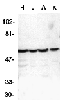
Left: Western blot analysis of caspase-10 in HeLa (H), Jurkat (J), A431 (A), K562 (K) whole cell lysates with Caspase-10 antibody at 1 µg/ml.


Source : Caspase-10 antibody was raised against a peptide corresponding to amino acids 505 to 521 of human FLICE2.

Purification : Antibody is DEAE purified

Clonality and Clone : This is a polyclonal antibody.

Host : Caspase-10 antibody was raised in rabbit. Please use anti-rabbit secondary antibodies.

Immunogen : Human Caspase-10 (C-Terminus) Peptides (Cat. No. 1128P)

Application : Caspase-10 antibody can be used for detection of caspase-10 by Western blot at 1:500 to 1:1000 dilution. A 59 kDa band can be detected. Antibody only recognizes FLICE2 form of caspase-10.

Tested Application(s) : E, WB

Buffer : Antibody is supplied in PBS containing 0.02% sodium azide.

Blocking Peptide : Cat. No. 1128P - Caspase-10 Peptide

Long-Term Storage : Caspase-10 antibody can be stored at 4ºC, stable for one year. As with all antibodies care should be taken to avoid repeated freeze thaw cycles. Antibodies should not be exposed to prolonged high temperatures.

Positive Control
1.Cat. No. 1201 - HeLa Cell Lysate
2.Cat. No. 1205 - Jurkat Whole Cell Lysate
3.Cat. No. 1202 - A431 Cell Lysate
4.Cat. No. 1204 - K562 Cell Lysate

Species Reactivity :H

GI Number : 4731237

Accession Number : AAD28402

Short Description : (CT) A new caspase

References

1.Vincenz.C. and Dixit,V.M. Fas-associated death domain protein interleukin-1beta-converting enzyme 2 (FLICE2), an ICE/Ced-3 homologue, is proximally involved in CD95- and p55-mediated death signaling. J. Biol. Chem. 1997;272:6578-6583
2.Fernandes-Alnemri T, Armstrong RC, Krebs J, Srinivasula SM, Wang L Bullrich F, Fritz LC, Trapani JA, Tomaselli KJ, Litwack G, Alnemri ES. In vitro activation of CPP32 and Mch3 by Mch4, a novel human apoptotic cysteine protease containing two FADD-like domains. Proc. Natl. Acad. Sci. USA. 1996;93:7464-7469
3.Cohen GM. Caspases: the executioners of apoptosis. Biochem J 1997;326:1-16 (RD1299)

For more detail regarding Peptide please visit here : Custom Peptide Synthesis

Caspase-1 Antibody

Caspase-1 Antibody

Catalog# : 3463

Caspases are a family of cysteine proteases that can be divided into the apoptotic and inflammatory caspase subfamilies. Unlike the apoptotic caspases, members of the inflammatory subfamily are generally not involved in cell death but are associated with the immune response to microbial pathogens (reviewed in 1 and 2). Members of this subfamily include caspase-1, -4, -5, and -12 and can activate proinflammatory cytokines such as IL-1? and IL-18 (3,4). Caspase-1 was initially identified as an IL-1beta-converting enzyme (5); later experiments revealed it to be a mammalian homolog of the C. elegans cell death gene ced-3 (6) whose overexpression can induce apoptosis in fibroblasts (7).

Additional Names : Caspase-1 (IN), ICE, IL-1beta converting enzyme

Description
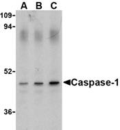
Left: Western blot analysis of Caspase-1 in HeLa cell lysate with Caspase-1 antibody (IN) at (A) 0.5, (B) 1, and (C) 2 µg/ml.

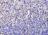
Below: Immunohistochemistry of caspase-1 in human spleen tissue with caspase-1 antibody at 10 µg/ml.

Other Product Images

Source : Caspase-1 antibody was raised against a 15 amino acid peptide from near the middle of human Caspase-1.

Purification : Affinity chromatography purified via peptide column

Clonality and Clone : This is a polyclonal antibody.

Host : Caspase-1 antibody was raised in rabbit. Please use anti-rabbit secondary antibodies.

Immunogen : Human Casp-1 / ICE / IL-1b converting enzyme (Intermediate Domain) Peptide (Cat. No. 3463P)

Application : Casp-1 antibody can be used for the detection of Caspase-1 by Western blot at 0.5 µg/ml.Depending on cell lines or tissues used, other cleavage products may be observed.

Tested Application(s) : E, WB, IHC

Buffer : Antibody is supplied in PBS containing 0.02% sodium azide.

Blocking Peptide : Cat. No. 3463P - Casp-1 Peptides

Long-Term Storage : Caspase-1 antibody can be stored at 4ºC, stable for one year. As with all antibodies care should be taken to avoid repeated freeze thaw cycles. Antibodies should not be exposed to prolonged high temperatures.

Positive Control
1.Cat. No. 1201 - HeLa Cell Lysate
2.Cat. No. 1306 - Human Spleen Tissue Lysate

Species Reactivity :H

GI Number : 15431328

Accession Number : NP_150634

Short Description : (IN) a caspase in the inflammatory subfamily

References
1.Martinon F and Tschopp J. Inflammatory caspases: linking an intracellular innate immune system to autoinflammatory diseases. CCell 2004; 117:561-74.
2.Zhivotovsky B and Orrenius S. Caspase-2 function in response to DNA damage. Biochim. Biophys. Res. Comm. 2005; 331:859-67.
3.Kuida K, Lippke JA, Ku G, et al. Altered cytokine export and apoptosis in mice deficient in interleukin-1 beta converting enzyme. Science 1995; 267:2000-3.
4.Gracie JA, Robertson SE, and McInnes IB. Interleukin-18. J. Leukoc. Biol. 2003; 73:213-224.

For more detail regarding Peptide please visit here : Custom Peptide Synthesis


Caspase-1 Antibody

Catalog# : 3459

Caspases are a family of cysteine proteases that can be divided into the apoptotic and inflammatory caspase subfamilies. Unlike the apoptotic caspases, members of the inflammatory subfamily are generally not involved in cell death but are associated with the immune response to microbial pathogens (reviewed in 1 and 2). Members of this subfamily include caspase-1, -4, -5, and -12 and can activate proinflammatory cytokines such as IL-1? and IL-18 (3,4). Caspase-1 was initially identified as an IL-1beta-converting enzyme (5); later experiments revealed it to be a mammalian homolog of the C. elegans cell death gene ced-3 (6) whose overexpression can induce apoptosis in fibroblasts (7).

Additional Names : Caspase-1 (CT), ICE, IL-1beta converting enzyme

Description
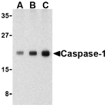
Left: Western blot analysis of Caspase-1 in human heart cell lysate with Caspase-1 antibody at (A) 0.5, (B) 1, and (C) 2 µg/ml.

Below: Immunohistochemical staining of human heart tissue using Caspase-1 antibody at 2 µg/ml.

Other Product Images

Source : Caspase-1 antibody was raised against a 16 amino acid peptide from near the carboxy-terminus of human Caspase-1.

Purification : Affinity chromatography purified via peptide column

Clonality and Clone : This is a polyclonal antibody.

Host : Caspase-1 antibody was raised in rabbit. Please use anti-rabbit secondary antibodies.

Immunogen : Human Casp-1 / ICE / IL-1b converting enzyme (C-Terminus) Peptide (Cat. No. 3459P)

Application : Casp-1 antibody can be used for the detection of Caspase-1 by Western blot at 0.5 µg/ml or

Immunohistochemistry. Depending on cell lines or tissues used, non-cleaved or other cleavage products may be observed.

Tested Application(s) : E, WB, IHC

Buffer : Antibody is supplied in PBS containing 0.02% sodium azide.

Blocking Peptide : Cat. No. 3459P - Casp-1 Peptide

Long-Term Storage : Caspase-1 antibody can be stored at 4ºC, stable for one year. As with all antibodies care should be taken to avoid repeated freeze thaw cycles. Antibodies should not be exposed to prolonged high temperatures.

Positive Control
1.Cat. No. 1301 - Human Heart Tissue Lysate

Species Reactivity :H

GI Number : 15431328

Accession Number : NP_150634

Short Description : (CT) a caspase in the inflammatory subfamily

References
1.Martinon F and Tschopp J. Inflammatory caspases: linking an intracellular innate immune system to autoinflammatory diseases. Cell 2004; 117:561-74.
2.Zhivotovsky B and Orrenius S. Caspase-2 function in response to DNA damage. Biochim. Biophys. Res. Comm. 2005; 331:859-67.
3.Kuida K, Lippke JA, Ku G, et al. Altered cytokine export and apoptosis in mice deficient in interleukin-1 beta converting enzyme. Science 1995; 267:2000-3.
4.Gracie JA, Robertson SE, and McInnes IB. Interleukin-18. J. Leukoc. Biol. 2003; 73:213-224.

For more detail regarding Peptide please visit here : Custom Peptide Synthesis


CARMA1 Antibody

CARMA1 Antibody

Catalog# : 3189

CARMA proteins belong to the membrane-associated guan-ylate kinase-like (MAGUK) family of proteins that can function as molecular scaffolds that assist assembly of signal transduction molecules (1). CARMA1, CARMA2, and CARMA3 share high degrees of sequence and functional homology, but their tissue-specific distribution suggests that they serve distinct biological functions in different cell types (2-4). Both CARMA1 and CARMA3 associate with NEMO, the regulatory subunit of the I?K complex, thereby regulating activation of the NF-kappaB transcription factor (5). Also, gene inactivation studies showed a complete block in T and B cell immunity as well as an impaired response to LPS, indicating that CARMA1 is a critical regulator in both the adaptive and innate immune systems (6).

Additional Names : CARMA1
Description
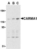
Left: Western blot analysis of CARMA1 expression in mouse thymus cell lysate with CARMA1 antibody at 0.5 (lane A), 1 (lane B), and 2 µg /ml (lane C), respectively.

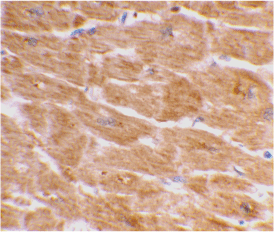
Below: Immunohistochemistry of CARMA1 in mouse thymus with CARMA1 antibody at 10 µg/ml.

Other Product Images


Source : CARMA1 antibody was raised against a synthetic peptide corresponding to amino acids at the C-terminus of human CARMA1.

Purification : Affinity chromatography purified via peptide column

Clonality and Clone : This is a polyclonal antibody.

Host : CARMA1 antibody was raised in rabbit. Please use anti-rabbit secondary antibodies.

Immunogen : Human CARMA1 Peptide (Cat. No. 3189 P)

Application : CARMA1 antibody can be used for detection of CARMA1 by Western blot at 0.5 to 2 µg/ml.A band at approximately 125 kDa can be detected.

Tested Application(s) : E, WB, IHC

Buffer : Antibody is supplied in PBS containing 0.02% sodium azide.

Blocking Peptide : Cat. No. 3189P - CARMA1 Peptide

Long-Term Storage : CARMA1 antibody can be stored at 4ºC, stable for one year. As with all antibodies care should be taken to avoid repeated freeze thaw cycles. Antibodies should not be exposed to prolonged high temperatures.

Positive Control
1.Cat. No. 1409 - Mouse Thymus Tissue Lysate

Species Reactivity :H, M

GI Number : 172046231

Accession Number : Q9BXL7

Short Description : lymphocyte activating CARD-containing protein

References
1.Fanning AS and Anderson JM. Protein modules as organizers of membrane structure.Curr. Opin. Cell Biol. 1999; 11:432-9.
2.Gaide O, Martinon F, Michau O, et al. Carma1, 1 CARD-containing binding partner of Bcl10, induces Bcl10 phosphorylation and NF-kappa B activation. FEBS Lett. 2001; 496:121-7.
3.Bertin J, Wang L, Guo Y, et al. CARD11 and CARD14 are novel caspase recruitment domain (CARD)/membrane-associated guanylate kinase (MAGUK) family members that interact with BCL10 and activate NF-kappa B. J. Biol. Chem. 2001; 276:11877-82.
4.McAllister-Lucas LM, Inohara N, Lucas PC, et al. Bimp1, a MAGUK family member linking protein kinase C activation to Bcl10-mediated NF-kappaB induction. J. Biol. Chem. 2001; 276:30589-97.

For more detail regarding Peptide please visit here : Custom Peptide Synthesis

CARD9 Antibody

CARD9 Antibody

Catalog# : 2515

Apoptosis is related to many diseases and development. Cell death signals are transduced by death domain (DD), death effector domain (DED), and caspase recruitment domain (CARD) containing molecules. CARD containing proteins include some caspases, Apaf-1, CARD4, IAPs, RICK, ARC, RAIDD, BCL-10, and ASC. A novel CARD-containing protein was recently identified and designated CARD9, which interacts with the CARD activation domain of BCL-10 (1). CARD9 associates with BCL-10 and forms a complex within cells. CARD9 induces apoptosis and activates NF-kappaB. CARD9 is an upstream activator of BCL-10 and NF-kappaB signaling.

Additional Names : CARD9
Description
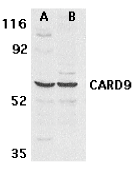
Left: Western blot analysis of CARD9 expression in human MDA-MB-361 (A) and PC-3 (B) cell lysate with CARD9 antibody at 2.5 µg /ml.

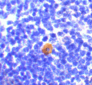
Below: Immunocytochemistry of CARD9 in K562 cells with CARD9 antibody at 10 μg/ml.

Other Product Images
Source : CARD9 antibody was raised against a synthetic peptide corresponding to amino acids 521 to 536 of human CARD9 The sequence is different from that of rat origin by two amino acids.

Purification : Affinity chromatography purified via peptide column

Clonality and Clone : This is a polyclonal antibody.

Host : CARD9 antibody was raised in rabbit. Please use anti-rabbit secondary antibodies.

Immunogen : Human CARD9 Peptide (Cat. No. 2515P)

Application : CARD9 antibody can be used for detection of CARD9 by Western blot at 0.5 to 1 µg/ml.A band at approximately 59 kDa can be detected.

Tested Application(s) : E, WB, ICC

Buffer : Antibody is supplied in PBS containing 0.02% sodium azide.

Blocking Peptide : Cat. No. 2515P - CARD9 Peptide

Long-Term Storage : CARD9 antibody can be stored at 4ºC, stable for one year. As with all antibodies care should be taken to avoid repeated freeze thaw cycles. Antibodies should not be exposed to prolonged high temperatures.

Positive Control
1.Cat. No. 1216 - PC-3 Cell Lysate
2.Cat. No. 1217 - MDA-MB-361 Cell Lysate
3.Cat. No. 1204 - K562 Cell Lysate

Species Reactivity :H

GI Number : 11066983

Accession Number : AF311287

Short Description : A new CARD-containing protein

References

1.Bertin J, Guo Y, Wang L, Srinivasula SM, Jacobson MD, Poyet JL, Merriam S, Du MQ, Dyer MJ, Robison KE, DiStefano PS, Alnemri ES. CARD9 is a novel caspase recruitment domain-containing protein that interacts with BCL10/CLAP and activates NF-kappa B. J Biol Chem. 2000;275(52):41082-6. (RD0202)

For more detail regarding Peptide please visit here : Custom Peptide Synthesis

CARD8 Antibody

CARD8 Antibody

Catalog# : 3187

Apoptosis is related to many diseases and development. Cell death signals are transduced by death domain (DD), death effector domain (DED), and caspase recruitment domain (CARD) containing molecules. CARD containing proteins include some caspases, Apaf-1, CARD4, IAPs, RICK, ARC, RAIDD, BCL-10, and ASC. A novel CARD-containing protein was recently identified and designated CARD8. This protein interacts with DRAL, a p53-responsive protein implicated in the induction of apoptosis (1,2), and caspase-1 and its related proteins ICEBERG and pesudo-ICE (3). Although there are conflicting reports on whether CARD8 acts a pro- or anti-apoptotic protein (2,4), it has been suggested that it functions as an adaptor molecule regulating caspase-1 and NF-kappaB activation.

Additional Names : CARD8, Cardinal/TUCAN

Description
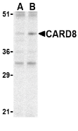
Left: Western blot analysis of CARD8 expression in K562 cell lysate with CARD8 antibody at (A) 2 and (B) 4 µg /ml.

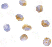
Below: Immunocytochemistry of CARD8 in K562 cells with CARD8 antibody at 10 µg/ml.

Other Product Images

Source : CARD8 antibody was raised against a synthetic peptide corresponding to amino acids at the C-terminus of human CARD8.

Purification : Affinity chromatography purified via peptide column

Clonality and Clone : This is a polyclonal antibody.

Host : CARD8 antibody was raised in rabbit. Please use anti-rabbit secondary antibodies.

Immunogen : Human CARD8 / Cardinal / TUCAN Peptide (Cat. No. 3187P)

Application : CARD8 antibody can be used for detection of CARD8 by Western blot at 2 µg/ml.An approximate 45 kDa can be detected.

Tested Application(s) : E, WB, ICC

Buffer : Antibody is supplied in PBS containing 0.02% sodium azide.

Blocking Peptide : Cat. No. 3187P - CARD8 Peptide

Long-Term Storage : CARD8 antibody can be stored at 4ºC, stable for one year. As with all antibodies care should be taken to avoid repeated freeze thaw cycles. Antibodies should not be exposed to prolonged high temperatures.

Positive Control
1.Cat. No. 1204 - K562 Cell Lysate

Species Reactivity :H

GI Number : 12247742

Accession Number : AAG50014

Short Description : A new CARD containing protein

References
1.Scholl FA, McLoghlin P, Ehler E, et al. DRAL is a p53-responsive gene whose four and a half LIM domain protein product induces apoptosis. J. Cell Biol. 2000; 151:495-506.
2.Stil R, Leonardi A, Formisano L, et al. TUCAN/CARDINAL and DRAL participate in a B activation. FEBS 2002;kcommon pathway for modulation of NF- 521:165-9.
3.Razmara M, Srinivasula SM, Wang L, et al. CARD-8 protein, a new CARD family member that regulates caspase-1 activation and apoptosis. J. Biol. Chem. 2002; 277:13952-8.
4.Pathan N, Marusawa H, Krajewska M, et al. TUCAN, an antiapoptotic caspase-assoicated recruitment domain family protein overexpressed in cancer. J. Biol. Chem. 2001; 276:32220-9.

For more detail regarding Peptide please visit here : Custom Peptide Synthesis

CARD10 Antibody

CARD10 Antibody

Catalog# : 3013

Apoptosis is related to many diseases and development. Cell death signals are transduced by death domain (DD), death effector domain (DED), and caspase recruitment domain (CARD) containing molecules. CARD containing proteins include some caspases, Apaf-1, CARD4, IAPs, RICK, ARC, RAIDD, Bcl-10, and ASC. A novel CARD-containing protein was recently identified and designated CARD10 (1). This protein belongs to the membrane-associated guanylate kinase-like (MAGUK) family of proteins that can function as molecular scaffolds that assist assembly of signal transduction molecules (2). CARD 10 interacts with Bcl-10, a Bcl protein that promotes apoptosis, caspase-9 maturation and activation of NF-kappaB (1,3). CARD 10 and the related protein CARMA1 also associate with NEMO, the regulatory subunit of the IkappaK complex (4), demonstrating its importance in the regulation of NF-kappaB transcription factor activation.

Additional Names : CARD10, Caspase recuitment domain family 10 / CARMA3 / Bimp1

Description
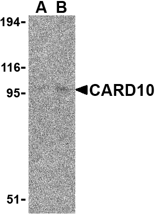
Left: Western blot analysis of CARD10 expression in EL4 cell lysate with CARD10 antibody at (A) 5 and (B) 10 µg /ml.

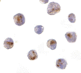
Below: Immunocytochemistry of CARD10 in EL4 cells with CARD10 antibody at 10 µg/ml.

Other Product Images


Source : CARD10 antibody was raised against a synthetic peptide corresponding to 14 amino acids at the C-terminus of human CARD10.

Purification : immunoaffinity purified

Clonality and Clone : This is a polyclonal antibody.

Host : CARD10 antibody was raised in rabbit. Please use anti-rabbit secondary antibodies.

Immunogen : Murine CARD10 / Caspase recuitment domain family 10 / CARMA3 / Bimp1 Peptide (Cat. No. 3013P)

Application : CARD10 antibody can be used for detection of CARD10 by Western blot at 5 - 10 µg/ml.

Tested Application(s) : E, WB, ICC

Buffer : Antibody is supplied in PBS containing 0.02% sodium azide.

Blocking Peptide : Cat. No. 3013P - CARD10 Peptide

Long-Term Storage : CARD10 antibody can be stored at 4ºC, stable for one year. As with all antibodies care should be taken to avoid repeated freeze thaw cycles. Antibodies should not be exposed to prolonged high temperatures.

Positive Control
1.Cat. No. 1287 - EL4 Cell Lysate

Species Reactivity :H, M

GI Number : 13488607

Accession Number : AAK26165

Short Description : Caspase recuitment domain family 10

References
1.Wang L, Guo Y, Huang W-J, et al. CARD10 is a novel caspase recruitment domain/membrane-associated guanylate kinase family member that interacts with BCL10 and activates NF-κB. J. Biol. Chem. 2001; 276:21405-9.
2.Fanning AS and Anderson JM. Protein modules as organizers of membrane structure. Curr. Opin. Cell Biol. 1999; 11:432-9.
3.Fischer KD, Tedford K, Wirth T. New roles for Bcl10 in B-cell development and LPS response. Trends Immunol. 2004; 25:113-6.
4.Stilo R, Liguoro D, Di Jeso B, et al. Physical and functional interaction of CARMA1 and CARMA3 with Iκ kinase κ-NF-κB essential modulator. J. Biol. Chem. 2004; 279:34323-31.

For more detail regarding Peptide please visit here : Custom Peptide Synthesis