Tuesday, July 19, 2011

IRAK-2 Antibody


Catalog#:2213

The pro-inflammatory cytokine IL-1 induces cellular response through two subunits of its receptor, IL-1 receptor I (IL-1RI) and IL-1 receptor accessory protein (IL-1RAcP). IL-1 receptor-associated kinase (IRAK) mediates activation of NF-kappaB , which is a pivotal transcription factor mediating inflammatory and immune response. A novel member in the IRAK/Pelle family was recently identified and designated IRAK2 (2). Both IRAK and IRAK2 recruit to the subunits of the IL-1R complex after IL-1 binding and lead to NF-kappaB activation. IRAKs also associate with Toll-like receptor (TLR) and the dominant negative mutants of IRAKs inhibit LPS-induced NF-kappaB activation (3,4). Members in IRAK/Pelle family play a central role in IL-1R and TLR mediated inflammatory responses to cytokine IL-1 and LPS. IRAK2 is expressed in a variety of human tissues.

Additional Names: IRAK-2 (CT), IRAK-2
Description

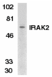
Left:
Western blot analysis of IRAK2 in K562 whole cell lysate with IRAK2 antibody at 1:500 dilution.


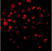
Below: Immunofluorescence of IRAK2 in HeLa cells with IRAK antibody at 10 µg/ml.

Other Product Images
Source:IRAK2 antibody was raised against a peptide corresponding to amino acids near the carboxy terminus of human IRAK2.
Purification: Affinity chromatography purified via peptide column
Clonality and Clone: This is a polyclonal antibody.
Host: IRAK-2 antibody was raised in rabbit.
Please use anti-rabbit secondary antibodies.
Immunogen: Human IRAK2 (C-Terminus) Peptide (Cat. No. 2213P)
Application: IRAK-2 antibody can be used for detection of IRAK2 by Western blot at 1:500 to 1:1000 dilution. A 65 kDa band can be detected. Anti-IRAK2 has no cross response to IRAK.
Tested Application(s): E, WB, IF
Buffer: Antibody is supplied in PBS containing 0.02% sodium azide.
Blocking Peptide:Cat. No. 2213P - IRAK-2 Peptide
Long-Term Storage: IRAK-2 antibody can be stored at 4ºC, stable for one year. As with all antibodies care should be taken to avoid repeated freeze thaw cycles. Antibodies should not be exposed to prolonged high temperatures.
Positive Control:
1.Cat. No. 1204 - K562 Cell Lysate
2. Cat. No. 1201 - HeLa Cell Lysate
Species Reactivity: H
GI Number: 37725373
Accession Number: AAO24761
Short Description: (CT) IL-1R Associated Kinase
References
1. Cao Z; Henzel WJ; Gao X. IRAK: a kinase associated with the interleukin-1 receptor. Science 1996;271:1128-31.
2. Muzio M, Ni J, Feng P, Dixit VM. IRAK (Pelle) family member IRAK-2 and MyD88 as proximal mediators of IL-1 signaling. Science 1997;278:1612-5
3. Zhang FX, Kirschning CJ, Mancinelli R, Xu XP, Jin Y, Faure E, Mantovani A, Rothe M, Muzio M, Arditi M. Bacterial lipopolysaccharide activates nuclear factor-kappaB through interleukin-1 signaling mediators in cultured human dermal endothelial cells and mononuclear phagocytes. J Biol Chem 1999;274:7611-4
4. Yang RB, Mark MR, Gurney AL , Godowski PJ. Signaling events induced by lipopolysaccharide-activated toll-like receptor 2. J Immunol 1999;163:639-43 (RD0300)

IRAK2 Antibody


Catalog#:3597

The pro-inflammatory cytokine IL-1 induces cellular response through two subunits of its receptor, IL-1 receptor I (IL-1RI) and IL-1 receptor accessory protein (IL-1RAcP). IL-1 receptor-associated kinase (IRAK) mediates activation of NF-kappaB, which is a pivotal transcription factor mediating inflammatory and immune response. A novel member in the IRAK/Pelle family has been identified and designated IRAK2 (1). Both IRAK and IRAK2 recruit to the subunits of the IL-1R complex after IL-1 binding and lead to NF-kappaB activation. IRAKs also associate with Toll like receptor (TLR) and the dominant negative mutants of IRAKs inhibit LPS-induced NF-?B activation (2,3). Members in IRAK/Pelle family play a central role in IL-1R and TLR mediated inflammatory response. Unlike human IRAK2, murine IRAK2 exists as four alternately spliced isoforms (IRAK2a-d), with two isoforms (IRAK2c and d) acting in an inhibitory fashion (4). IRAK2 is expressed in a variety of tissues.

Additional Names: IRAK2 (C2), mIRAK2


Description

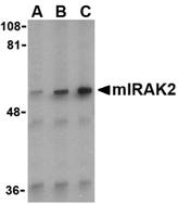
Left: Western blot analysis of IRAK2 in A-20 whole cell lysate with IRAK2 antibody (C2) at (A) 0.5, (B) 1, and (C) 2 µg/ml.


Source:RAK2 antibody was raised against a peptide corresponding to 16 amino acids near the carboxy-terminus of mouse IRAK2a which is common to all four isoforms. Anti-IRAK2 is mouse specific.
Purification: Affinity chromatography purified via peptide column
Clonality and Clone: This is a polyclonal antibody.
Host: IRAK2 antibody was raised in rabbit.
Please use anti-rabbit secondary antibodies.
Immunogen: Human IRAK2 (C-Terminus 2) Peptide (Cat. No. 3597P)
Application: IRAK2 antibody can be used for detection of mouse IRAK2 by Western blot at 0.5 - 1 µg/ml.Anti-IRAK2 has no cross response to IRAK.
Tested Application(s): E, WB
Buffer: Antibody is supplied in PBS containing 0.02% sodium azide.
Blocking Peptide:Cat. No. 3597P - IRAK2 Peptide
Long-Term Storage: IRAK2 antibody can be stored at 4ºC, stable for one year. As with all antibodies care should be taken to avoid repeated freeze thaw cycles. Antibodies should not be exposed to prolonged high temperatures.
Positive Control:
1. Cat. No. 1288 - A-20 Cell Lysate
Species Reactivity: M
GI Number: 37725373
Accession Number: AAO24761
Short Description: (C2) mouse IL-1R associated kinase
References
1. Muzio M, Ni J, Feng P, et al. IRAK (Pelle) family member IRAK-2 and MyD88 as proximal mediators of IL-1 signaling. Science 1997; 278:1612-5.
2. Zhang FX, Kirschning CJ, Mancinelli R, et al. Bacterial lipopolysaccharide activates nuclear factor-kappaB through interleukin-1 signaling mediators in cultured human dermal endothelial cells and mononuclear phagocytes. J. Biol. Chem. 1999; 274:7611-4.
3. Yang RB, Mark MR, Gurney AL, et al. Signaling events induced by lipopolysaccharide-activated toll-like receptor 2. J. Immunol. 1999; 163:639-43.
4. Hardy MP and O’Neill LAJ. The murine IRAK2 gene encodes four alternately spliced isoforms, two of which are inhibitory. J. Biol. Chem. 2004; 279:27699-708.

IRAK2 Antibody


Catalog#:3595

The pro-inflammatory cytokine IL-1 induces cellular response through two subunits of its receptor, IL-1 receptor I (IL-1RI) and IL-1 receptor accessory protein (IL-1RAcP). IL-1 receptor-associated kinase (IRAK) mediates activation of NF-kappaB, which is a pivotal transcription factor mediating inflammatory and immune response. A novel member in the IRAK/Pelle family has been identified and designated IRAK2 (1). Both IRAK and IRAK2 recruit to the subunits of the IL-1R complex after IL-1 binding and lead to NF-kappaB activation. IRAKs also associate with Toll like receptor (TLR) and the dominant negative mutants of IRAKs inhibit LPS-induced NF-?B activation (2,3). Members in IRAK/Pelle family play a central role in IL-1R and TLR mediated inflammatory response. Unlike human IRAK2, murine IRAK2 exists as four alternately spliced isoforms (IRAK2a-d), with two isoforms (IRAK2c and d) acting in an inhibitory fashion (4). IRAK2 is expressed in a variety of tissues.

Additional Names: IRAK2 (CT), mIRAK2
Description
Left:
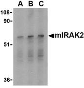
Western blot analysis of IRAK2 in RAW264.7 whole cell lysate with mIRAK2 antibody at (A) 0.5, (B) 1, and (C) 2 µg/ml.


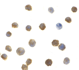
Below: Immunocytochemistry of IRAK2 in A-20 cells with IRAK2 antibody at 1 µg/ml.

Other Product Images
Source:IRAK2 antibody was raised against a peptide corresponding to 17 amino acids near the carboxy-terminus of mouse IRAK2a which is common to all four isoforms. mIRAK2 antibody will also recognize human IRAK2.
Purification: Affinity chromatography purified via peptide column
Clonality and Clone: This is a polyclonal antibody.
Host: IRAK2 antibody was raised in rabbit.
Please use anti-mouse secondary antibodies.
Immunogen: Human IRAK2 (C-Terminus) Peptide (Cat. No. 3595P)
Application: IRAK2 antibody can be used for detection of mouse IRAK2 by Western blot at 0.5 - 1 µg/ml.A 65 kDa band should be detected. Anti-IRAK2 has no cross response to IRAK. Anti-IRAK2 will also recognize human IRAK2.
Tested Application(s): E, WB, ICC
Buffer: Antibody is supplied in PBS containing 0.02% sodium azide.
Blocking Peptide:Cat. No. 3595P - IRAK2 Peptide
Long-Term Storage: IRAK2 antibody can be stored at 4ºC, stable for one year. As with all antibodies care should be taken to avoid repeated freeze thaw cycles. Antibodies should not be exposed to prolonged high temperatures.
Positive Control:
1. Cat. No. 1283 - RAW264.7 Cell Lysate
2. Cat. No. 1288 - A-20 Cell Lysate
Species Reactivity: H, M
GI Number: 37725373
Accession Number: AAO24761
Short Description: (CT) mouse IL-1R associated kinase
References
1. Muzio M, Ni J, Feng P, et al. IRAK (Pelle) family member IRAK-2 and MyD88 as proximal mediators of IL-1 signaling. Science 1997; 278:1612-5.
2. Zhang FX, Kirschning CJ, Mancinelli R, et al. Bacterial lipopolysaccharide activates nuclear factor-kappaB through interleukin-1 signaling mediators in cultured human dermal endothelial cells and mononuclear phagocytes. J. Biol. Chem. 1999; 274:7611-4.
3. Yang RB, Mark MR, Gurney AL, et al. Signaling events induced by lipopolysaccharide-activated toll-like receptor 2. J. Immunol. 1999; 163:639-43.
4. Hardy MP and O’Neill LAJ. The murine IRAK2 gene encodes four alternately spliced isoforms, two of which are inhibitory. J. Biol. Chem. 2004; 279:27699-708.

Wednesday, July 13, 2011

Extinct Biological Hazard Organisms


Biosynthesis DNA Testing (Ancestry/DNA Identity Profile) provides you with a simple and objective description of your deep ancestral origins as well as a certificate of your unique genetic fingerprint. The test gives you an estimated percentage of ancestry from the four major historical population groups.

Biological Hazard

A biological hazard or biohazard is an organism, or substance derived from an organism, that poses a threat to (primarily) human health. This can include medical waste or samples of a microorganism, virus or toxin (from a biological source) that can impact human health. It can also include substances harmful to animals. The term and its associated symbol is generally used as a warning, so that those potentially exposed to the substances will know to take precautions. There is also a biohazard HCS/WHMIS logo which utilizes the same symbol.

Biological Agents

Biological agents include prions, viruses, microorganisms (bacteria and fungi) and some unicellular and multicellular eukaryotes (for example parasites) and their associated toxins. They have the ability to adversely affect human health in a variety of ways, ranging from allergic reactions that are usually relatively mild, to serious medical conditions, even death. These organisms are ubiquitous in the natural environment; they are found in water, soil, plants, and animals.

For more information visit: www.biosyn.com

IRAK Monoclonal Antibody


Catalog#:PM-5317

Nuclear factor kappa B (NF-kappaB) is a ubiquitous transcription factor and an essential mediator of gene expression during activation of immune and inflammatory responses. NF-kappaB mediates the expression of a great variety of genes in response to extracellular stimuli including IL-1, TNFalpha and LPS. A serine/threonine protein kinase associated with IL-1 receptor (IRAK) and its homologue mouse pelle-like protein kinase (mPLK) were identified recently. IRAK is associated with the IL-1 receptor subunits IL-1RI and IL-1RAcP after IL-1 binding and serves as a signaling molecule to mediate IL-1 response. IRAK mediates a signaling cascade leading to NF-kB activation by members in IL-1 family including IL-1 and a novel cytokine IL-18 (also termed IGIF).

Additional Names:
IRAK (8F1A7),IL-1-Receptor Associated Kinase

Description

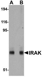
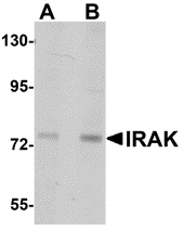
Left: Western blot analysis of IRAK in mouse liver lysate with IRAK antibody at (A) 1 and (B) 2 µg/ml.


Source:Mouse monoclonal IRAK antibody was raised against a recombinant protein corresponding to amino acids 618 - 712 of human IRAK.
Purification: Immunoaffinity chromotography purified IgG
Clonality and Clone: This is a monoclonal antibody. (Clone 8F1A7)
Host: IRAK monoclonal antibody was raised in mouse.
Please use anti-mouse secondary antibodies.
Immunogen: Recombinant protein corresponding to amino acids 618 - 712 of human IRAK.
Application: IRAK antibody can be used for Western blot at 1 - 2 µg/ml.
Tested Application(s): E, WB
Buffer: Antibody is supplied in PBS containing 0.02% sodium azide.
Blocking Peptide:
Long-Term Storage: IRAK monoclonal antibody can be stored at 4ºC, stable for one year.
Positive Control:
1. Cat. No. 95-112 - IRAK Recombinant Protein
Species Reactivity: H, M
Short Description: IL-1-Receptor Associated Kinase
References
1. Cao Z,; Henzel WJ, and Gao X. IRAK: a kinase associated with the interleukin-1 receptor. Science 1996; 271:1128-31.
2. Trofimova M, Sprenkle AB, Green M; et al. Developmental and tissue-specific expression of mouse pelle-like protein kinase. J. Bio. Chem. 1996; 271: 17609-12.
3. Jianing Huang, Xiong Gao, Shyun Li, et al. Recruitment of IRAK to the interleukin 1 receptor complex requires interleukin-1 receptor accessory protein. Proc. Natl. Acad. Sci. USA 1997; 94:12829-32.
4. Robinson D, Shibuya K, Mui A, et al. IGIF does not drive Th1 development but synergizes with IL-12 for interferon-gamma production and activates IRAK and NF-kB. Immunity 1997; 7:571-81.

IRAK Monoclonal Antibody


Catalog#:PM-5315

Nuclear factor kappa B (NF-kappaB) is a ubiquitous transcription factor and an essential mediator of gene expression during activation of immune and inflammatory responses. NF-kappaB mediates the expression of a great variety of genes in response to extracellular stimuli including IL-1, TNFalpha and LPS. A serine/threonine protein kinase associated with IL-1 receptor (IRAK) and its homologue mouse pelle-like protein kinase (mPLK) were identified recently. IRAK is associated with the IL-1 receptor subunits IL-1RI and IL-1RAcP after IL-1 binding and serves as a signaling molecule to mediate IL-1 response. IRAK mediates a signaling cascade leading to NF-kB activation by members in IL-1 family including IL-1 and a novel cytokine IL-18 (also termed IGIF).

Additional Names: IRAK (1F12C1),IL-1-Receptor Associated Kinase


Description

Left: Western blot analysis of IRAK in HeLa lysates with IRAK antibody at (A) 1 and (B) 2 µg/ml.


Source:Mouse monoclonal IRAK antibody was raised against a recombinant protein corresponding to amino acids 618 - 712 of human IRAK.
Purification: Immunoaffinity chromotography purified IgG
Clonality and Clone: This is a monoclonal antibody. (Clone 1F12C1)
Host: IRAK monoclonal antibody was raised in mouse.
Please use anti-mouse secondary antibodies.
Immunogen: Recombinant protein corresponding to amino acids 618 - 712 of human IRAK.
Application: RAK antibody can be used for Western blot at 1 µg/ml.
Tested Application(s): E, WB
Buffer: Antibody is supplied in PBS containing 0.02% sodium azide.
Blocking Peptide:
Long-Term Storage: IRAK monoclonal antibody can be stored at 4ºC, stable for one year.
Positive Control:
1. Cat. No. 95-112 - IRAK Recombinant Protein
Species Reactivity: H, M
Short Description: IL-1-Receptor Associated Kinase
References
1. Cao Z,; Henzel WJ, and Gao X. IRAK: a kinase associated with the interleukin-1 receptor. Science 1996; 271:1128-31.
2. Trofimova M, Sprenkle AB, Green M; et al. Developmental and tissue-specific expression of mouse pelle-like protein kinase. J. Bio. Chem. 1996; 271: 17609-12.
3. Jianing Huang, Xiong Gao, Shyun Li, et al. Recruitment of IRAK to the interleukin 1 receptor complex requires interleukin-1 receptor accessory protein. Proc. Natl. Acad. Sci. USA 1997; 94:12829-32.
4. Robinson D, Shibuya K, Mui A, et al. IGIF does not drive Th1 development but synergizes with IL-12 for interferon-gamma production and activates IRAK and NF-kB. Immunity 1997; 7:571-81.

IRAK Antibody


Catalog#:1007

Nuclear factor kappa B (NF-kappaB) is a ubiquitous transcription factor and an essential mediator of gene expression during activation of immune and inflammatory responses. NF-kappaB mediates the expression of a great variety of genes in response to extracellular stimuli including IL-1, TNFalpha and LPS. A serine/threonine protein kinase associated with IL-1 receptor (IRAK) and its homologue mouse pelle-like protein kinase (mPLK) were identified recently. IRAK is associated with the IL-1 receptor subunits IL-1RI and IL-1RAcP after IL-1 binding and serves as a signaling molecule to mediate IL-1 response. IRAK mediates a signaling cascade leading to NF-kappaB activation by members in IL-1 family including IL-1 and a novel cytokine IL-18 (also termed IGIF).

Additional Names: IRAK (CT), IL-1-Receptor Associated Kinase


Description

Left:
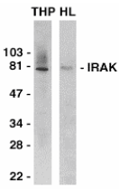
Western blot analysis of IRAK in THP-1 (THP) and HeLa (HL) whole cell lysates with IRAK antibody at 1:2000 dilution.


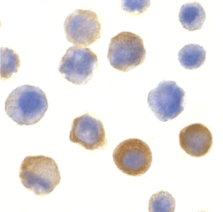
Below: Immunocytochemistry of IRAK in HeLa cells with IRAK antibody at 10 µg/ml.

Other Product Images

Source:IRAK antibody was raised against a peptide corresponding to amino acids near the carboxy terminus of human IRAK.
Purification: Antibody is DEAE purified
Clonality and Clone: This is a polyclonal antibody.
Host: IRAK antibody was raised in rabbit.
Please use anti-rabbit secondary antibodies.
Immunogen: Human IRAK (C-Terminus) Peptide (Cat. No. 1007P)
Application: IRAK antibody can be used for Western blot at 1:1000 dilution and for immunoprecipitation with 2 to 4 ug per sample. An 80 kDa band should be detected in the lysate from non-activated cells. It has no cross response to IRAK2.
Tested Application(s): E, WB, ICC, IP
Buffer: Antibody is supplied in PBS containing 0.02% sodium azide.
Blocking Peptide:Cat. No. 1007P - IRAK Peptide
Long-Term Storage: IRAK antibody can be stored at 4ºC, stable for one year. As with all antibodies care should be taken to avoid repeated freeze thaw cycles. Antibodies should not be exposed to prolonged high temperatures.
Positive Control:
1. Cat. No. 1201 - HeLa Cell Lysate
2. Cat. No. 1208 - THP-1 Whole Cell Lysate
Species Reactivity: H, M, R
GI Number: 8928535
Accession Number: P51617
Short Description: (CT) IL-1R associated kinase
References
1. Cao Z; Henzel WJ; Gao X. IRAK: a kinase associated with the interleukin-1 receptor. Science 1996;271:1128-31.
2. Trofimova M; Sprenkle AB; Green M; Sturgill TW; Goebl MG; Harrington MA. Developmental and tissue-specific expression of mouse pelle-like protein kinase. J Bio Chem 1996; 271: 17609-1
3. Jianing Huang, Xiong Gao, Shyun Li, and Zhaodan Cao. Recruitment of IRAK to the interleukin 1 receptor complex requires interleukin-1 receptor accessory protein. Proc Natl Acad Sci USA 1997;94:12829-12832
4. Robinson D, Shibuya K, Mui A, Zonin F, Murphy E, Sana T, Hartley SB, Menon S, Kastelein R, Bazan F, O'Garra A. IGIF does not drive Th1 development but synergizes with IL-12 for interferon-gamma production and activates IRAK and NF-kappaB. Immunity 1997;7:571-581

Wednesday, July 6, 2011

DNA Sequence


BIO-SYNTHESIS is second to none in its ability to meet our customer's needs, often providing modified DNA with wide variety of selections. BSI offer BNA/DNA chimera synthesis at competitive prices and quick delivery. Bio-Synthesis continued to provide quality DNA products and services for the research community, but it has also become a world leader in providing custom peptide products and services.

DNA Sequence

A DNA sequence or genetic sequence is a succession of letters representing the primary structure of a real or hypothetical DNA molecule or strand, with the capacity to carry information. The possible letters are A, C, G, and T, representing the four nucleotide subunits of a DNA strand - adenine, cytosine, guanine, thymine bases covalently linked to phospho-backbone. In the typical case, the sequences are printed abutting one another without gaps, as in the sequence AAAGTCTGAC, going from 5' to 3' from left to right. A succession of any number of nucleotides greater than four is liable to be called a sequence.

DNA

The concept was to mimic an oligonucleotide binding to double stranded DNA via Hoogsteen base pairing in the major groove. Thus the nucleobases of DNA were retained, but the deoxyribose phosphodiester backbone of DNA was replaced by a pseudo-peptide backbone that according to computer model building was homomorphous with the DNA backbone.

Deoxyribonucleic Acid

Deoxyribonucleic acid (DNA) is a nucleic acid that contains the genetic instructions used in the development and functioning of all known living organisms and some viruses. The main role of DNA molecules is the long-term storage of information.

Catalog#:4781

Interleukin-34 (IL-34) was identified through a functional screening of a library of secreted proteins after transfection of an embryonic kidney cell line with recombinant cDNAs. Flow cytometry and monocyte viability assays demonstrated increased survival and/or proliferation of monocytes induced by IL-34 and specific binding of IL-34 to CD14+ monocytes. IL-34 is a second ligand to the colony stimulating factor (CSF)-1 receptor in addition to CSF1. Like CSF1, IL-34 stimulates phosphorylation of ERK1 and ERK2 in human monocytes and induces differentiation of bone marrow cells into macrophages. Despite its predicted molecular weight, IL-34 will often run at higher molecular weight in SDS-PAGE.

Additional Names: IL-34 (IN), Interleukin-34


Description

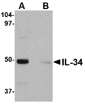
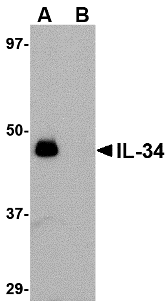
Left: Western blot analysis of IL-34 in human brain tissue lysate with IL-34 antibody at 0.25 µg/ml in (A) the absence and (B) the presence of blocking peptide.


Source:IL-34 antibody was raised against a 13 amino acid peptide from near the center of human IL-34.
Purification: Affinity chromatography purified via peptide column
Clonality and Clone: This is a polyclonal antibody.
Host: IL-34 antibody was raised in rabbit.
Please use anti-rabbit secondary antibodies.
Application: IL-34 antibody can be used for the detection of IL-34 by Western blot at 0.25 – 0.5 µg/ml.
Tested Application(s): E, WB
Buffer: Antibody is supplied in PBS containing 0.02% sodium azide.
Blocking Peptide:Cat.No. 4781P - IL-34 Peptide
Long-Term Storage: IL-34 antibody can be stored at 4ºC, stable for one year. As with all antibodies care should be taken to avoid repeated freeze thaw cycles. Antibodies should not be exposed to prolonged high temperatures.
Positive Control:
1.Cat. No. 1303 - Human Brain Tissue Lysate
Species Reactivity: H, M, R
GI Number: 187610438
Accession Number: ACD13474
Short Description: (IN) Interleukin 34
References
1. Lin H, Lee E, Hestir K, et al. Discovery of a cytokine and its receptor by functional screening of the extracellular proteome. Science 2008; 320:807-11.

Catalog#:4779

Interleukin-34 (IL-34) was identified through a functional screening of a library of secreted proteins after transfection of an embryonic kidney cell line with recombinant cDNAs. Flow cytometry and monocyte viability assays demonstrated increased survival and/or proliferation of monocytes induced by IL-34 and specific binding of IL-34 to CD14+ monocytes. IL-34 is a second ligand to the colony stimulating factor (CSF)-1 receptor in addition to CSF1. Like CSF1, IL-34 stimulates phosphorylation of ERK1 and ERK2 in human monocytes and induces differentiation of bone marrow cells into macrophages. Despite its predicted molecular weight, IL-34 will often run at higher molecular weight in SDS-PAGE.

Additional Names: IL-34 (NT), Interleukin-34
Description
Left: Western blot analysis of IL-34 in human brain tissue lysate with IL-34 antibody at 0.25 µg/ml in (A) the absence and (B) the presence of blocking peptide.


Source:IL-34 antibody was raised against a 16 amino acid peptide from near the amino terminus of human IL-34.
Purification: Affinity chromatography purified via peptide column
Clonality and Clone: This is a polyclonal antibody.
Host: IL-34 antibody was raised in rabbit.
Please use anti-rabbit secondary antibodies.
Application: IL-34 antibody can be used for the detection of IL-34 by Western blot at 0.25 – 0.5 µg/ml.
Tested Application(s): E, WB
Buffer: Antibody is supplied in PBS containing 0.02% sodium azide.
Blocking Peptide:Cat.No. 4779P - IL-34 Peptide
Long-Term Storage: IL-34 antibody can be stored at 4ºC, stable for one year. As with all antibodies care should be taken to avoid repeated freeze thaw cycles. Antibodies should not be exposed to prolonged high temperatures.
Positive Control:
1. Cat. No. 1303 - Human Brain Tissue Lysate
Species Reactivity: H, M, R
GI Number: 187610438
Accession Number: ACD13474
Short Description: (NT) Interleukin 34
References
1. Lin H, Lee E, Hestir K, et al. Discovery of a cytokine and its receptor by functional screening of the extracellular proteome. Science 2008; 320:807-11.