Monday, June 20, 2011

GBL Antibody


Catalog#:3495

GbetaL (G protein beta protein subunit-like) is a member of a signaling pathway that regulates mammalian cell growth in response to the presence of nutrients and growth factors (1). It binds to the kinase domain of TOR (Target of rapamycin, also known as mTOR), an evolutionarily conserved serine/threonine kinase that regulates cell growth and cell cycle through its ability to integrate signals from nutrient levels and growth factors (reviewed in 2). Rapamycin inhibits TOR resulting in reduced cell growth and reduced rates of cell cycle and cell proliferation (reviewed in 3). TOR is normally associated with GbetaL and an additional regulatory protein RAPTOR, allowing TOR to control protein biosynthesis (3). The binding of GbetaL to TOR stimulates TOR’s kinase activity towards downstream proteins such as RPS6K (ribosomal protein S6 kinase) and the translation factor 4E-BP1 which leads to increased protein translation and cell growth (3).

Additional Names: GBL, G beta protein subunit-like
Description

Left:
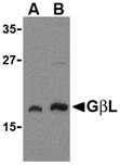
Western blot analysis of GbL in human brain cell lysate with GbL antibody at (A) 1 and (B) 2 µg/ml.


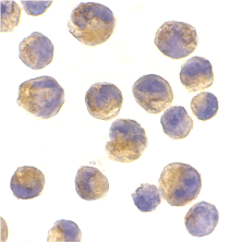
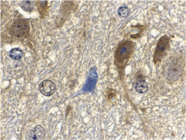
Below: Immunohistochemistry of GbL in mouse brain tissue with GbL antibody at 10 µg/ml.

Other Product Images
Source:GbL antibody was raised against a 14 amino acid peptide from near the carboxy-terminus of human GbL.
Purification: Affinity chromatography purified via peptide column
Clonality and Clone: This is a polyclonal antibody.
Host: GBL antibody was raised in rabbit.
Please use anti-rabbit secondary antibodies.
Immunogen: Human GbL / G beta protein subunit-like Peptide (Cat. No. 3495P)
Application: GbL antibody can be used for the detection of GbL by Western blot at 1 µg/ml.
Tested Application(s): E, WB, IHC
Buffer: Antibody is supplied in PBS containing 0.02% sodium azide.
Blocking Peptide:Cat. No. 3495P - GBL Peptide
Long-Term Storage: GBL antibody can be stored at 4ºC, stable for one year. As with all antibodies care should be taken to avoid repeated freeze thaw cycles. Antibodies should not be exposed to prolonged high temperatures.
Positive Control:
1. Cat. No. 1303 - Human Brain Tissue Lysate
2. Cat. No. 1403 - Mouse Brain Tissue Lysate
Species Reactivity: H, M, R
GI Number: 30411038
Accession Number: AAH52292
Short Description: a member of the TOR signaling pathway
References
1. Kim D-H, Sarbassov DD, Ali SM, et al. GbetaL, a positive regulator of the Rapamycin-sensitive pathway required for the nutrient-sensitive interaction between Raptor and mTOR. Mol. Cell 2003; 11:895-904.
2. Shamji AF, Ngheim P, and Schreiber SL. Integration of growth factor and nutrient signaling: implications for cancer biology. Mol. Cell 2003; 12:271-80.
3. Fingar DC and Blenis J. Target of rapamycin (TOR): an integrator of nutrient and growth factor signals and coordinator of cell growth and cell cycle progression. Oncogene 2004; 23:3151-71.

Friday, June 17, 2011

DNA Synthesis Primer


For over 20 years, BioSynthesis has provided custom oligonucleotide production services worldwide, including researcher at university, biotechnology and pharmaceutical institutions. We are not only synthesizing standard DNA primer and probes but are also specialized in production and purification of modified oligomers. Base, ribose and backbone modifications and reporter groups like dyes and haptens are routinely incorporated at small discovery research scale to multi-gram ASR/cGMP for clinical diagnostic production. As always, quality is guaranteed!

DNA Synthesis

DNA replication is the process of copying a double-stranded DNA molecule to form two double-stranded molecules. The process of DNA replication is a fundamental process used by all living organisms as it is the basis for biological inheritance. As each DNA strand holds the same genetic information, both strands can serve as templates for the reproduction of the opposite strand. The template strand is preserved in its entirety and the new strand is assembled from nucleotides.

DNA Polymerase

A DNA polymerase is an enzyme that assists in DNA replication. Such enzymes catalyze the polymerization of deoxyribonucleotides alongside a DNA strand, which they "read" and use as a template. The newly-polymerized molecule is complementary to the template strand and identical to the template's partner strand.

Variation Across Species

DNA polymerases have highly-conserved structure, which means that their overall catalytic subunits vary, on a whole, very little from species to species. Conserved structures usually indicate important, irreplicable functions of the cell, the maintenance of which provides evolutionary advantages.

For more information visit: www.biosyn.com


GAPDH Antibody

Catalog#:3783

Glyceraldehyde-3-phosphate dehydrogenase (GAPDH) catalyzes the reversible oxidative phosphorylation of glyceraldehyde-3-phosphate in the presence of inorganic phosphate and nicotinamide adenine dinucleotide (NAD), an important energy-yielding step in carbohydrate metabolism. Recent evidence suggests that it also is involved in a number of cellular processes such as membrane fusion, phosphotransferase activity, DNA replication and repair, and nuclear RNA export (1). GAPDH has also been implicated in playing a role in different pathologies such as cancer progression, apoptosis, and neuronal diseases such as Alzheimer’s and Huntington’s disease (2). GAPDH is constitutively expressed at high levels in almost all tissues and cell lines making it ideal for use as a loading control marker in immunoblots.

Additional Names: GAPDH (NT), Glyceraldehyde-3-phosphate dehydrogenase, G3PDH, GAPD


Description

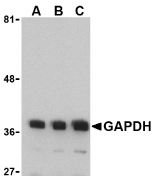
Left: Western blot analysis of GAPDH in HeLa cell lysate with GAPDH antibody at (A) 0.125, (B) 0.25 and (C) 0.5 µg/ml.


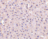
Below: Immunohistochemistry of GAPDH in human liver tissue with GAPDH antibody at 10 µg/ml.

Other Product Images
Source:
GAPDH antibody was raised against a 16 amino acid peptide from near the amino-terminus of human GAPDH.
Purification: Affinity chromatography purified via peptide column
Clonality and Clone: This is a polyclonal antibody.
Host: GAPDH antibody was raised in rabbit.

Please use anti-rabbit secondary antibodies.
Immunogen: Human GAPDH / Glyceraldehyde-3-phosphate dehydrogenase / G3PDH / GAPD (N-Terminus) Peptide (Cat. No.
Application: GAPDH antibody can be used for the detection of GAPDH by Western blot at 0.5 - 1 µg/ml.
Tested Application(s): E, WB, IHC
Buffer: Antibody is supplied in PBS containing 0.02% sodium azide.
Blocking Peptide:Cat. No. 3783P - GAPDH Peptide
Long-Term Storage: GAPDH antibody can be stored at 4ºC, stable for one year. As with all antibodies care should be taken to avoid repeated freeze thaw cycles. Antibodies should not be exposed to prolonged high temperatures.
Positive Control:
1. Cat. No. 1201 - HeLa Cell Lysate
Species Reactivity: H, M, R
GI Number: 120649
Accession Number: P04406
Short Description: (NT) An eznyme in the glycolytic pathway
References

1. Sirover MA. New nuclear functions of the glycolytic protein, glyceraldehyde-3-phosphate dehydrogenase, in mammalian cells. J. Cell. Biochem. 2005; 95:45-52.
2. Glyceraldehyde-3-phosphate dehydrogenase, apoptosis, and neurodegenerative diseases. Annu. Rev. Pharmacol. Toxicol. 2005; 45:269-90.

GAPDH Antibody


Catalog#:3781

Glyceraldehyde-3-phosphate dehydrogenase (GAPDH) catalyzes the reversible oxidative phosphorylation of glyceraldehyde-3-phosphate in the presence of inorganic phosphate and nicotinamide adenine dinucleotide (NAD), an important energy-yielding step in carbohydrate metabolism. Recent evidence suggests that it also is involved in a number of cellular processes such as membrane fusion, phosphotransferase activity, DNA replication and repair, and nuclear RNA export (1). GAPDH has also been implicated in playing a role in different pathologies such as cancer progression, apoptosis, and neuronal diseases such as Alzheimer’s and Huntington’s disease (2). GAPDH is constitutively expressed at high levels in almost all tissues and cell lines making it ideal for use as a loading control marker in immunoblots.

Additional Names: GAPDH, Glyceraldehyde-3-phosphate dehydrogenase, G3PDH, GAPD


Description

Left:
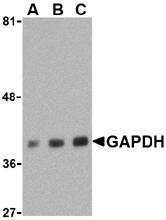
Western blot analysis of GAPDH in HeLa cell lysate with GAPDH antibody at (A) 0.5, (B) 1 and (C) 2 µg/ml.


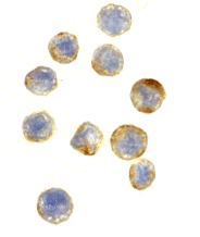
Below: Immunohistochemical staining of GAPDH in HeLa cells at a dilution of 10 µg/ml.


Other Product Images
Source:GAPDH antibody was raised against a 16 amino acid peptide from near the carboxy-terminus of human GAPDH.
Purification: Affinity chromatography purified via peptide column
Clonality and Clone: This is a polyclonal antibody.
Host: GAPDH antibody was raised in rabbit.
Please use anti-rabbit secondary antibodies.
Immunogen: Human GAPDH / Glyceraldehyde-3-phosphate dehydrogenase / G3PDH / GAPD (C-Terminus) Peptide (Cat. No.
Application: GAPDH antibody can be used for the detection of GAPDH by Western blot at 0.5 – 1 µg/ml.
Tested Application(s): E, WB, IHC
Buffer: Antibody is supplied in PBS containing 0.02% sodium azide.
Blocking Peptide:Cat. No. 3781P - GAPDH Peptide
Long-Term Storage: GAPDH antibody can be stored at 4ºC, stable for one year. As with all antibodies care should be taken to avoid repeated freeze thaw cycles. Antibodies should not be exposed to prolonged high temperatures.
Positive Control:
1.Cat. No. 1201 - HeLa Cell Lysate
Species Reactivity: H, M, R
GI Number: 120649
Accession Number: P04406
Short Description: (CT) an enzyme in the glycolytic pathway
References
1. Sirover MA. New nuclear functions of the glycolytic protein, glyceraldehyde-3-phosphate dehydrogenase, in mammalian cells. J. Cell. Biochem. 2005; 95:45-52.
2. Glyceraldehyde-3-phosphate dehydrogenase, apoptosis, and neurodegenerative diseases. Annu. Rev. Pharmacol. Toxicol. 2005; 45:269-90.

FGF4 Antibody


Catalog#:4433

Fibroblast growth factor 4 (FGF4) is a member of the fibroblast growth factor (FGF) family that possess broad mitogenic and cell survival activities and play key roles in growth and survival of stem cells during embryogenesis, tissue regeneration, and carcinogenesis. FGF4 was identified by its strong oncogenic transforming activity and is a potent angiogenic factor, expressed in several highly vascularized tumors and also in adult mouse testis, intestine, and brain. Studies on the mouse homolog suggests a function in bone morphogenesis and limb development through the sonic hedgehog (SHH) signaling pathway. Furthermore, FGF4 regulates neural progenitor cell proliferation and neuronal differentiation. Recent studies show a growth-promoting role for FGF4 in human embryonic stem cells and a putative feedback inhibition mechanism by a novel FGF4 splice isoform that may serve to promote differentiation at a later stages of development.

Additional Names: FGF4, Fibroblast growth factor 4, Heparin secretory-transforming protein, HST-1, HST, Transforming protein KS3, HBGF-4, Kaposi sarcoma oncogene


Description

Left:
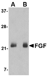
Western blot analysis of FGF4 in NIH 3T3 cell lysate with FGF4 antibody at (A) 0.5 and (B) 1 µg/ml.


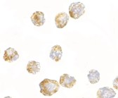
Below: Immunohistochemistry of CXCR4Lo in HeLa cells with CXCR4Lo antibody at 2 µg/ml.


Other Product Images


Source:FGF4 antibody was raised against a 18 amino acid peptide near the carboxy terminus of the human FGF4.
Purification: Affinity chromatography purified via peptide column
Clonality and Clone: This is a polyclonal antibody.
Host: FGF4 antibody was raised in rabbit.
Please use anti-rabbit secondary antibodies.
Application: FGF4 antibody can be used for detection of FGF4 by Western blot at 0.5 – 1 µg/ml.
Tested Application(s): E, WB, ICC
Buffer: Antibody is supplied in PBS containing 0.02% sodium azide.
Blocking Peptide:Cat.No. 4433P - FGF4 Peptide
Long-Term Storage: FGF4 antibody can be stored at 4ºC, stable for one year. As with all antibodies care should be taken to avoid repeated freeze thaw cycles. Antibodies should not be exposed to prolonged high temperatures.
Positive Control:
1. Cat. No. 1282 - 3T3 (NIH) Cell Lysate
Species Reactivity: H, M
GI Number: 122750
Accession Number: P08620
Short Description: Fibroblast growth factor 4
References
1. Powers CJ, McLeskey SW, and Wellstein A. Fibroblast growth factors, their receptors and signaling. Endocr. Relat. Cancer 2000; 7:165-97.
2. Delli-Bovi P, Curatola AM, Kern FG, et al. An oncogene isolated by transfection of Kaposi's sarcoma DNA encodes a growth factor that is a member of the FGF family. Cell 1987; 50:729-37.
3. Yoshida T, Ishimaru K, Sakamoto H, et al. Angiogenic activity of the recombinant hst-1 protein. Cancer Lett. 1994; 83:261-268.
4. Laufer E, Nelson CE, Johnson RL, et al. Sonic hedgehog and Fgf-4 act through a signaling cascade and feedback loop to integrate growth and patterning of the developing limb bud. Cell 1994; 79:993-1003.

Thursday, June 16, 2011

RNA Electrophoresis


Biosyn Serving molecular diagnostic community since 1984. It provides great resource of RNA Research Analysis, RNA catalysis and RNA processing. We are providing comprehensive Custom RNA synthesis and modification such as RNA Chimeras and RNA Conjugations. From RNA probe selection to chemical synthesis. Our team of molecular biologists is dedicated in serving clients for their RNA research.

Electrophoresis

Electrophoresis permits assessment of RNA by size and amount. In general, electrophoresis of RNA is done as a step prior to Northern analysis.

RNA Processing

Post transcriptional modification is a genetic process in cell biology by which, in eukaryotic cells, primary transcript RNA is converted into mature RNA. It is most often associated with the conversion of precursor messenger RNA into mature messenger RNA (mRNA), which is also known as splicing, during the larger process of protein synthesis.

RNA Research Analysis

Today RNA biology is becoming important in all areas of biomedical research. This is extremely useful for researchers who design molecules for RNA interference, and need to statistically test complex structure hypotheses.

For more information visit: www.biosyn.com

EphA10 Antibody


Catalog#:5253

Eph receptors, the largest subfamily of receptor tyrosine kinases (RTKs), and their ephrin ligands are important mediators of cell–cell communication regulating cell attachment, shape, and mobility of neuronal and endothelial cells in central nervous system function and in development. Eph receptors can be divided into two subgroups: EphA and EphB. In mammals, the EphA class consists of eight members (EphA 1-7 and 10) that in general bind to ephrin-A members linked to the cell membrane through a glycosylphosphatidylinositol linkage. The EphB class consists of six members (EphB 1-6) that in general bind ephrin-B members that transverse the cell membrane. The Ephrin / EPH signaling pathway networks with the WNT signaling pathway during embryogenesis, tissue regeneration, and carcinogenesis. Recent studies show that Eph/EFN might be relevant in normal B-cell biology and could represent new potential prognostic markers and therapeutic targets for CLL.

Additional Names: EphA10, EPH receptor A10, Ephrin type-A receptor 10, FLJ16103


Description

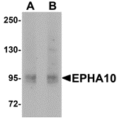
Left: Western blot analysis of EphA10 in 293 cell lysate with EphA10 antibody at (A) 1 µg/ml and (B) 2 µg/ml.


Source:EphA10 antibody was raised against a 14 amino acid peptide of near the amino terminus of human EphA10.
Purification: Affinity chromatography purified via peptide column
Clonality and Clone: This is a polyclonal antibody.
Host: EphA10 antibody was raised in rabbit.
Please use anti-rabbit secondary antibodies.
Application: EphA10 antibody can be used for detection of EphA10 by Western blot at 1 - 2 µg/ml.
Tested Application(s): E, WB
Buffer: Antibody is supplied in PBS containing 0.02% sodium azide.
Blocking Peptide:Cat.No. 5253P - EphA10 Peptide
Long-Term Storage: EphA10 antibody can be stored at 4ºC, stable for one year. As with all antibodies care should be taken to avoid repeated freeze thaw cycles. Antibodies should not be exposed to prolonged high temperatures.
Positive Control:
1. Cat. No. 1210 - 293 Cell Lysate
Species Reactivity: H, M, R
GI Number: 150456460
Accession Number: NP_001092909
Short Description: EPH receptor A10
References
1. Flanagan JG and Vanderhaeghen P. The ephrins and Eph receptors in neural development. Annu. Rev. Neurosci. 1998;.21:309–45.
2. Frisen J, Holmberg J, and Barbacid M. Ephrins and their Eph receptors: multitalented directors of embryonic development. EMBO J. 1999; 18:5159–65.
3. Eph Nomenclature Committee. Unified nomenclature for Eph family receptors and their ligands, the ephrins. Cell 1997; 90:403–4.
4. Holder N and Klein R. Eph receptors and ephrins: effectors of morphogenesis, Development 1999; 126:2033–44.

Catalog#:1107

Signals from most growth factors and cytokines are transduced by receptor tyrosine kinases or non-receptor tyrosine kinases. Activated tyrosine kinases phosphorylate their substrates, which mediate the cellular response to extracellular stimuli. A long-sought major substrate termed p62dok (downstream of tyrosine kinase) for many tyrosine kinases including c-kit, v-abl, v-Fps, v-Src, v-Fms, and activated EGF, PDGF, IGF, VEGF and insulin receptors was identified recently from human and mouse by several laboratories. Upon phosphorylation, p62dok forms a complex with the ras GTPase-activating protein (RasGAP). p62dok represents a new family with very recently identified p56dok.

Additional Names: DOK1 (CT), DOK1


Description

Left:
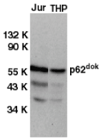
Western blot analysis of DOK1 in Jurkat (Jur) and THP-1 (THP) cell lysates with DOK1 antibody at 1 µg/ml.


Below: Immunocytochemistry of DOK1 in K562 cells with DOK1 antibody at 2 µg/ml.

Other Product Images
Source:DOK1 antibody was raised against a peptide corresponding to amino acids near the carboxy terminus of human DOK1.
Purification: Affinity chromatography purified via peptide column
Clonality and Clone: This is a polyclonal antibody.
Host: DOK1 antibody was raised in rabbit.
Please use anti-rabbit secondary antibodies.
Immunogen: Human DOK Peptide (Cat. No. 1107P)
Application: DOK1 antibody can be used for detection of DOK1 expression by Western blot at 0.5 - 1 µg/ml.A 62 kDa band should be detected.
Tested Application(s): E, WB, ICC
Buffer: Antibody is supplied in PBS containing 0.02% sodium azide.
Blocking Peptide:Cat. No. 1107P - DOK1 Peptide
Long-Term Storage: DOK1 antibody can be stored at 4ºC, stable for one year. As with all antibodies care should be taken to avoid repeated freeze thaw cycles. Antibodies should not be exposed to prolonged high temperatures.
Positive Control:
1. Cat. No. 1205 - Jurkat Cell Lysate
2. Cat. No. 1208 - THP-1 Cell Lysate
3. Cat. No. 1204 - K562 Cell Lysate
Species Reactivity: H
GI Number: 1848277
Accession Number: AAC51127
Short Description: (CT) Tyrosine kinase substrate DOK
References
1. Carpino N, Wisniewski D, Strife A, Marshak D, Kobayashi R, Stillman B, Clarkson B p62(dok): a constitutively tyrosine-phosphorylated, GAP-associated protein in chronic myelogenous leukemia progenitor cells. Cell 1997;88:197-204.
2. Yamanashi Y, Baltimore D Identification of the Abl- and rasGAP-associated 62 KDa protein as a docking protein, Dok. Cell 1997;88:205-211.
3. Holland SJ, Gale NW, Gish GD, Roth RA, Songyang Z, Cantley LC, Henkemeyer M, Yancopoulos GD, Pawson T. Juxtamembrane tyrosine residues couple the Eph family receptor EphB2/Nuk to specific SH2 domain proteins in neuronal cells. EMBO J 1997;16:3877-3888.
4. Di Cristofano A, Carpino N, Dunant N, Friedland G, Kobayashi R, Strife A, Wisniewski D, Clarkson B, Pandolfi PP, Resh MD. Molecular cloning and characterization of p56(dok-2) defines a new family of RasGAP-binding proteins. J Biol Chem 1998;273:4827-4830.

Disp2 Antibody


atalog#:4883

Disp2 is the second of three known homologs of the D. melanogaster protein Dispatched. It is a multi-transmembrane protein containing two PTCH/DISP domains and is thought to be involved in the release of lipid-anchored Hedgehog from producing cells. Hedgehog is a major player in signaling pathways during embryogenesis, tissue regeneration, and carcinogenesis and the Disp proteins have been implicated in these pathways. Recently, it has been shown that Disp2 is translationally regulated by the microRNA miR-214 in zebrafish. Expression of this miRNA decreased Disp2 promoter activity in vitro and its overexpression in zebrafish resulted in a phenotype identical to that observed by Disp2 mutants. At least two isoforms of Disp2 are known to exist. This Disp2 antibody is predicted to not cross-react with Disp1 or Disp3.

Additional Names: Disp2 (CT), Dispatched homolog 2, DISPB


Description

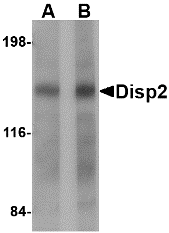
Left: Western blot analysis of Disp2 in rat brain tissue lysate with Disp2 antibody at (A) 1 and (B) 2 µg/ml.


Source:Disp2 antibody was raised against a 19 amino acid peptide from near the carboxy terminus of human Disp2.
Purification: Affinity chromatography purified via peptide column
Clonality and Clone: This is a polyclonal antibody.
Host: Disp2 antibody was raised in rabbit.
Please use anti-rabbit secondary antibodies.
Application: Disp2 antibody can be used for the detection of Disp2 by Western blot at 1 – 2 µg/ml.
Tested Application(s): E, WB
Buffer: Antibody is supplied in PBS containing 0.02% sodium azide.
Blocking Peptide:Cat.No. 4883P - Disp2 Peptide
Long-Term Storage: Disp2 antibody can be stored at 4ºC, stable for one year. As with all antibodies care should be taken to avoid repeated freeze thaw cycles. Antibodies should not be exposed to prolonged high temperatures.
Positive Control:
1. Cat. No. 1463 - Rat Brain Tissue Lysate
Species Reactivity: H, M, R
GI Number: 160380692
Accession Number: A7MBM2
Short Description: (CT) Dispatched homolog 2
References

1. Katoh Y and Katoh M. Identification and characterization of DISP3 gene in silico. Int. J. Oncol. 2005; 26:551-6.
2. Katoh Y and Katoh M. Hedgehog signaling pathway and gastric cancer. Can. Biol. & Ther. 2005; 4:1050-4.
3. Li N, Flynt AS, Kim HR, et al. Dispatched homolog 2 is targeted by miR-214 through a combination of three week microRNA recognition sites. Nuc. Acids Res. 2008; 36:4277-85.

Wednesday, June 15, 2011

Free DNA Paternity Test


A maternity or paternity identification test is conducted to establish whether a person is the biological parent of another person. A test to prove paternity (whether a man is someone's father) is known as a paternity test; a test to prove maternity (whether a woman is someone's mother) is called a maternity test.

DNA Synthesis

At bio synthesis any synthesis of dna that happens outside the phase of the cell cycle. The term is to describe the replication of dna during the nucleotide excision repair of dna damage and thus distinct from the semi-conservative replication of dna which is confined to the s-phase of the eukaryotic cell cycle.

DNA Testing

The DNA of an individual is almost exactly the same in each and every somatic (non reproductive) cell. Sexual reproduction brings the DNA of both parents together randomly to create a unique combination of genetic material in a new cell, so the genetic material of an individual is derived from the genetic material of both their parents in roughly equal amounts. This genetic material is known as the nuclear genome of the individual, because it is found in the nucleus.

DNA Test

A genealogical DNA test examines the nucleotides at specific locations on a person's DNA for genetic genealogy purposes. The test results are meant to have no informative medical value and do not determine specific genetic diseases or disorders (see possible exceptions in Medical information below); they are intended only to give genealogical information.

For more information visit: www.biosyn.com