Biopolymer Small Molecule Labeling
Small molecules can have a variety of biological functions, serving as cell signalling molecules, as tools in molecular biology, as drugs in medicine, and in countless other roles. These compounds can be natural (such as secondary metabolites) or artificial (such as antiviral drugs); they may have a beneficial effect against a disease (such as drugs) or may be detrimental (such as teratogens and carcinogens). We can design, sythesize and conjugate these small molecule with other biopolymer based on the client's requirements. Molecules we work with include antibodies and their fragments, proteins (soluble and membrane proteins), enzymes, nucleic acids and their analogs, peptides and peptidomimetics, fluorescent compounds, biotin and avidin/streptavidin, solid support media such as gold, glass, bead membrane, and other biologically active molecules.
Small Molecules:
Fluorescent compound
Chemiluminescent compound,
Biotin
Hapten
Custom made compound
Commerical chemicals with reactive functional group(s)
Biopolyer:
Peptides and peptidomimetics
Oligonucleotides: DNA, RNA, BNA and analogs
Protein
Antibodies and antibody fragments
Alkaline phosphatase, horseradish peroxidase
Lipid, carbohydrate
To obtain further information regarding custom bioconjugation, contact our National Customer Services Center at 800.227.0627 or contact us online with each inquiry to assist in meeting customer specifications.
Monday, May 31, 2010
Sunday, May 30, 2010
Custom Oligonucleotide Microarrays
Custom Oligonucleotide Microarrays
Bio-Synthesis offers custom oligonucleotide microarray services using synthetic DNA, RNA, LNA, BNA, client-supplied products or other biomolecules. Using state-of-the-art printing technology and staff experience in chip printing technology, hybridization, confocal scanning and data analysis, BSI ensures reliable and expedient project execution. Contact us at biosyn@biosyn.com
Custom Spotted Oligonucleotide Full Array Services
This is a complete suite of microarray design services, including experimental design consultation, DNA sequence quality control, optimal probe selection, microarray layout and design, array chip printing, post array processing, target sample labeling, hybriziation, real-time PCR, scanning and data analysis.
Custom Array Chip Printing
Customers provide their own set of oligos and we design, test and, if required, modify the microarray to achieve customers' satisfaction. The arrayed material such as DNA, RNA, LNA BNA, peptides and proteins, and the target substrate may be coated on glass slides, porous nylon membranes, or microplates.
Array Processing
The array processing involves target sample labeling, printing, hybridization, scanning, and data analysis by using various commerical microarray platforms such as Affymetric, Illumina, Agilent, etc.
Array Related Services
Additional array related services such as DNA/RNA Extraction, RNA quality check, Real-Time PCR, western blot, data analysis and consultations.
Thursday, May 27, 2010
Custom oligonucleotide synthesis
Since 1984, Bio-Synthesis, the first company to provide commercial custom oligonucleotide synthesis services, has remained a major force in advancing biotechnology research both as leading supplier and developer of new technologies for oligo synthesis. Combining unique know-how, constantly evolving technology and wide experience in oligo manufacturing and services, BSI expanded its oligo synthesis from custom DNA, RNA, LNA to BNA (peptide nucleic acid) synthesis with SPEED and QUALITY. All of our custom oligo synthesis includes following features: Features:
- Deprotected, desalted and ready to use
- Multi-scale capacities
- Purification by PAGE or RP-HPLC upon request
- QC analysis by MALDI-TOF Mass spectrometry, HPLC, PAGE analysis
- Fast delivery with high throughput capacity
Wednesday, May 26, 2010
GMP Peptide Manufacturing
GMP Peptide Manufacturing
Bio-Synthesis has been providing peptide synthesis since 1984. Our unique and comprehensive experience has enable us in providing peptides from discovery research project to highly regulated multiple kilogram per batch GMP peptides. Our goal is to be your single peptide partner at every stage in the peptide drug development continuum from design of peptide libraries, through to GMP manufacturing of active pharmaceutical ingredients (APIs) and commercialization. Our consistency, viability, and delivery are guaranteed. Your Success is Our Business Our commitment to customer satisfaction is at the heart of our corporate philosophy. That's why our clients find so many advantages in working with us, including:
Bio-Synthesis has been providing peptide synthesis since 1984. Our unique and comprehensive experience has enable us in providing peptides from discovery research project to highly regulated multiple kilogram per batch GMP peptides. Our goal is to be your single peptide partner at every stage in the peptide drug development continuum from design of peptide libraries, through to GMP manufacturing of active pharmaceutical ingredients (APIs) and commercialization. Our consistency, viability, and delivery are guaranteed. Your Success is Our Business Our commitment to customer satisfaction is at the heart of our corporate philosophy. That's why our clients find so many advantages in working with us, including:
- Extensive expertise in peptide chemistry
- FDA-compliant GMP facilities
- Flexible strategies for development and production to meet your needs
- A commitment to finding the best project execution
- Sensitivity to time demands
- Cost-effective full-scale manufacture
- Client-dedicated project management teams
- Comprehensive regulatory support
- Regulatory Agencies Rapport
- Long-term stability for security of supply

Monday, May 24, 2010
cIAP Antibody
cIAP Antibody
Catalog# : 3325
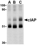
Apoptosis, or programmed cell death, is related to many diseases, such as cancer. Apoptosis is triggered by a variety of stimuli including members in the TNF family and can be prevented by the inhibitor of apoptosis (IAP) proteins. IAP proteins form a conserved gene family that binds to and inhibits cell death proteases (1 for review). The two isoforms of c-IAP (c-IAP1 and c-IAP2) are structurally related to XIAP, containing 3 baculoviral IAP repeat (BIR) motifs that are essential and sufficient for the binding and inhibition of caspases–3, –7, (2,3). The c-IAPs can associate with the death receptor TNF-R2, and mediate the ubiquitinization of TRAF2 following the binding of TNF-alpha by its receptor (2,4). Omi, a negative regulator of c-IAP, inhibits its activity by catalytically cleaving c-IAP (5). Another negative regulator, Smac/DIABLO, acts by enhancing the auto-ubiquitization activity of c-IAP (6).
Additional Names : cIAP (CT), cIAP
 Description
Left: Western blot analysis of c-IAP in human lung lysate with c-IAP antibody at 1 (lane A), 2 (lane B), and 4 (lane C) µg/ml, respectively.
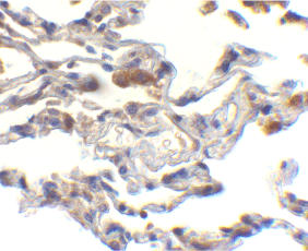
Below: Immunohistochemistry of cIAP in human lung cells with cIAP antibody at 10 µg/ml.
Other Product Images
 Source : c-IAP antibody was raised against a synthetic peptide corresponding to 14 amino acids at the C-terminus of human c-IAP1 c-IAP antibody detects both c-IAP1 and c-IAP2.
Purification : Affinity chromatography purified via peptide column
Clonality and Clone : This is a polyclonal antibody.
Host : cIAP antibody was raised in rabbit. Please use anti-rabbit secondary antibodies.
Immunogen : Human cIAP (C-Terminus) Peptide (Cat. No. 3325P)
Application : cIAP antibody can be used for the detection of c-IAP by Western blot at 1 to 2 µg/ml.
Tested Application(s) : E, WB, IHC
Buffer : Antibody is supplied in PBS containing 0.02% sodium azide.
Blocking Peptide : Cat. No. 3325P - cIAP Peptide
Long-Term Storage : cIAP antibody can be stored at 4ºC, stable for one year. As with all antibodies care should be taken to avoid repeated freeze thaw cycles. Antibodies should not be exposed to prolonged high temperatures.
Positive Control
1.Cat. No. 1331 - Human Lung Tissue Lysate
Species Reactivity :H, M
GI Number : 4502141
Accession Number : NP_001157
Short Description : (CT) An apoptosis inhibitor protein
References
1.Schimmer AD. Inhibitor of apoptosis proteins: translating basic knowledge into clinical practice. Cancer Res. 2004; 64:7183-90.
2.Rothe M, Pan M-G, Henzel WJ, et al. The TNFR2-TRAF signaling complex contains two novel proteins related to baculoviral inhibitor of apoptosis proteins. Cell 1995; 83:1243-52.
3.Deveraux QL, Leo E, Stennicke HR, et al. IAPs block apoptotic events induced by caspase-8 and cytochrome c by direct inhibition of distinct caspases. EMBO J. 1998; 17:2215-23.
4.Li X, Yang Y, Ashwell JD. TNF-RII and c-IAP1 mediate ubiquitinization and degradation of TRAF 2. Nature 2002; 416:345-7.
Catalog# : 3325
Apoptosis, or programmed cell death, is related to many diseases, such as cancer. Apoptosis is triggered by a variety of stimuli including members in the TNF family and can be prevented by the inhibitor of apoptosis (IAP) proteins. IAP proteins form a conserved gene family that binds to and inhibits cell death proteases (1 for review). The two isoforms of c-IAP (c-IAP1 and c-IAP2) are structurally related to XIAP, containing 3 baculoviral IAP repeat (BIR) motifs that are essential and sufficient for the binding and inhibition of caspases–3, –7, (2,3). The c-IAPs can associate with the death receptor TNF-R2, and mediate the ubiquitinization of TRAF2 following the binding of TNF-alpha by its receptor (2,4). Omi, a negative regulator of c-IAP, inhibits its activity by catalytically cleaving c-IAP (5). Another negative regulator, Smac/DIABLO, acts by enhancing the auto-ubiquitization activity of c-IAP (6).
Additional Names : cIAP (CT), cIAP
 Description
Left: Western blot analysis of c-IAP in human lung lysate with c-IAP antibody at 1 (lane A), 2 (lane B), and 4 (lane C) µg/ml, respectively.
Below: Immunohistochemistry of cIAP in human lung cells with cIAP antibody at 10 µg/ml.
Other Product Images
 Source : c-IAP antibody was raised against a synthetic peptide corresponding to 14 amino acids at the C-terminus of human c-IAP1 c-IAP antibody detects both c-IAP1 and c-IAP2.
Purification : Affinity chromatography purified via peptide column
Clonality and Clone : This is a polyclonal antibody.
Host : cIAP antibody was raised in rabbit. Please use anti-rabbit secondary antibodies.
Immunogen : Human cIAP (C-Terminus) Peptide (Cat. No. 3325P)
Application : cIAP antibody can be used for the detection of c-IAP by Western blot at 1 to 2 µg/ml.
Tested Application(s) : E, WB, IHC
Buffer : Antibody is supplied in PBS containing 0.02% sodium azide.
Blocking Peptide : Cat. No. 3325P - cIAP Peptide
Long-Term Storage : cIAP antibody can be stored at 4ºC, stable for one year. As with all antibodies care should be taken to avoid repeated freeze thaw cycles. Antibodies should not be exposed to prolonged high temperatures.
Positive Control
1.Cat. No. 1331 - Human Lung Tissue Lysate
Species Reactivity :H, M
GI Number : 4502141
Accession Number : NP_001157
Short Description : (CT) An apoptosis inhibitor protein
References
1.Schimmer AD. Inhibitor of apoptosis proteins: translating basic knowledge into clinical practice. Cancer Res. 2004; 64:7183-90.
2.Rothe M, Pan M-G, Henzel WJ, et al. The TNFR2-TRAF signaling complex contains two novel proteins related to baculoviral inhibitor of apoptosis proteins. Cell 1995; 83:1243-52.
3.Deveraux QL, Leo E, Stennicke HR, et al. IAPs block apoptotic events induced by caspase-8 and cytochrome c by direct inhibition of distinct caspases. EMBO J. 1998; 17:2215-23.
4.Li X, Yang Y, Ashwell JD. TNF-RII and c-IAP1 mediate ubiquitinization and degradation of TRAF 2. Nature 2002; 416:345-7.
Chk2 Antibody
Catalog# : 2391
The p53 tumor-suppressor gene integrates numerous signals that control cell life and death. Several novel molecules involved in p53 signaling, including Chk2 , p53R2 (2), p53AIP1 (3), Noxa (4), PIDD (5), and PID/MTA2 (6), were recently discovered. The checkpoint kinase Chk2 is the mammalian homologue of yeast Cds1/Rad53. In response to DNA damage, the checkpoint kinase ATM phosphorylates and activates Chk2, which in turn directly phosphorylates and activates p53 (7,8). Chk2 serves as ATM downstream effector to mediate activation of p53. Chk2 also phosphorylates and activates BRCA1, the product of a tumor suppressor gene that is mutated in breast and ovarian cancer (9).
Additional Names : Chk2 (NT), Chk2
 Description
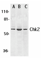
Left: Western blot analysis of Chk2 expression in K562 (A), Jurkat (B), and HL-60 (C) whole cell lysates with Chk2 antibody at 1 µg /ml. .
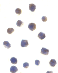
Below: Immunocytochemistry of Chk2 in Jurkat cells with Chk2 antibody at 1 µg/ml.
Other Product Images
 Source : Chk2 antibody was raised against a synthetic peptide corresponding to amino acids near the amino terminus of human Chk2 .
Purification : Affinity chromatography purified via peptide column
Clonality and Clone : This is a polyclonal antibody.
Host : Chk2 antibody was raised in rabbit. Please use anti-rabbit secondary antibodies.
Immunogen : Human Chk2 (N-Terminus) Peptide (Cat. No. 2391P)
Application : Chk2 antibody can be used for detection of Chk2 by Western blot at 0.5 to 1 µg/ml.A 60 kDa band can be detected.
Tested Application(s) : E, WB, ICC
Buffer : Antibody is supplied in PBS containing 0.02% sodium azide.
Blocking Peptide : Cat. No. 2391P - Chk2 Peptide
Long-Term Storage : Chk2 antibody can be stored at 4ºC, stable for one year. As with all antibodies care should be taken to avoid repeated freeze thaw cycles. Antibodies should not be exposed to prolonged high temperatures.
Positive Control
1.Cat. No. 1205 - Jurkat Cell Lysate
2.Cat. No. 1204 - K562 Cell Lysate
Species Reactivity :H, M, R
GI Number : 6005850
Accession Number : NP_009125
Short Description : (NT) A novel check point kinase
References
1.Matsuoka S, Huang M, Elledge SJ. Linkage of ATM to cell cycle regulation by the Chk2 protein kinase. Science. 1998;282:1893-7.
2.Tanaka H, Arakawa H, Yamaguchi T, Shiraishi K, Fukuda S, Matsui K, Takei Y, Nakamura Y. A ribonucleotide reductase gene involved in a p53-dependent cell-cycle checkpoint for DNA damage. Nature. 2000;404:42-9.
3.Oda E, Ohki R, Murasawa H, Nemoto J, Shibue T, Yamashita T, Tokino T, Taniguchi T, Tanaka N. Noxa, a BH3-only member of the Bcl-2 family and candidate mediator of p53-induced apoptosis. Science. 2000;288(5468):1053-8.
4.Oda K, Arakawa H, Tanaka T, Matsuda K, Tanikawa C, Mori T, Nishimori H, Tamai K, Tokino T, Nakamura Y, Taya Y. p53AIP1, a potential mediator of p53-dependent apoptosis, and its regulation by Ser-46-phosphorylated p53. Cell. 2000 Sep 15;102(6):849-62.
Catalog# : 2391
The p53 tumor-suppressor gene integrates numerous signals that control cell life and death. Several novel molecules involved in p53 signaling, including Chk2 , p53R2 (2), p53AIP1 (3), Noxa (4), PIDD (5), and PID/MTA2 (6), were recently discovered. The checkpoint kinase Chk2 is the mammalian homologue of yeast Cds1/Rad53. In response to DNA damage, the checkpoint kinase ATM phosphorylates and activates Chk2, which in turn directly phosphorylates and activates p53 (7,8). Chk2 serves as ATM downstream effector to mediate activation of p53. Chk2 also phosphorylates and activates BRCA1, the product of a tumor suppressor gene that is mutated in breast and ovarian cancer (9).
Additional Names : Chk2 (NT), Chk2
 Description
Left: Western blot analysis of Chk2 expression in K562 (A), Jurkat (B), and HL-60 (C) whole cell lysates with Chk2 antibody at 1 µg /ml. .
Below: Immunocytochemistry of Chk2 in Jurkat cells with Chk2 antibody at 1 µg/ml.
Other Product Images
 Source : Chk2 antibody was raised against a synthetic peptide corresponding to amino acids near the amino terminus of human Chk2 .
Purification : Affinity chromatography purified via peptide column
Clonality and Clone : This is a polyclonal antibody.
Host : Chk2 antibody was raised in rabbit. Please use anti-rabbit secondary antibodies.
Immunogen : Human Chk2 (N-Terminus) Peptide (Cat. No. 2391P)
Application : Chk2 antibody can be used for detection of Chk2 by Western blot at 0.5 to 1 µg/ml.A 60 kDa band can be detected.
Tested Application(s) : E, WB, ICC
Buffer : Antibody is supplied in PBS containing 0.02% sodium azide.
Blocking Peptide : Cat. No. 2391P - Chk2 Peptide
Long-Term Storage : Chk2 antibody can be stored at 4ºC, stable for one year. As with all antibodies care should be taken to avoid repeated freeze thaw cycles. Antibodies should not be exposed to prolonged high temperatures.
Positive Control
1.Cat. No. 1205 - Jurkat Cell Lysate
2.Cat. No. 1204 - K562 Cell Lysate
Species Reactivity :H, M, R
GI Number : 6005850
Accession Number : NP_009125
Short Description : (NT) A novel check point kinase
References
1.Matsuoka S, Huang M, Elledge SJ. Linkage of ATM to cell cycle regulation by the Chk2 protein kinase. Science. 1998;282:1893-7.
2.Tanaka H, Arakawa H, Yamaguchi T, Shiraishi K, Fukuda S, Matsui K, Takei Y, Nakamura Y. A ribonucleotide reductase gene involved in a p53-dependent cell-cycle checkpoint for DNA damage. Nature. 2000;404:42-9.
3.Oda E, Ohki R, Murasawa H, Nemoto J, Shibue T, Yamashita T, Tokino T, Taniguchi T, Tanaka N. Noxa, a BH3-only member of the Bcl-2 family and candidate mediator of p53-induced apoptosis. Science. 2000;288(5468):1053-8.
4.Oda K, Arakawa H, Tanaka T, Matsuda K, Tanikawa C, Mori T, Nishimori H, Tamai K, Tokino T, Nakamura Y, Taya Y. p53AIP1, a potential mediator of p53-dependent apoptosis, and its regulation by Ser-46-phosphorylated p53. Cell. 2000 Sep 15;102(6):849-62.
CDIP Antibody
CDIP Antibody
Catalog# : 5047
The p53 tumor-suppressor gene integrates numerous signals that control cell life and death; loss of its functions contributes to the development of most cancers. CDIP is a novel pro-apoptotic target gene whose inhibition abrogates p53-mediated apoptotic responses. Overexpression of CDIP induced apoptosis in transfected cells while siRNA suppression of caspase-8 mRNA blocked this CDIP-induced apoptosis, indicating that the CDIP-dependent apoptosis pathway proceeds through extrinsic cell death pathway. CDIP may thus represent a novel target for drug design to maximize p53 response and sensitize tumor cells to cancer therapy. Multiple isoforms of CDIP are known to exist.
Additional Names : CDIP (IN1), Cell death involved p53-target
 Description
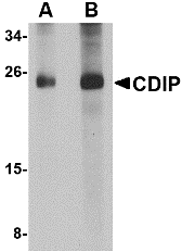
Left: Western blot analysis of CDIP in human brain lysate with CDIP antibody at (A) 1 and (B) 2 µg/ml.
Source : CDIP antibody was raised against a 16 amino acid peptide near the center of human CDIP.
Purification : Affinity chromatography purified via peptide column
Clonality and Clone : This is a polyclonal antibody.
Host : CDIP antibody was raised in rabbit. Please use anti-rabbit secondary antibodies.
Application : CDIP antibody can be used for detection of CDIP by Western blot at 1 – 2 µg/ml.
Tested Application(s) : E, WB
Buffer : Antibody is supplied in PBS containing 0.02% sodium azide.
Blocking Peptide : Cat.No. 5047P - CDIP Peptide
Long-Term Storage : CDIP antibody can be stored at 4ºC, stable for one year. As with all antibodies care should be taken to avoid repeated freeze thaw cycles. Antibodies should not be exposed to prolonged high temperatures.
Positive Control
1.Cat. No. 1303 - Human Brain Tissue Lysate
Species Reactivity :H, M
GI Number : 118344450
Accession Number : NP_037531
Short Description : (IN1) Cell death involved p53-target
References
1.Guimaraes DP and Hainaut P. TP53: a key gene in human cancer. Biochimie 2002; 84:83-93.
2.Brown L, Ongusaha PP, Kim H-H, et al. CDIP, a novel pro-apoptotic gene, regulates TNFa-mediated apoptosis in a p53-dependent manner. EMBO J. 2007; 26:3410-22.
Catalog# : 5047
The p53 tumor-suppressor gene integrates numerous signals that control cell life and death; loss of its functions contributes to the development of most cancers. CDIP is a novel pro-apoptotic target gene whose inhibition abrogates p53-mediated apoptotic responses. Overexpression of CDIP induced apoptosis in transfected cells while siRNA suppression of caspase-8 mRNA blocked this CDIP-induced apoptosis, indicating that the CDIP-dependent apoptosis pathway proceeds through extrinsic cell death pathway. CDIP may thus represent a novel target for drug design to maximize p53 response and sensitize tumor cells to cancer therapy. Multiple isoforms of CDIP are known to exist.
Additional Names : CDIP (IN1), Cell death involved p53-target
 Description
Left: Western blot analysis of CDIP in human brain lysate with CDIP antibody at (A) 1 and (B) 2 µg/ml.
Source : CDIP antibody was raised against a 16 amino acid peptide near the center of human CDIP.
Purification : Affinity chromatography purified via peptide column
Clonality and Clone : This is a polyclonal antibody.
Host : CDIP antibody was raised in rabbit. Please use anti-rabbit secondary antibodies.
Application : CDIP antibody can be used for detection of CDIP by Western blot at 1 – 2 µg/ml.
Tested Application(s) : E, WB
Buffer : Antibody is supplied in PBS containing 0.02% sodium azide.
Blocking Peptide : Cat.No. 5047P - CDIP Peptide
Long-Term Storage : CDIP antibody can be stored at 4ºC, stable for one year. As with all antibodies care should be taken to avoid repeated freeze thaw cycles. Antibodies should not be exposed to prolonged high temperatures.
Positive Control
1.Cat. No. 1303 - Human Brain Tissue Lysate
Species Reactivity :H, M
GI Number : 118344450
Accession Number : NP_037531
Short Description : (IN1) Cell death involved p53-target
References
1.Guimaraes DP and Hainaut P. TP53: a key gene in human cancer. Biochimie 2002; 84:83-93.
2.Brown L, Ongusaha PP, Kim H-H, et al. CDIP, a novel pro-apoptotic gene, regulates TNFa-mediated apoptosis in a p53-dependent manner. EMBO J. 2007; 26:3410-22.
CDIP Antibody
CDIP Antibody
Catalog# : 5059
The p53 tumor-suppressor gene integrates numerous signals that control cell life and death; loss of its functions contributes to the development of most cancers. CDIP is a novel pro-apoptotic target gene whose inhibition abrogates p53-mediated apoptotic responses. Overexpression of CDIP induced apoptosis in transfected cells while siRNA suppression of caspase-8 mRNA blocked this CDIP-induced apoptosis, indicating that the CDIP-dependent apoptosis pathway proceeds through extrinsic cell death pathway. CDIP may thus represent a novel target for drug design to maximize p53 response and sensitize tumor cells to cancer therapy. Multiple isoforms of CDIP are known to exist.
Additional Names : CDIP (IN2), Cell death involved p53-target
 Description
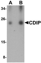
Left: Western blot analysis of CDIP in human brain lysate with CDIP antibody at (A) 0.25 and (B) 0.5 µg/ml.
Source : CDIP antibody was raised against a 19 amino acid peptide near the center of human CDIP.
Purification : Affinity chromatography purified via peptide column
Clonality and Clone : This is a polyclonal antibody.
Host : CDIP antibody was raised in rabbit. Please use anti-rabbit secondary antibodies.
Application : CDIP antibody can be used for detection of CDIP by Western blot at 0.25 – 0.5 µg/ml.
Tested Application(s) : E, WB
Buffer : Antibody is supplied in PBS containing 0.02% sodium azide.
Blocking Peptide : Cat.No. 5059P - CDIP Peptide
Long-Term Storage : CDIP antibody can be stored at 4ºC, stable for one year. As with all antibodies care should be taken to avoid repeated freeze thaw cycles. Antibodies should not be exposed to prolonged high temperatures.
Positive Control
1.Cat. No. 1303 - Human Brain Tissue Lysate
Species Reactivity :H, M
GI Number : 118344450
Accession Number : NP_037531
Short Description : (IN2) Cell death involved p53-target
References
1.Guimaraes DP and Hainaut P. TP53: a key gene in human cancer. Biochimie 2002; 84:83-93.
2.Brown L, Ongusaha PP, Kim H-H, et al. CDIP, a novel pro-apoptotic gene, regulates TNFa-mediated apoptosis in a p53-dependent manner. EMBO J. 2007; 26:3410-22.
Catalog# : 5059
The p53 tumor-suppressor gene integrates numerous signals that control cell life and death; loss of its functions contributes to the development of most cancers. CDIP is a novel pro-apoptotic target gene whose inhibition abrogates p53-mediated apoptotic responses. Overexpression of CDIP induced apoptosis in transfected cells while siRNA suppression of caspase-8 mRNA blocked this CDIP-induced apoptosis, indicating that the CDIP-dependent apoptosis pathway proceeds through extrinsic cell death pathway. CDIP may thus represent a novel target for drug design to maximize p53 response and sensitize tumor cells to cancer therapy. Multiple isoforms of CDIP are known to exist.
Additional Names : CDIP (IN2), Cell death involved p53-target
 Description
Left: Western blot analysis of CDIP in human brain lysate with CDIP antibody at (A) 0.25 and (B) 0.5 µg/ml.
Source : CDIP antibody was raised against a 19 amino acid peptide near the center of human CDIP.
Purification : Affinity chromatography purified via peptide column
Clonality and Clone : This is a polyclonal antibody.
Host : CDIP antibody was raised in rabbit. Please use anti-rabbit secondary antibodies.
Application : CDIP antibody can be used for detection of CDIP by Western blot at 0.25 – 0.5 µg/ml.
Tested Application(s) : E, WB
Buffer : Antibody is supplied in PBS containing 0.02% sodium azide.
Blocking Peptide : Cat.No. 5059P - CDIP Peptide
Long-Term Storage : CDIP antibody can be stored at 4ºC, stable for one year. As with all antibodies care should be taken to avoid repeated freeze thaw cycles. Antibodies should not be exposed to prolonged high temperatures.
Positive Control
1.Cat. No. 1303 - Human Brain Tissue Lysate
Species Reactivity :H, M
GI Number : 118344450
Accession Number : NP_037531
Short Description : (IN2) Cell death involved p53-target
References
1.Guimaraes DP and Hainaut P. TP53: a key gene in human cancer. Biochimie 2002; 84:83-93.
2.Brown L, Ongusaha PP, Kim H-H, et al. CDIP, a novel pro-apoptotic gene, regulates TNFa-mediated apoptosis in a p53-dependent manner. EMBO J. 2007; 26:3410-22.
Caspase-9 Antibody
Caspase-9 Antibody
Catalog# : 2073
Apoptosis is related to many diseases and induced by a family of cell death receptors and their ligands. Cell death signals are transduced by death domain containing adapter molecules and members of the caspase family of proteases. A novel member in the caspase family was recently identified and designated ICE-LAP6, Mch6, and Apaf-3 (1-3). Caspase-9 and Apaf-1 bind to each other, which leads to caspase-9 activation (3). Caspase-9 is also activated by granzyme B and CPP32 (1,2). Activated caspase-9 cleaves and activates caspase-3 that is one of the key proteases, being responsible for the proteolytic cleavage of many key proteins in apoptosis (3). Caspase-9 play a central role in cell death induced by a wide variety of apoptosis activators including TNFalpha, TRAIL, anti-CD-95, FADD, and TRADD (4). Caspase-9 is expressed in a variety of human tissues (1,2).
Additional Names : Caspase-9 (IN2), Mch6/ Apaf-3, Casp-9
 Description
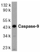
Left: Western blot analysis of caspase-9 in HeLa whole cell lysate with Caspase-9 antibody at 1:1000 dilution
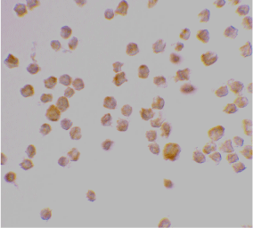
Below: Immunocytochemistry staining of K562 cells using Caspase-9 antibody at 2 µg/ml.
Other Product Images
Source : Caspase-9 antibody was raised against a peptide corresponding to amino acids 299 to 318 of human caspase-9 .
Purification : Affinity chromatography purified via peptide column
Clonality and Clone : This is a polyclonal antibody.
Host : Caspase-9 antibody was raised in rabbit. Please use anti-rabbit secondary antibodies.
Immunogen : Human Caspase-9 (Intermediate Domain 2) Peptide (Cat. No. 2073P)
Application : Caspase-9 antibody can be used for detection of caspase-9 by Western blot at 1:1000 to 1:2000 dilution. A 46 kDa band should be detected. It has no cross response to other members in caspase family.
Tested Application(s) : E, WB, ICC
Buffer : Antibody is supplied in PBS containing 0.02% sodium azide.
Blocking Peptide : Cat. No. 2073P - Caspase-9 Peptide
Long-Term Storage : Caspase-9 antibody can be stored at 4ºC, stable for one year. As with all antibodies care should be taken to avoid repeated freeze thaw cycles. Antibodies should not be exposed to prolonged high temperatures.
Positive Control
1.Cat. No. 1201 - HeLa Cell Lysate
Species Reactivity :H
GI Number : 28558771
Accession Number : P55211
Short Description : (IN2) A new caspase
References
1.Duan H, Orth K, Chinnaiyan AM, Poirier GG, Froelich CJ, He WW, Dixit VM. ICE-LAP6, a novel member of the ICE/Ced-3 gene family, is activated by the cytotoxic T cell protease granzyme B. J Biol Chem 1996;271:16720-4
Catalog# : 2073
Apoptosis is related to many diseases and induced by a family of cell death receptors and their ligands. Cell death signals are transduced by death domain containing adapter molecules and members of the caspase family of proteases. A novel member in the caspase family was recently identified and designated ICE-LAP6, Mch6, and Apaf-3 (1-3). Caspase-9 and Apaf-1 bind to each other, which leads to caspase-9 activation (3). Caspase-9 is also activated by granzyme B and CPP32 (1,2). Activated caspase-9 cleaves and activates caspase-3 that is one of the key proteases, being responsible for the proteolytic cleavage of many key proteins in apoptosis (3). Caspase-9 play a central role in cell death induced by a wide variety of apoptosis activators including TNFalpha, TRAIL, anti-CD-95, FADD, and TRADD (4). Caspase-9 is expressed in a variety of human tissues (1,2).
Additional Names : Caspase-9 (IN2), Mch6/ Apaf-3, Casp-9
 Description
Left: Western blot analysis of caspase-9 in HeLa whole cell lysate with Caspase-9 antibody at 1:1000 dilution
Below: Immunocytochemistry staining of K562 cells using Caspase-9 antibody at 2 µg/ml.
Other Product Images

Source : Caspase-9 antibody was raised against a peptide corresponding to amino acids 299 to 318 of human caspase-9 .
Purification : Affinity chromatography purified via peptide column
Clonality and Clone : This is a polyclonal antibody.
Host : Caspase-9 antibody was raised in rabbit. Please use anti-rabbit secondary antibodies.
Immunogen : Human Caspase-9 (Intermediate Domain 2) Peptide (Cat. No. 2073P)
Application : Caspase-9 antibody can be used for detection of caspase-9 by Western blot at 1:1000 to 1:2000 dilution. A 46 kDa band should be detected. It has no cross response to other members in caspase family.
Tested Application(s) : E, WB, ICC
Buffer : Antibody is supplied in PBS containing 0.02% sodium azide.
Blocking Peptide : Cat. No. 2073P - Caspase-9 Peptide
Long-Term Storage : Caspase-9 antibody can be stored at 4ºC, stable for one year. As with all antibodies care should be taken to avoid repeated freeze thaw cycles. Antibodies should not be exposed to prolonged high temperatures.
Positive Control
1.Cat. No. 1201 - HeLa Cell Lysate
Species Reactivity :H
GI Number : 28558771
Accession Number : P55211
Short Description : (IN2) A new caspase
References
1.Duan H, Orth K, Chinnaiyan AM, Poirier GG, Froelich CJ, He WW, Dixit VM. ICE-LAP6, a novel member of the ICE/Ced-3 gene family, is activated by the cytotoxic T cell protease granzyme B. J Biol Chem 1996;271:16720-4
Caspase-9 Antibody
Caspase-9 Antibody
Catalog# : 2071
Apoptosis is related to many diseases and induced by a family of cell death receptors and their ligands. Cell death signals are transduced by death domain containing adapter molecules and members of the caspase family of proteases. A novel member in the caspase family was recently identified and designated ICE-LAP6, Mch6, and Apaf-3 (1-3). Caspase-9 and Apaf-1 bind to each other, which leads to caspase-9 activation (3). Caspase-9 is also activated by granzyme B and CPP32 (1,2). Activated caspase-9 cleaves and activates caspase-3 that is one of the key proteases, being responsible for the proteolytic cleavage of many key proteins in apoptosis (3). Caspase-9 play a central role in cell death induced by a wide variety of apoptosis activators including TNFalpha, TRAIL, anti-CD-95, FADD, and TRADD (4). Caspase-9 is expressed in a variety of human tissues (1,2).
Additional Names : Caspase-9 (IN1), Mch6/ Apaf-3, Casp-9
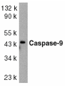 Description
Left: Western blot analysis of caspase-9 in HeLa whole cell lysate with caspase-9 antibody at 1:1000 dilution.
Source : Caspase-9 antibody was raised against a peptide corresponding to amino acids 41 to 56 of human caspase-9 .
Purification : Affinity chromatography purified via peptide column
Clonality and Clone : This is a polyclonal antibody.
Host : Caspase-9 antibody was raised in rabbit. Please use anti-rabbit secondary antibodies.
Immunogen : Human Caspase-9 (Intermediate Domain 1) Peptide (Cat. No. 2071P)
Application : Caspase-9 antibody can be used for detection of caspase-9 by Western blot at 1:1000 to 1:2000 dilution. A 46 kDa band should be detected. It has no cross response to other members in caspase family.
Tested Application(s) : E, WB
Buffer : Antibody is supplied in PBS containing 0.02% sodium azide.
Blocking Peptide : Cat. No. 2071P - Caspase-9 Peptide
Long-Term Storage : Caspase-9 antibody can be stored at 4ºC, stable for one year. As with all antibodies care should be taken to avoid repeated freeze thaw cycles. Antibodies should not be exposed to prolonged high temperatures.
Positive Control
1.Cat. No. 1201 - HeLa Cell Lysate
Species Reactivity :H, M, R
GI Number : 28558771
Accession Number : P55211
Short Description : (IN1) A new caspase
References
1.Duan H, Orth K, Chinnaiyan AM, Poirier GG, Froelich CJ, He WW, Dixit VM. ICE-LAP6, a novel member of the ICE/Ced-3 gene family, is activated by the cytotoxic T cell protease granzyme B. J Biol Chem 1996;271:16720-4
2.Srinivasula SM, Fernandes-Alnemri T, Zangrilli J, Robertson N, Armstrong RC, Wang L, Trapani JA, Tomaselli KJ, Litwack G, Alnemri ES. The Ced-3/interleukin 1beta converting enzyme-like homolog Mch6 and the lamin-cleaving enzyme Mch2alpha are substrates for the apoptotic mediator CPP32. J Biol Chem 1996;271:27099-106
3.Li P, Nijhawan D, Budihardjo I, Srinivasula SM, Ahmad M, Alnemri ES, Wang X. Cytochrome c and dATP-dependent formation of Apaf-1/caspase-9 complex initiates an apoptotic protease cascade. Cell 1997;91:479-89
4.Pan G, O'Rourke K, Dixit VM. Caspase-9, Bcl-XL, and Apaf-1 form a ternary complex. J Biol Chem 1998;273:5841-5 (RD1299)
Catalog# : 2071
Apoptosis is related to many diseases and induced by a family of cell death receptors and their ligands. Cell death signals are transduced by death domain containing adapter molecules and members of the caspase family of proteases. A novel member in the caspase family was recently identified and designated ICE-LAP6, Mch6, and Apaf-3 (1-3). Caspase-9 and Apaf-1 bind to each other, which leads to caspase-9 activation (3). Caspase-9 is also activated by granzyme B and CPP32 (1,2). Activated caspase-9 cleaves and activates caspase-3 that is one of the key proteases, being responsible for the proteolytic cleavage of many key proteins in apoptosis (3). Caspase-9 play a central role in cell death induced by a wide variety of apoptosis activators including TNFalpha, TRAIL, anti-CD-95, FADD, and TRADD (4). Caspase-9 is expressed in a variety of human tissues (1,2).
Additional Names : Caspase-9 (IN1), Mch6/ Apaf-3, Casp-9
 Description
Left: Western blot analysis of caspase-9 in HeLa whole cell lysate with caspase-9 antibody at 1:1000 dilution.
Source : Caspase-9 antibody was raised against a peptide corresponding to amino acids 41 to 56 of human caspase-9 .
Purification : Affinity chromatography purified via peptide column
Clonality and Clone : This is a polyclonal antibody.
Host : Caspase-9 antibody was raised in rabbit. Please use anti-rabbit secondary antibodies.
Immunogen : Human Caspase-9 (Intermediate Domain 1) Peptide (Cat. No. 2071P)
Application : Caspase-9 antibody can be used for detection of caspase-9 by Western blot at 1:1000 to 1:2000 dilution. A 46 kDa band should be detected. It has no cross response to other members in caspase family.
Tested Application(s) : E, WB
Buffer : Antibody is supplied in PBS containing 0.02% sodium azide.
Blocking Peptide : Cat. No. 2071P - Caspase-9 Peptide
Long-Term Storage : Caspase-9 antibody can be stored at 4ºC, stable for one year. As with all antibodies care should be taken to avoid repeated freeze thaw cycles. Antibodies should not be exposed to prolonged high temperatures.
Positive Control
1.Cat. No. 1201 - HeLa Cell Lysate
Species Reactivity :H, M, R
GI Number : 28558771
Accession Number : P55211
Short Description : (IN1) A new caspase
References
1.Duan H, Orth K, Chinnaiyan AM, Poirier GG, Froelich CJ, He WW, Dixit VM. ICE-LAP6, a novel member of the ICE/Ced-3 gene family, is activated by the cytotoxic T cell protease granzyme B. J Biol Chem 1996;271:16720-4
2.Srinivasula SM, Fernandes-Alnemri T, Zangrilli J, Robertson N, Armstrong RC, Wang L, Trapani JA, Tomaselli KJ, Litwack G, Alnemri ES. The Ced-3/interleukin 1beta converting enzyme-like homolog Mch6 and the lamin-cleaving enzyme Mch2alpha are substrates for the apoptotic mediator CPP32. J Biol Chem 1996;271:27099-106
3.Li P, Nijhawan D, Budihardjo I, Srinivasula SM, Ahmad M, Alnemri ES, Wang X. Cytochrome c and dATP-dependent formation of Apaf-1/caspase-9 complex initiates an apoptotic protease cascade. Cell 1997;91:479-89
4.Pan G, O'Rourke K, Dixit VM. Caspase-9, Bcl-XL, and Apaf-1 form a ternary complex. J Biol Chem 1998;273:5841-5 (RD1299)
Caspase-8 Antibody
Caspase-8 Antibody
Catalog# : 3475
Caspases are a family of cysteine proteases that can be divided into the apoptotic and inflammatory caspase subfamilies. Unlike the apoptotic caspases, members of the inflammatory subfamily are generally not involved in cell death but are associated with the immune response to microbial pathogens (reviewed in 1 and 2). The apoptotic subfamily can be further divided into initiator caspases, which are activated in response to death signals, and executioner caspases, which are activated by the initiator caspases and are responsible for cleavage of cellular substrates that ultimately lead to cell death (reviewed in 3). Caspase-8 is an initiator caspase that was identified as a member of the Fas/APO-1 death-inducing signaling complex (4). The adaptor molecule FADD couples procaspase-8 to the Fas receptor death domain; subsequent oligomerization promotes procaspase-8 autoactivation (reviewed in 5). FLIP, a catalytically inactive caspase-8-like molecule inhibits these interactions and thus can inhibit apoptosis (6). This antibody will only detect isoform E of caspase-8.
Additional Names : Caspase-8 (E), FLICE, MACH ,Mch5
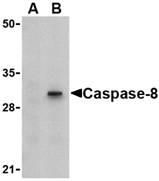
Description
Left: Western blot analysis of Caspase-8 in HT-29 cell lysate with Caspase-8 antibody (IN) at 1 µg/ml in (A) the presence or (B) the absence of blocking peptide.
Source : Caspase-8 antibody was raised against a 15 amino acid peptide from near the carboxy terminus human caspase-8 isoform E.
Purification : Affinity chromatography purified via peptide column
Clonality and Clone : This is a polyclonal antibody.
Host : Caspase-8 antibody was raised in rabbit. Please use anti-rabbit secondary antibodies.
Immunogen : Virus Casp-8 / FLICE / MACH / Mch5 (E) Peptide (Cat. No. 3475P)
Application : Casp-8 antibody can be used for the detection of Caspase-8 by Western blot at 0.5 µg/ml.
Tested Application(s) : E, WB
Buffer : Antibody is supplied in PBS containing 0.02% sodium azide.
Blocking Peptide : Cat. No. 3475P - Casp-8 Peptide
Long-Term Storage : Caspase-8 antibody can be stored at 4ºC, stable for one year. As with all antibodies care should be taken to avoid repeated freeze thaw cycles. Antibodies should not be exposed to prolonged high temperatures.
Positive Control
1.HT-29 Cell Lysate
Species Reactivity :H, M
GI Number : 15718704
Accession Number : NP_001219
Short Description : (E) an initiator caspase
References
1.Martinon F and Tschopp J. Inflammatory caspases: linking an intracellular innate immune system to autoinflammatory diseases. Cell 2004; 117:561-74.
2.Zhivotovsky B and Orrenius S. Caspase-2 function in response to DNA damage. Biochim. Biophys. Res. Comm. 2005; 331:859-67.
3.Wolf BB and Green DR. Suicidal tendencies: apoptotic cell death by caspase family proteinases. J. Biol. Chem. 1999; 274:20049-52.
4.Muzio M, Chinnaiyan AM, Kischkel FC, et al. FLICE, a novel FADD-homologous ICE/CED-3-like protease, is recruited to the CD95 (Fas/APO-1) death-inducing signaling complex. Cell 1996; 85:817-27.
Catalog# : 3475
Caspases are a family of cysteine proteases that can be divided into the apoptotic and inflammatory caspase subfamilies. Unlike the apoptotic caspases, members of the inflammatory subfamily are generally not involved in cell death but are associated with the immune response to microbial pathogens (reviewed in 1 and 2). The apoptotic subfamily can be further divided into initiator caspases, which are activated in response to death signals, and executioner caspases, which are activated by the initiator caspases and are responsible for cleavage of cellular substrates that ultimately lead to cell death (reviewed in 3). Caspase-8 is an initiator caspase that was identified as a member of the Fas/APO-1 death-inducing signaling complex (4). The adaptor molecule FADD couples procaspase-8 to the Fas receptor death domain; subsequent oligomerization promotes procaspase-8 autoactivation (reviewed in 5). FLIP, a catalytically inactive caspase-8-like molecule inhibits these interactions and thus can inhibit apoptosis (6). This antibody will only detect isoform E of caspase-8.
Additional Names : Caspase-8 (E), FLICE, MACH ,Mch5

Description
Left: Western blot analysis of Caspase-8 in HT-29 cell lysate with Caspase-8 antibody (IN) at 1 µg/ml in (A) the presence or (B) the absence of blocking peptide.
Source : Caspase-8 antibody was raised against a 15 amino acid peptide from near the carboxy terminus human caspase-8 isoform E.
Purification : Affinity chromatography purified via peptide column
Clonality and Clone : This is a polyclonal antibody.
Host : Caspase-8 antibody was raised in rabbit. Please use anti-rabbit secondary antibodies.
Immunogen : Virus Casp-8 / FLICE / MACH / Mch5 (E) Peptide (Cat. No. 3475P)
Application : Casp-8 antibody can be used for the detection of Caspase-8 by Western blot at 0.5 µg/ml.
Tested Application(s) : E, WB
Buffer : Antibody is supplied in PBS containing 0.02% sodium azide.
Blocking Peptide : Cat. No. 3475P - Casp-8 Peptide
Long-Term Storage : Caspase-8 antibody can be stored at 4ºC, stable for one year. As with all antibodies care should be taken to avoid repeated freeze thaw cycles. Antibodies should not be exposed to prolonged high temperatures.
Positive Control
1.HT-29 Cell Lysate
Species Reactivity :H, M
GI Number : 15718704
Accession Number : NP_001219
Short Description : (E) an initiator caspase
References
1.Martinon F and Tschopp J. Inflammatory caspases: linking an intracellular innate immune system to autoinflammatory diseases. Cell 2004; 117:561-74.
2.Zhivotovsky B and Orrenius S. Caspase-2 function in response to DNA damage. Biochim. Biophys. Res. Comm. 2005; 331:859-67.
3.Wolf BB and Green DR. Suicidal tendencies: apoptotic cell death by caspase family proteinases. J. Biol. Chem. 1999; 274:20049-52.
4.Muzio M, Chinnaiyan AM, Kischkel FC, et al. FLICE, a novel FADD-homologous ICE/CED-3-like protease, is recruited to the CD95 (Fas/APO-1) death-inducing signaling complex. Cell 1996; 85:817-27.
Caspase-8 Antibody
Caspase-8 Antibody
Catalog# : 3473
Caspases are a family of cysteine proteases that can be divided into the apoptotic and inflammatory caspase subfamilies. Unlike the apoptotic caspases, members of the inflammatory subfamily are generally not involved in cell death but are associated with the immune response to microbial pathogens (reviewed in 1 and 2). The apoptotic subfamily can be further divided into initiator caspases, which are activated in response to death signals, and executioner caspases, which are activated by the initiator caspases and are responsible for cleavage of cellular substrates that ultimately lead to cell death (reviewed in 3). Caspase-8 is an initiator caspase that was identified as a member of the Fas/APO-1 death-inducing signaling complex (4). The adaptor molecule FADD couples procaspase-8 to the Fas receptor death domain; subsequent oligomerization promotes procaspase-8 autoactivation (reviewed in 5). FLIP, a catalytically inactive caspase-8-like molecule inhibits these interactions and thus can inhibit apoptosis (6).
Additional Names : Caspase-8 (CT), FLICE, MACH ,Mch5
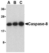 Description
Left: Western blot analysis of caspase-8 in Jurkat cell lysate with caspase-8 antibody at (A) 0.5, (B) 1, and (C) 2 µg/ml.
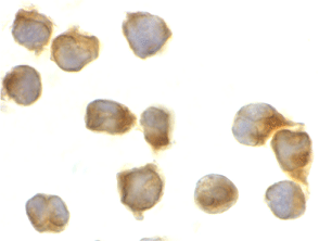
Below: Immunocytochemistry of caspase-8 in Jurkat cells with caspase-8 antibody at 2 µg/ml.
Other Product Images
 Source : Caspase-8 antibody was raised against a 16 amino acid peptide from near the carboxy-terminus of human Caspase-8 isoform A.
Purification : Affinity chromatography purified via peptide column
Clonality and Clone : This is a polyclonal antibody.
Host : Caspase-8 antibody was raised in rabbit. Please use anti-rabbit secondary antibodies.
Immunogen : Virus Casp-8 / FLICE / MACH / Mch5 (C-Terminus) Peptide (Cat. No. 3473P)
Application : Caspase-8 antibody can be used for the detection of caspase-8 by Western blot at 0.5 µg/ml.Depending on cell lines or tissues used, either full-length or other cleavage products may be observed.
Tested Application(s) : E, WB, ICC
Buffer : Antibody is supplied in PBS containing 0.02% sodium azide.
Blocking Peptide : Cat. No. 3473P - Casp-8 Peptide
Long-Term Storage : Caspase-8 antibody can be stored at 4ºC, stable for one year. As with all antibodies care should be taken to avoid repeated freeze thaw cycles. Antibodies should not be exposed to prolonged high temperatures.
Positive Control
1.Cat. No. 1205 - Jurkat Cell Lysate
Species Reactivity :H, M, R
GI Number : 15718704
Accession Number : NP_001219
Short Description : (CT) an initiator caspase
References
1.Martinon F and Tschopp J. Inflammatory caspases: linking an intracellular innate immune system to autoinflammatory diseases. Cell 2004; 117:561-74.
2.Zhivotovsky B and Orrenius S. Caspase-2 function in response to DNA damage. Biochim. Biophys. Res. Comm. 2005; 331:859-67.
3.Wolf BB and Green DR. Suicidal tendencies: apoptotic cell death by caspase family proteinases. J. Biol. Chem. 1999; 274:20049-52.
4.Muzio M, Chinnaiyan AM, Kischkel FC, et al. FLICE, a novel FADD-homologous ICE/CED-3-like protease, is recruited to the CD95 (Fas/APO-1) death-inducing signaling complex. Cell 1996; 85:817-27.
Catalog# : 3473
Caspases are a family of cysteine proteases that can be divided into the apoptotic and inflammatory caspase subfamilies. Unlike the apoptotic caspases, members of the inflammatory subfamily are generally not involved in cell death but are associated with the immune response to microbial pathogens (reviewed in 1 and 2). The apoptotic subfamily can be further divided into initiator caspases, which are activated in response to death signals, and executioner caspases, which are activated by the initiator caspases and are responsible for cleavage of cellular substrates that ultimately lead to cell death (reviewed in 3). Caspase-8 is an initiator caspase that was identified as a member of the Fas/APO-1 death-inducing signaling complex (4). The adaptor molecule FADD couples procaspase-8 to the Fas receptor death domain; subsequent oligomerization promotes procaspase-8 autoactivation (reviewed in 5). FLIP, a catalytically inactive caspase-8-like molecule inhibits these interactions and thus can inhibit apoptosis (6).
Additional Names : Caspase-8 (CT), FLICE, MACH ,Mch5
 Description
Left: Western blot analysis of caspase-8 in Jurkat cell lysate with caspase-8 antibody at (A) 0.5, (B) 1, and (C) 2 µg/ml.
Below: Immunocytochemistry of caspase-8 in Jurkat cells with caspase-8 antibody at 2 µg/ml.
Other Product Images
 Source : Caspase-8 antibody was raised against a 16 amino acid peptide from near the carboxy-terminus of human Caspase-8 isoform A.
Purification : Affinity chromatography purified via peptide column
Clonality and Clone : This is a polyclonal antibody.
Host : Caspase-8 antibody was raised in rabbit. Please use anti-rabbit secondary antibodies.
Immunogen : Virus Casp-8 / FLICE / MACH / Mch5 (C-Terminus) Peptide (Cat. No. 3473P)
Application : Caspase-8 antibody can be used for the detection of caspase-8 by Western blot at 0.5 µg/ml.Depending on cell lines or tissues used, either full-length or other cleavage products may be observed.
Tested Application(s) : E, WB, ICC
Buffer : Antibody is supplied in PBS containing 0.02% sodium azide.
Blocking Peptide : Cat. No. 3473P - Casp-8 Peptide
Long-Term Storage : Caspase-8 antibody can be stored at 4ºC, stable for one year. As with all antibodies care should be taken to avoid repeated freeze thaw cycles. Antibodies should not be exposed to prolonged high temperatures.
Positive Control
1.Cat. No. 1205 - Jurkat Cell Lysate
Species Reactivity :H, M, R
GI Number : 15718704
Accession Number : NP_001219
Short Description : (CT) an initiator caspase
References
1.Martinon F and Tschopp J. Inflammatory caspases: linking an intracellular innate immune system to autoinflammatory diseases. Cell 2004; 117:561-74.
2.Zhivotovsky B and Orrenius S. Caspase-2 function in response to DNA damage. Biochim. Biophys. Res. Comm. 2005; 331:859-67.
3.Wolf BB and Green DR. Suicidal tendencies: apoptotic cell death by caspase family proteinases. J. Biol. Chem. 1999; 274:20049-52.
4.Muzio M, Chinnaiyan AM, Kischkel FC, et al. FLICE, a novel FADD-homologous ICE/CED-3-like protease, is recruited to the CD95 (Fas/APO-1) death-inducing signaling complex. Cell 1996; 85:817-27.
Caspase-7 Antibody
Caspase-7 Antibody
Catalog# : 3467
Caspases are a family of cysteine proteases that can be divided into the apoptotic and inflammatory caspase subfamilies. Unlike the apoptotic caspases, members of the inflammatory subfamily are generally not involved in cell death but are associated with the immune response to microbial pathogens (reviewed in 1 and 2). The apoptotic subfamily can be further divided into initiator caspases, which are activated in response to death signals, and executioner caspases, which are activated by the initiator caspases and are responsible for cleavage of cellular substrates that ultimately lead to cell death (reviewed in 3). Caspase-7 is an executioner caspase that was identified based on its homology with caspases 1 and 3, as well as the C. elegans cell death protein CED-3 (4). Alternative splicing of Caspase-7 mRNA results in the production of 3 distinct isoforms (4). Caspase-7 activity can be directly inhibited by XIAP expression (5).
Additional Names : Caspase-7 (NT), ICELAP3 ,Mch3
 Description
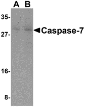
Left: Western blot analysis of Caspase-7 in mouse skeletal muscle cell lysate with Caspase-7 antibody at (A) 0.5 and (B) 1 µg/ml.
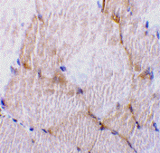
Below: Immunohistochemistry of caspase-7 in rat skeletal muscle tissue with caspase-7 antibody at 10 µg/ml.
Other Product Images
 Source : Caspase-7 antibody was raised against a 16 amino acid peptide from near the amino-terminus of human Caspase-7.
Purification : Affinity chromatography purified via peptide column
Clonality and Clone : This is a polyclonal antibody.
Host : Caspase-7 antibody was raised in rabbit. Please use anti-rabbit secondary antibodies.
Immunogen : Human Casp-7 / ICELAP3 / Mch3 (N-Terminus) Peptide (Cat. No. 3467P)
Application : Casp-7 antibody can be used for the detection of Caspase-7 by Western blot at 0.5 µg/ml.Depending on cell lines or tissues used, either full-length or other cleavage products may be observed.
Tested Application(s) : E, WB, IHC
Buffer : Antibody is supplied in PBS containing 0.02% sodium azide.
Blocking Peptide : Cat. No. 3467P - Casp-7 Peptide
Long-Term Storage : Caspase-7 antibody can be stored at 4ºC, stable for one year. As with all antibodies care should be taken to avoid repeated freeze thaw cycles. Antibodies should not be exposed to prolonged high temperatures.
Positive Control
1.Cat. No. 1475 - Mouse Skeletal Muscle Tissue Lysate
2.Cat. No. 1467 - Rat Skeletal Muscle Tissue Lysate
Species Reactivity :H, M, R
GI Number : 1730092
Accession Number : P55210
Short Description : (NT) An executioner caspase
References
1.Martinon F and Tschopp J. Inflammatory caspases: linking an intracellular innate immune system to autoinflammatory diseases. Cell 2004; 117:561-74.
2.Zhivotovsky B and Orrenius S. Caspase-2 function in response to DNA damage. Biochim. Biophys. Res. Comm. 2005; 331:859-67.
3.Wolf BB and Green DR. Suicidal tendencies: apoptotic cell death by caspase family proteinases. J. Biol. Chem. 1999; 274:20049-52.
4.Juan TSC, McNiece IK, Argento JM, et al. Identification and mapping of Casp7, a cysteine protease resembling CPP32b, Interleukin-1b converting enzyme, and CED-3. Genomics 1997; 40:86-93.
Catalog# : 3467
Caspases are a family of cysteine proteases that can be divided into the apoptotic and inflammatory caspase subfamilies. Unlike the apoptotic caspases, members of the inflammatory subfamily are generally not involved in cell death but are associated with the immune response to microbial pathogens (reviewed in 1 and 2). The apoptotic subfamily can be further divided into initiator caspases, which are activated in response to death signals, and executioner caspases, which are activated by the initiator caspases and are responsible for cleavage of cellular substrates that ultimately lead to cell death (reviewed in 3). Caspase-7 is an executioner caspase that was identified based on its homology with caspases 1 and 3, as well as the C. elegans cell death protein CED-3 (4). Alternative splicing of Caspase-7 mRNA results in the production of 3 distinct isoforms (4). Caspase-7 activity can be directly inhibited by XIAP expression (5).
Additional Names : Caspase-7 (NT), ICELAP3 ,Mch3
 Description
Left: Western blot analysis of Caspase-7 in mouse skeletal muscle cell lysate with Caspase-7 antibody at (A) 0.5 and (B) 1 µg/ml.
Below: Immunohistochemistry of caspase-7 in rat skeletal muscle tissue with caspase-7 antibody at 10 µg/ml.
Other Product Images
 Source : Caspase-7 antibody was raised against a 16 amino acid peptide from near the amino-terminus of human Caspase-7.
Purification : Affinity chromatography purified via peptide column
Clonality and Clone : This is a polyclonal antibody.
Host : Caspase-7 antibody was raised in rabbit. Please use anti-rabbit secondary antibodies.
Immunogen : Human Casp-7 / ICELAP3 / Mch3 (N-Terminus) Peptide (Cat. No. 3467P)
Application : Casp-7 antibody can be used for the detection of Caspase-7 by Western blot at 0.5 µg/ml.Depending on cell lines or tissues used, either full-length or other cleavage products may be observed.
Tested Application(s) : E, WB, IHC
Buffer : Antibody is supplied in PBS containing 0.02% sodium azide.
Blocking Peptide : Cat. No. 3467P - Casp-7 Peptide
Long-Term Storage : Caspase-7 antibody can be stored at 4ºC, stable for one year. As with all antibodies care should be taken to avoid repeated freeze thaw cycles. Antibodies should not be exposed to prolonged high temperatures.
Positive Control
1.Cat. No. 1475 - Mouse Skeletal Muscle Tissue Lysate
2.Cat. No. 1467 - Rat Skeletal Muscle Tissue Lysate
Species Reactivity :H, M, R
GI Number : 1730092
Accession Number : P55210
Short Description : (NT) An executioner caspase
References
1.Martinon F and Tschopp J. Inflammatory caspases: linking an intracellular innate immune system to autoinflammatory diseases. Cell 2004; 117:561-74.
2.Zhivotovsky B and Orrenius S. Caspase-2 function in response to DNA damage. Biochim. Biophys. Res. Comm. 2005; 331:859-67.
3.Wolf BB and Green DR. Suicidal tendencies: apoptotic cell death by caspase family proteinases. J. Biol. Chem. 1999; 274:20049-52.
4.Juan TSC, McNiece IK, Argento JM, et al. Identification and mapping of Casp7, a cysteine protease resembling CPP32b, Interleukin-1b converting enzyme, and CED-3. Genomics 1997; 40:86-93.
Caspase-7 Antibody
Caspase-7 Antibody
Catalog# : 3465
Caspases are a family of cysteine proteases that can be divided into the apoptotic and inflammatory caspase subfamilies. Unlike the apoptotic caspases, members of the inflammatory subfamily are generally not involved in cell death but are associated with the immune response to microbial pathogens (reviewed in 1 and 2). The apoptotic subfamily can be further divided into initiator caspases, which are activated in response to death signals, and executioner caspases, which are activated by the initiator caspases and are responsible for cleavage of cellular substrates that ultimately lead to cell death (reviewed in 3). Caspase-7 is an executioner caspase that was identified based on its homology with caspases 1 and 3, as well as the C. elegans cell death protein CED-3 (4). Alternative splicing of Caspase-7 mRNA results in the production of 3 distinct isoforms (4). Caspase-7 activity can be directly inhibited by XIAP expression (5).
Additional Names : Caspase-7 (CT), ICELAP3 ,Mch3
 Description
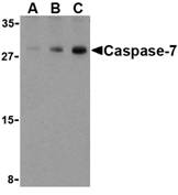
Left: Western blot analysis of Caspase-7 in human skeletal muscle cell lysate with Caspase-7 antibody at (A) 0.5, (B) 1, and (C) 2 µg/ml.
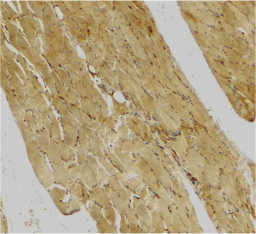
Below: Immunohistochemical staining of human skeletal muscle using Caspase-7 antibody at 2 µg/ml.
Other Product Images
Source : Caspase-7 antibody was raised against a 16 amino acid peptide from near the carboxy-terminus of human Caspase-7.
Purification : Affinity chromatography purified via peptide column
Clonality and Clone : This is a polyclonal antibody.
Host : Caspase-7 antibody was raised in rabbit. Please use anti-rabbit secondary antibodies.
Immunogen : Human Casp-7 / ICELAP3 / Mch3 (C-Terminus) Peptide (Cat. No. 3465P)
Application : Casp-7 antibody can be used for the detection of Caspase-7 by Western blot at 0.5 µg/ml.Depending on cell lines or tissues used, either full-length or other cleavage products may be observed.
Tested Application(s) : E, WB, IHC
Buffer : Antibody is supplied in PBS containing 0.02% sodium azide.
Blocking Peptide : Cat. No. 3465P - Casp-7 Peptide
Long-Term Storage : Caspase-7 antibody can be stored at 4ºC, stable for one year. As with all antibodies care should be taken to avoid repeated freeze thaw cycles. Antibodies should not be exposed to prolonged high temperatures.
Positive Control
1.Cat. No. 1375 - Human Skeletal Muscle Tissue Lysate
Species Reactivity :H, M, R
GI Number : 1730092
Accession Number : P55210
Short Description : (CT) an executioner caspase
References
1.Martinon F and Tschopp J. Inflammatory caspases: linking an intracellular innate immune system to autoinflammatory diseases. Cell 2004; 117:561-74.
2.Zhivotovsky B and Orrenius S. Caspase-2 function in response to DNA damage. Biochim. Biophys. Res. Comm. 2005; 331:859-67.
3.Wolf BB and Green DR. Suicidal tendencies: apoptotic cell death by caspase family proteinases. J. Biol. Chem. 1999; 274:20049-52.
4.Juan TSC, McNiece IK, Argento JM, et al. Identification and mapping of Casp7, a cysteine protease resembling CPP32b, Interleukin-1b converting enzyme, and CED-3. Genomics 1997; 40:86-93.
Catalog# : 3465
Caspases are a family of cysteine proteases that can be divided into the apoptotic and inflammatory caspase subfamilies. Unlike the apoptotic caspases, members of the inflammatory subfamily are generally not involved in cell death but are associated with the immune response to microbial pathogens (reviewed in 1 and 2). The apoptotic subfamily can be further divided into initiator caspases, which are activated in response to death signals, and executioner caspases, which are activated by the initiator caspases and are responsible for cleavage of cellular substrates that ultimately lead to cell death (reviewed in 3). Caspase-7 is an executioner caspase that was identified based on its homology with caspases 1 and 3, as well as the C. elegans cell death protein CED-3 (4). Alternative splicing of Caspase-7 mRNA results in the production of 3 distinct isoforms (4). Caspase-7 activity can be directly inhibited by XIAP expression (5).
Additional Names : Caspase-7 (CT), ICELAP3 ,Mch3
 Description
Left: Western blot analysis of Caspase-7 in human skeletal muscle cell lysate with Caspase-7 antibody at (A) 0.5, (B) 1, and (C) 2 µg/ml.
Below: Immunohistochemical staining of human skeletal muscle using Caspase-7 antibody at 2 µg/ml.
Other Product Images

Source : Caspase-7 antibody was raised against a 16 amino acid peptide from near the carboxy-terminus of human Caspase-7.
Purification : Affinity chromatography purified via peptide column
Clonality and Clone : This is a polyclonal antibody.
Host : Caspase-7 antibody was raised in rabbit. Please use anti-rabbit secondary antibodies.
Immunogen : Human Casp-7 / ICELAP3 / Mch3 (C-Terminus) Peptide (Cat. No. 3465P)
Application : Casp-7 antibody can be used for the detection of Caspase-7 by Western blot at 0.5 µg/ml.Depending on cell lines or tissues used, either full-length or other cleavage products may be observed.
Tested Application(s) : E, WB, IHC
Buffer : Antibody is supplied in PBS containing 0.02% sodium azide.
Blocking Peptide : Cat. No. 3465P - Casp-7 Peptide
Long-Term Storage : Caspase-7 antibody can be stored at 4ºC, stable for one year. As with all antibodies care should be taken to avoid repeated freeze thaw cycles. Antibodies should not be exposed to prolonged high temperatures.
Positive Control
1.Cat. No. 1375 - Human Skeletal Muscle Tissue Lysate
Species Reactivity :H, M, R
GI Number : 1730092
Accession Number : P55210
Short Description : (CT) an executioner caspase
References
1.Martinon F and Tschopp J. Inflammatory caspases: linking an intracellular innate immune system to autoinflammatory diseases. Cell 2004; 117:561-74.
2.Zhivotovsky B and Orrenius S. Caspase-2 function in response to DNA damage. Biochim. Biophys. Res. Comm. 2005; 331:859-67.
3.Wolf BB and Green DR. Suicidal tendencies: apoptotic cell death by caspase family proteinases. J. Biol. Chem. 1999; 274:20049-52.
4.Juan TSC, McNiece IK, Argento JM, et al. Identification and mapping of Casp7, a cysteine protease resembling CPP32b, Interleukin-1b converting enzyme, and CED-3. Genomics 1997; 40:86-93.
Sunday, May 23, 2010
Peptide Arrays
Bio-Synthesis of large numbers of extremely affordable custom peptides has never been easier ...
Bio-Synthesis has developed high throughput custom peptide synthesis to meet the growing demands for synthetic peptides in the proteomics boom. We offer parallel synthesis of small quantities of peptide libraries with the fastest and most efficient high throughput, delivering 96 different peptides, unbound, in a 96-well format. Each plate is individually tested for accuracy and can be used for epitope mapping, libraries, protein characterization and much more. This technique provides researchers the ability to order large numbers of peptides at extremely affordable prices and delivered in a short time.
A major reason for the demand of high throughput peptide synthesis has been the realization that synthetic peptides have become invaluable tools in elucidating important contiguous epitopes that are essential for functional and structural domains in proteins. In particular, the systematic screening with a series of overlapping fragments, called epitope mapping or substitution analogues, needs short peptides in large numbers although, in general, micro molar quantities are sufficient. In this regard, the value of using synthetic peptides as probes in biochemistry and pharmacology is comparable to the value of oligonucleotides in molecular biology. The systematic use of peptides, however, has been limited by the cost, time and special expertise or equipment required to provide a significant number of peptides for evaluation. Therefore, BSI over the past several years has developed synthesis platforms that are capable of the simultaneous synthesis of many peptides with FLEXIBILITY, SPEED, and HIGH THROUGHPUT at an economic price, resulting in a great value to our customers.
FEATURES:
- Quantity: 0.5-2 mgs
- Length: 8-20 aa average 75% pure
- 96 crude peptides per plate in 96-well format
- Optimized chemistries
- No cross contamination
- QC: MALDI-TOF Mass Spectrometry on all peptides
- Including: Electronic PDF files of QC data ( 2D bar code)
- Delivery: 7-10 days
- N-terminal biotinylation
- N-terminal acetylation
- Non-standard residue incorporation
- C-terminal amidation
- Disulfide bridge formation (cyclization)
- Epitope mapping
- Alanine walking
- Single amino acid mutation screening
- Protein-protein interaction studies
- Kinase motif discovery
- Protease motif discovery
Tuesday, May 18, 2010
Peptide Libary Design Tool
Bio-Synthesis has implemented peptide libary tools to its rapid high-throughput parallel peptide synthesis platform. These libraries can be used to screen highly active compounds such as antigenic peptides, receptor ligands, antimicrobial compounds, and enzyme inhibitors. Some of the applications of peptide screening tools are as follows:
Alanine Peptide Scanning Library: The generation of peptide library in which alanine (Ala, A) is systematically substituted into each of the amino acids which can be used to identify epitope activity.
Positional Peptide Library: A selected position in a peptide sequence is systematically replaced with different amino acid to show the effect on the substitute amino acid at certain position.
Truncation Pepitde Library: The truncation libary is been used to predict the minimum amino acid length required for optimum epitope activity.
Random Peptide Library: It's constructed by substituting selected positions on the original peptide randomly and simultaneously with all other natural amino acids in a shot gun approach with a purpose to elucidate potential alternatives for enhanced peptide activity.
Scramble Peptide Library: Scramble Library is constructed by carrying out permutation on the original peptide's sequence. It has the potential to give all possible alternatives and offers and represents the highest degree of variability for peptide library.
- Epitope mapping studies.
- Vaccine research.
- High-throughput protein interaction analysis.
- Customized peptide microarray production.
- Kinase assays.
Alanine Peptide Scanning Library: The generation of peptide library in which alanine (Ala, A) is systematically substituted into each of the amino acids which can be used to identify epitope activity.
Positional Peptide Library: A selected position in a peptide sequence is systematically replaced with different amino acid to show the effect on the substitute amino acid at certain position.
Truncation Pepitde Library: The truncation libary is been used to predict the minimum amino acid length required for optimum epitope activity.
Random Peptide Library: It's constructed by substituting selected positions on the original peptide randomly and simultaneously with all other natural amino acids in a shot gun approach with a purpose to elucidate potential alternatives for enhanced peptide activity.
Scramble Peptide Library: Scramble Library is constructed by carrying out permutation on the original peptide's sequence. It has the potential to give all possible alternatives and offers and represents the highest degree of variability for peptide library.
Wednesday, May 12, 2010
Caspase-6 Antibody
Caspase-6 Antibody
Catalog# : 3471
Caspases are a family of cysteine proteases that can be divided into the apoptotic and inflammatory caspase subfamilies. Unlike the apoptotic caspases, members of the inflammatory subfamily are generally not involved in cell death but are associated with the immune response to microbial pathogens (reviewed in 1 and 2). The apoptotic subfamily can be further divided into initiator caspases, which are activated in response to death signals, and executioner caspases, which are activated by the initiator caspases and are responsible for cleavage of cellular substrates that ultimately lead to cell death (reviewed in 3). Caspase-6 is an executioner caspase that was idientifed based on its homology with human caspases 2 and 3 as well as the C. elegans cell death protein CED-3 (4). It possesses two isoforms, of which only the longer form possesses protease activity (4). Caspase-6 is highly expressed in adult brain and may play a role in several neuronal pathologies (5).
Additional Names : Caspase-6 (NT), Mch2
 Description
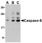
Left: Western blot analysis of caspase-6 in Jurkat cell lysate with caspase-6 antibody at (A) 0.5, (B) 1, and (C) 2 µg/ml.
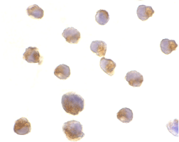
Below: Immunocytochemistry of caspase-6 in Jurkat cells with caspase-6 antibody at 2 µg/ml.
Other Product Images
Source : Caspase-6 antibody was raised against a 15 amino acid peptide from near the amino-terminus of human Caspase-6.
Purification : Affinity chromatography purified via peptide column
Clonality and Clone : This is a polyclonal antibody.
Host : Caspase-6 antibody was raised in rabbit. Please use anti-rabbit secondary antibodies.
Immunogen : Virus Casp-6 / Mch2 (N-Terminus) Peptide (Cat. No. 3471P)
Application : Caspase-6 antibody can be used for the detection of caspase-6 by Western blot at 0.5 µg/ml.Depending on cell lines or tissues used, either full-length or other cleavage products may be observed.
Tested Application(s) : E, WB, ICC
Buffer : Antibody is supplied in PBS containing 0.02% sodium azide.
Blocking Peptide : Cat. No. 3471P - Casp-6 Peptide
Long-Term Storage : Caspase-6 antibody can be stored at 4ºC, stable for one year. As with all antibodies care should be taken to avoid repeated freeze thaw cycles. Antibodies should not be exposed to prolonged high temperatures.
Positive Control
1.Cat. No. 1205 - Jurkat Cell Lysate
Species Reactivity :H
GI Number : 14916483
Accession Number : NP_001217
Short Description : (NT) an executioner caspase
References
1.Martinon F and Tschopp J. Inflammatory caspases: linking an intracellular innate immune system to autoinflammatory diseases. Cell 2004; 117:561-74.
2.Zhivotovsky B and Orrenius S. Caspase-2 function in response to DNA damage. Biochim. Biophys. Res. Comm. 2005; 331:859-67.
3.Wolf BB and Green DR. Suicidal tendencies: apoptotic cell death by caspase family proteinases. J. Biol. Chem. 1999; 274:20049-52.
4.Fernandes-Alnemri T, Litwack G, and Alnemri ES. Mch2, a new member of the apoptotic Ced-3/Ice cysteine protease gene family. Cancer Res. 1995; 55:2737-42.
Catalog# : 3471
Caspases are a family of cysteine proteases that can be divided into the apoptotic and inflammatory caspase subfamilies. Unlike the apoptotic caspases, members of the inflammatory subfamily are generally not involved in cell death but are associated with the immune response to microbial pathogens (reviewed in 1 and 2). The apoptotic subfamily can be further divided into initiator caspases, which are activated in response to death signals, and executioner caspases, which are activated by the initiator caspases and are responsible for cleavage of cellular substrates that ultimately lead to cell death (reviewed in 3). Caspase-6 is an executioner caspase that was idientifed based on its homology with human caspases 2 and 3 as well as the C. elegans cell death protein CED-3 (4). It possesses two isoforms, of which only the longer form possesses protease activity (4). Caspase-6 is highly expressed in adult brain and may play a role in several neuronal pathologies (5).
Additional Names : Caspase-6 (NT), Mch2
 Description
Left: Western blot analysis of caspase-6 in Jurkat cell lysate with caspase-6 antibody at (A) 0.5, (B) 1, and (C) 2 µg/ml.
Below: Immunocytochemistry of caspase-6 in Jurkat cells with caspase-6 antibody at 2 µg/ml.
Other Product Images

Source : Caspase-6 antibody was raised against a 15 amino acid peptide from near the amino-terminus of human Caspase-6.
Purification : Affinity chromatography purified via peptide column
Clonality and Clone : This is a polyclonal antibody.
Host : Caspase-6 antibody was raised in rabbit. Please use anti-rabbit secondary antibodies.
Immunogen : Virus Casp-6 / Mch2 (N-Terminus) Peptide (Cat. No. 3471P)
Application : Caspase-6 antibody can be used for the detection of caspase-6 by Western blot at 0.5 µg/ml.Depending on cell lines or tissues used, either full-length or other cleavage products may be observed.
Tested Application(s) : E, WB, ICC
Buffer : Antibody is supplied in PBS containing 0.02% sodium azide.
Blocking Peptide : Cat. No. 3471P - Casp-6 Peptide
Long-Term Storage : Caspase-6 antibody can be stored at 4ºC, stable for one year. As with all antibodies care should be taken to avoid repeated freeze thaw cycles. Antibodies should not be exposed to prolonged high temperatures.
Positive Control
1.Cat. No. 1205 - Jurkat Cell Lysate
Species Reactivity :H
GI Number : 14916483
Accession Number : NP_001217
Short Description : (NT) an executioner caspase
References
1.Martinon F and Tschopp J. Inflammatory caspases: linking an intracellular innate immune system to autoinflammatory diseases. Cell 2004; 117:561-74.
2.Zhivotovsky B and Orrenius S. Caspase-2 function in response to DNA damage. Biochim. Biophys. Res. Comm. 2005; 331:859-67.
3.Wolf BB and Green DR. Suicidal tendencies: apoptotic cell death by caspase family proteinases. J. Biol. Chem. 1999; 274:20049-52.
4.Fernandes-Alnemri T, Litwack G, and Alnemri ES. Mch2, a new member of the apoptotic Ced-3/Ice cysteine protease gene family. Cancer Res. 1995; 55:2737-42.
Caspase-6 Antibody
Caspase-6 Antibody
Catalog# : 3469
Caspases are a family of cysteine proteases that can be divided into the apoptotic and inflammatory caspase subfamilies. Unlike the apoptotic caspases, members of the inflammatory subfamily are generally not involved in cell death but are associated with the immune response to microbial pathogens (reviewed in 1 and 2). The apoptotic subfamily can be further divided into initiator caspases, which are activated in response to death signals, and executioner caspases, which are activated by the initiator caspases and are responsible for cleavage of cellular substrates that ultimately lead to cell death (reviewed in 3). Caspase-6 is an executioner caspase that was identified based on its homology with human caspases 2 and 3 as well as the C. elegans cell death protein CED-3 (4). It possesses two isoforms, of which only the longer form possesses protease activity (4). Caspase-6 is highly expressed in adult brain and may play a role in several neuronal pathologies (5).
Additional Names : Caspase-6 (IN), Mch2
 Description
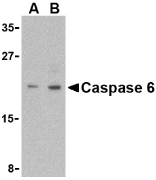
Left: Western blot analysis of Caspase-6 in MCF7 cell lysate with Caspase-6 antibody (IN) at (A) 1 and (B) 2 µg/ml.
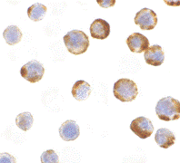
Below: Immunocytochemistry of caspase-6 in MCF7 with caspase-6 antibody at 1 µg/ml.
Other Product Images
 Source : Caspase-6 antibody was raised against a 15 amino acid peptide from near the center of human Caspase-6.
Purification : immunoaffinity purified
Clonality and Clone : This is a polyclonal antibody.
Host : Caspase-6 antibody was raised in rabbit. Please use anti-rabbit secondary antibodies.
Immunogen : Human Casp-6 / Mch2 (Intermediate Domain) Peptide (Cat. No. 3469P)
Application : Casp-6 antibody can be used for the detection of Caspase-6 by Western blot at 1 µg/ml.MCF7 cell lysate can be used as positive control. Anti-Caspase-6 is human specific. Depending on cell lines or tissues used, either full-length or other cleavage products may be observed.
Tested Application(s) : E, WB, ICC
Buffer : Antibody is supplied in PBS containing 0.02% sodium azide.
Blocking Peptide : Cat. No. 3469P - Casp-6 Peptide
Long-Term Storage : Caspase-6 antibody can be stored at 4ºC, stable for one year. As with all antibodies care should be taken to avoid repeated freeze thaw cycles. Antibodies should not be exposed to prolonged high temperatures.
Positive Control
1.Cat. No. 1219 - MCF7 Cell Lysate
Species Reactivity :H
GI Number : 14916483
Accession Number : NP_001217
Short Description : (IN) an executioner caspase
References
1.Martinon F and Tschopp J. Inflammatory caspases: linking an intracellular innate immune system to autoinflammatory diseases. Cell 2004; 117:561-74.
2.Zhivotovsky B and Orrenius S. Caspase-2 function in response to DNA damage. Biochim. Biophys. Res. Comm. 2005; 331:859-67.
3.Wolf BB and Green DR. Suicidal tendencies: apoptotic cell death by caspase family proteinases. J. Biol. Chem. 1999; 274:20049-52.
4.Fernandes-Alnemri T, Litwack G, and Alnemri ES. Mch2, a new member of the apoptotic Ced-3/Ice cysteine protease gene family. Cancer Res. 1995; 55:2737-42.
Catalog# : 3469
Caspases are a family of cysteine proteases that can be divided into the apoptotic and inflammatory caspase subfamilies. Unlike the apoptotic caspases, members of the inflammatory subfamily are generally not involved in cell death but are associated with the immune response to microbial pathogens (reviewed in 1 and 2). The apoptotic subfamily can be further divided into initiator caspases, which are activated in response to death signals, and executioner caspases, which are activated by the initiator caspases and are responsible for cleavage of cellular substrates that ultimately lead to cell death (reviewed in 3). Caspase-6 is an executioner caspase that was identified based on its homology with human caspases 2 and 3 as well as the C. elegans cell death protein CED-3 (4). It possesses two isoforms, of which only the longer form possesses protease activity (4). Caspase-6 is highly expressed in adult brain and may play a role in several neuronal pathologies (5).
Additional Names : Caspase-6 (IN), Mch2
 Description
Left: Western blot analysis of Caspase-6 in MCF7 cell lysate with Caspase-6 antibody (IN) at (A) 1 and (B) 2 µg/ml.
Below: Immunocytochemistry of caspase-6 in MCF7 with caspase-6 antibody at 1 µg/ml.
Other Product Images
 Source : Caspase-6 antibody was raised against a 15 amino acid peptide from near the center of human Caspase-6.
Purification : immunoaffinity purified
Clonality and Clone : This is a polyclonal antibody.
Host : Caspase-6 antibody was raised in rabbit. Please use anti-rabbit secondary antibodies.
Immunogen : Human Casp-6 / Mch2 (Intermediate Domain) Peptide (Cat. No. 3469P)
Application : Casp-6 antibody can be used for the detection of Caspase-6 by Western blot at 1 µg/ml.MCF7 cell lysate can be used as positive control. Anti-Caspase-6 is human specific. Depending on cell lines or tissues used, either full-length or other cleavage products may be observed.
Tested Application(s) : E, WB, ICC
Buffer : Antibody is supplied in PBS containing 0.02% sodium azide.
Blocking Peptide : Cat. No. 3469P - Casp-6 Peptide
Long-Term Storage : Caspase-6 antibody can be stored at 4ºC, stable for one year. As with all antibodies care should be taken to avoid repeated freeze thaw cycles. Antibodies should not be exposed to prolonged high temperatures.
Positive Control
1.Cat. No. 1219 - MCF7 Cell Lysate
Species Reactivity :H
GI Number : 14916483
Accession Number : NP_001217
Short Description : (IN) an executioner caspase
References
1.Martinon F and Tschopp J. Inflammatory caspases: linking an intracellular innate immune system to autoinflammatory diseases. Cell 2004; 117:561-74.
2.Zhivotovsky B and Orrenius S. Caspase-2 function in response to DNA damage. Biochim. Biophys. Res. Comm. 2005; 331:859-67.
3.Wolf BB and Green DR. Suicidal tendencies: apoptotic cell death by caspase family proteinases. J. Biol. Chem. 1999; 274:20049-52.
4.Fernandes-Alnemri T, Litwack G, and Alnemri ES. Mch2, a new member of the apoptotic Ced-3/Ice cysteine protease gene family. Cancer Res. 1995; 55:2737-42.
Caspase-5 Antibody
Caspase-5 Antibody
Catalog# : 3457
Caspases are a family of cysteine proteases that can be divided into the apoptotic and inflammatory caspase subfamilies. Unlike the apoptotic caspases, members of the inflammatory subfamily are generally not involved in cell death but are associated with the immune response to microbial pathogens (reviewed in 1). Members of this subfamily include caspase-1, -4, -5, and -12. Activation of these caspases results in the cleavage and activation of proinflammatory cytokines such as IL-1beta and IL-18 (2,3). Caspase-5 can interact with caspase-1; both are constituents of the NALP1 inflammasome, a complex that can trigger the cleavage of pro-IL-1beta (4). Expression of caspase-5 can be regulated by lipopolysaccharide (LPS) and IFN-ϒ (5).
Additional Names : Caspase-5 (IN), ICH-3 , ICE rel III
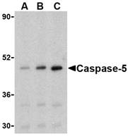 Description
Left: Western blot analysis of caspase-5 in Ramos cells with caspase-5 antibody at (A) 0.5, (B) 1, and (C) 2 µg/ml.
Below: Immunocytochemistry of caspase-5 in P815 cells with caspase-5 antibody at 2 µg/ml.
Source : Caspase-5 antibody was raised against a 14 amino acid peptide from the center of human Caspase-5.
Purification : Affinity chromatography purified via peptide column
Clonality and Clone : This is a polyclonal antibody.
Host : Caspase-5 antibody was raised in rabbit. Please use anti-rabbit secondary antibodies.
Immunogen : Human Casp-5 / (ICH-3 / ICE rel III (Intermediate Domain) Peptide (Cat. No. 3457P)
Application : Casp-5 antibody can be used for the detection of caspase-5 by Western blot at 0.5 - 1 µg/mlDepending on cell lines or tissues used, other cleavage products may be observed.
Tested Application(s) : E, WB, ICC
Buffer : Antibody is supplied in PBS containing 0.02% sodium azide.
Blocking Peptide : Cat. No. 3457P - Casp-5 Peptide
Long-Term Storage : Caspase-5 antibody can be stored at 4ºC, stable for one year. As with all antibodies care should be taken to avoid repeated freeze thaw cycles. Antibodies should not be exposed to prolonged high temperatures.
Positive Control
1.Cat. No. 1225 - Ramos Cell Lysate
2.Cat. No. 1286 - P815 Cell Lysate
Species Reactivity :H
GI Number : 212276423
Accession Number : P51878
Short Description : (IN) a caspase in the inflammatory subfamily
References
1.Martinon F and Tschopp J. Inflammatory caspases: linking an intracellular innate immune system to autoinflammatory diseases. Cell 2004; 117:561-74.
2.Kuida K, Lippke JA, Ku G, et al. Altered cytokine export and apoptosis in mice deficient in interleukin-1 beta converting enzyme. Science 1995; 267:2000-3.
3.Gracie JA, Robertson SE, and McInnes IB. Interleukin-18. J. Leukoc. Biol. 2003; 73:213-224.
4.Martinon F, Burns K, and Tschopp J. The inflammasome: a molecular platform triggering activation of the inflammatory caspases and processing of proIL-1. Mol. Cell. 2002; 10:417-26.
Catalog# : 3457
Caspases are a family of cysteine proteases that can be divided into the apoptotic and inflammatory caspase subfamilies. Unlike the apoptotic caspases, members of the inflammatory subfamily are generally not involved in cell death but are associated with the immune response to microbial pathogens (reviewed in 1). Members of this subfamily include caspase-1, -4, -5, and -12. Activation of these caspases results in the cleavage and activation of proinflammatory cytokines such as IL-1beta and IL-18 (2,3). Caspase-5 can interact with caspase-1; both are constituents of the NALP1 inflammasome, a complex that can trigger the cleavage of pro-IL-1beta (4). Expression of caspase-5 can be regulated by lipopolysaccharide (LPS) and IFN-ϒ (5).
Additional Names : Caspase-5 (IN), ICH-3 , ICE rel III
 Description
Left: Western blot analysis of caspase-5 in Ramos cells with caspase-5 antibody at (A) 0.5, (B) 1, and (C) 2 µg/ml.
Below: Immunocytochemistry of caspase-5 in P815 cells with caspase-5 antibody at 2 µg/ml.
Source : Caspase-5 antibody was raised against a 14 amino acid peptide from the center of human Caspase-5.
Purification : Affinity chromatography purified via peptide column
Clonality and Clone : This is a polyclonal antibody.
Host : Caspase-5 antibody was raised in rabbit. Please use anti-rabbit secondary antibodies.
Immunogen : Human Casp-5 / (ICH-3 / ICE rel III (Intermediate Domain) Peptide (Cat. No. 3457P)
Application : Casp-5 antibody can be used for the detection of caspase-5 by Western blot at 0.5 - 1 µg/mlDepending on cell lines or tissues used, other cleavage products may be observed.
Tested Application(s) : E, WB, ICC
Buffer : Antibody is supplied in PBS containing 0.02% sodium azide.
Blocking Peptide : Cat. No. 3457P - Casp-5 Peptide
Long-Term Storage : Caspase-5 antibody can be stored at 4ºC, stable for one year. As with all antibodies care should be taken to avoid repeated freeze thaw cycles. Antibodies should not be exposed to prolonged high temperatures.
Positive Control
1.Cat. No. 1225 - Ramos Cell Lysate
2.Cat. No. 1286 - P815 Cell Lysate
Species Reactivity :H
GI Number : 212276423
Accession Number : P51878
Short Description : (IN) a caspase in the inflammatory subfamily
References
1.Martinon F and Tschopp J. Inflammatory caspases: linking an intracellular innate immune system to autoinflammatory diseases. Cell 2004; 117:561-74.
2.Kuida K, Lippke JA, Ku G, et al. Altered cytokine export and apoptosis in mice deficient in interleukin-1 beta converting enzyme. Science 1995; 267:2000-3.
3.Gracie JA, Robertson SE, and McInnes IB. Interleukin-18. J. Leukoc. Biol. 2003; 73:213-224.
4.Martinon F, Burns K, and Tschopp J. The inflammasome: a molecular platform triggering activation of the inflammatory caspases and processing of proIL-1. Mol. Cell. 2002; 10:417-26.
Caspase-5 Antibody
Caspase-5 Antibody
Catalog# : 3455
Caspases are a family of cysteine proteases that can be divided into the apoptotic and inflammatory caspase subfamilies. Unlike the apoptotic caspases, members of the inflammatory subfamily are generally not involved in cell death but are associated with the immune response to microbial pathogens (reviewed in 1). Members of this subfamily include caspase-1, -4, -5, and -12. Activation of these caspases results in the cleavage and activation of proinflammatory cytokines such as IL-1beta and IL-18 (2,3). Caspase-5 can interact with caspase-1; both are constituents of the NALP1 inflammasome, a complex that can trigger the cleavage of pro-IL-1beta (4). Expression of caspase-5 can be regulated by lipopolysaccharide (LPS) and IFN-ϒ (5).
Additional Names : Caspase-5 (NT), ICH-3 , ICE rel III
Caspase-5 Antibody
Caspase-5 Antibody
Catalog# : 3455
Caspases are a family of cysteine proteases that can be divided into the apoptotic and inflammatory caspase subfamilies. Unlike the apoptotic caspases, members of the inflammatory subfamily are generally not involved in cell death but are associated with the immune response to microbial pathogens (reviewed in 1). Members of this subfamily include caspase-1, -4, -5, and -12. Activation of these caspases results in the cleavage and activation of proinflammatory cytokines such as IL-1beta and IL-18 (2,3). Caspase-5 can interact with caspase-1; both are constituents of the NALP1 inflammasome, a complex that can trigger the cleavage of pro-IL-1beta (4). Expression of caspase-5 can be regulated by lipopolysaccharide (LPS) and IFN-ϒ (5).
Additional Names : Caspase-5 (NT), ICH-3 , ICE rel III
Caspase-4 Antibody
Caspase-4 Antibody
Catalog# : 3453
Caspases are a family of cysteine proteases that can be divided into the apoptotic and inflammatory caspase subfamilies. Unlike the apoptotic caspases, members of the inflammatory subfamily are generally not involved in cell death but are associated with the immune response to microbial pathogens (reviewed in 1). Members of this subfamily include caspase-1, -4, -5, and -12. Activation of these caspases results in the cleavage and activation of proinflammatory cytokines such as IL-1beta and IL-18 (2,3). Caspase-4 was initially identified as a homologous protein to Caspase-1 and the C. elegans Ced-3 which could induce apoptosis in transfected cells (4). More recent studies have shown that it can be activated by ER stress and has been suggested to be involved in multiple neuronal pathologies such as Alzheimer’s disease (5).
Additional Names : Caspase-4 (IN), ICH-2, ICE relII , Mih1
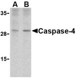 Description
Left: Western blot analysis of caspase-4 in human spleen cells with caspase-4 antibody at (A) 1 and (B) 2 µg/ml.
Below: Immunohistochemical staining of human spleen tissue using caspase-4 antibody at 2 µg/ml.
Other Product Images
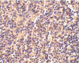 Source : Caspase-4 antibody was raised against a 15 amino acid peptide from near the center of human Caspase-4.
Purification : Affinity chromatography purified via peptide column
Clonality and Clone : This is a polyclonal antibody.
Host : Caspase-4 antibody was raised in rabbit. Please use anti-rabbit secondary antibodies.
Immunogen : Human Casp-4 / ICH-2 / ICE rel II / Mih1 (Intermediate Domain) Peptide (Cat. No. 3453P)
Application : Casp-4 antibody can be used for the detection of Caspase-4 by Western blot at 2 µg/ml.Depending on cell lines or tissues used, other cleavage products may be observed.
Tested Application(s) : E, WB, IHC
Buffer : Antibody is supplied in PBS containing 0.02% sodium azide.
Blocking Peptide : Cat. No. 3453P - Casp-4 Peptide
Long-Term Storage : Caspase-4 antibody can be stored at 4ºC, stable for one year. As with all antibodies care should be taken to avoid repeated freeze thaw cycles. Antibodies should not be exposed to prolonged high temperatures.
Positive Control
1.Cat. No. 1306 - Human Spleen Tissue Lysate
Species Reactivity :H
GI Number : 886050
Accession Number : AAA86890
Short Description : (IN) a caspase induced by ER stress
References
1.Martinon F and Tschopp J. Inflammatory caspases: linking an intracellular innate immune system to autoinflammatory diseases. Cell 2004; 117:561-74.
2.Kuida K, Lippke JA, Ku G, et al. Altered cytokine export and apoptosis in mice deficient in interleukin-1 beta converting enzyme. Science 1995; 267:2000-3.
3.Gracie JA, Robertson SE, and McInnes IB. Interleukin-18. J. Leukoc. Biol. 2003; 73:213-224.
4.Kamens J, Paskind M, Hugunin M, et al. Identification and characterization of ICH-2, a novel member of the interleukin-1 beta-converting enzyme family of cysteine proteases. J. Biol. Chem. 1995; 270:15250-6.
Catalog# : 3453
Caspases are a family of cysteine proteases that can be divided into the apoptotic and inflammatory caspase subfamilies. Unlike the apoptotic caspases, members of the inflammatory subfamily are generally not involved in cell death but are associated with the immune response to microbial pathogens (reviewed in 1). Members of this subfamily include caspase-1, -4, -5, and -12. Activation of these caspases results in the cleavage and activation of proinflammatory cytokines such as IL-1beta and IL-18 (2,3). Caspase-4 was initially identified as a homologous protein to Caspase-1 and the C. elegans Ced-3 which could induce apoptosis in transfected cells (4). More recent studies have shown that it can be activated by ER stress and has been suggested to be involved in multiple neuronal pathologies such as Alzheimer’s disease (5).
Additional Names : Caspase-4 (IN), ICH-2, ICE relII , Mih1
 Description
Left: Western blot analysis of caspase-4 in human spleen cells with caspase-4 antibody at (A) 1 and (B) 2 µg/ml.
Below: Immunohistochemical staining of human spleen tissue using caspase-4 antibody at 2 µg/ml.
Other Product Images
 Source : Caspase-4 antibody was raised against a 15 amino acid peptide from near the center of human Caspase-4.
Purification : Affinity chromatography purified via peptide column
Clonality and Clone : This is a polyclonal antibody.
Host : Caspase-4 antibody was raised in rabbit. Please use anti-rabbit secondary antibodies.
Immunogen : Human Casp-4 / ICH-2 / ICE rel II / Mih1 (Intermediate Domain) Peptide (Cat. No. 3453P)
Application : Casp-4 antibody can be used for the detection of Caspase-4 by Western blot at 2 µg/ml.Depending on cell lines or tissues used, other cleavage products may be observed.
Tested Application(s) : E, WB, IHC
Buffer : Antibody is supplied in PBS containing 0.02% sodium azide.
Blocking Peptide : Cat. No. 3453P - Casp-4 Peptide
Long-Term Storage : Caspase-4 antibody can be stored at 4ºC, stable for one year. As with all antibodies care should be taken to avoid repeated freeze thaw cycles. Antibodies should not be exposed to prolonged high temperatures.
Positive Control
1.Cat. No. 1306 - Human Spleen Tissue Lysate
Species Reactivity :H
GI Number : 886050
Accession Number : AAA86890
Short Description : (IN) a caspase induced by ER stress
References
1.Martinon F and Tschopp J. Inflammatory caspases: linking an intracellular innate immune system to autoinflammatory diseases. Cell 2004; 117:561-74.
2.Kuida K, Lippke JA, Ku G, et al. Altered cytokine export and apoptosis in mice deficient in interleukin-1 beta converting enzyme. Science 1995; 267:2000-3.
3.Gracie JA, Robertson SE, and McInnes IB. Interleukin-18. J. Leukoc. Biol. 2003; 73:213-224.
4.Kamens J, Paskind M, Hugunin M, et al. Identification and characterization of ICH-2, a novel member of the interleukin-1 beta-converting enzyme family of cysteine proteases. J. Biol. Chem. 1995; 270:15250-6.
Caspase-4 Antibody
Caspase-4 Antibody
Catalog# : 3451
Caspases are a family of cysteine proteases that can be divided into the apoptotic and inflammatory caspase subfamilies. Unlike the apoptotic caspases, members of the inflammatory subfamily are generally not involved in cell death but are associated with the immune response to microbial pathogens (reviewed in 1). Members of this subfamily include caspase-1, -4, -5, and -12. Activation of these caspases results in the cleavage and activation of proinflammatory cytokines such as IL-1beta and IL-18 (2,3). Caspase-4 was initially identified as a homologous protein to Caspase-1 and the C. elegans Ced-3 which could induce apoptosis in transfected cells (4). More recent studies have shown that it can be activated by ER stress and has been suggested to be involved in multiple neuronal pathologies such as Alzheimer’s disease (5).
Additional Names : Caspase-4 (NT), ICH-2, ICE rel II , Mih1
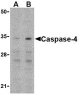 Description
Left: Western blot analysis of caspase-4 in Ramos cells with caspase-4 antibody at (A) 0.5 and (B) 1 µgg/ml.
Below: Immunohistochemical staining of mouse spleen using caspase-4 antibody at 2 µg/ml.
Other Product Images
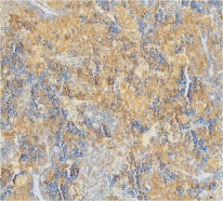 Source : Caspase-4 antibody was raised against a 16 amino acid peptide from the amino-terminus of human Caspase-4.
Purification : Affinity chromatography purified via peptide column
Clonality and Clone : This is a polyclonal antibody.
Host : Caspase-4 antibody was raised in rabbit. Please use anti-rabbit secondary antibodies.
Immunogen : Human Casp-4 / ICH-2 / ICE rel II / Mih1 (N-Terminus) Peptide (Cat. No. 3451P)
Application : Casp-4 antibody can be used for the detection of Caspase-4 by Western blot at 1 µg/ml.Depending on cell lines or tissues used, other cleavage products may be observed.
Tested Application(s) : E, WB, IHC
Buffer : Antibody is supplied in PBS containing 0.02% sodium azide.
Blocking Peptide : Cat. No. 3451P - Casp-4 Peptide
Long-Term Storage : Caspase-4 antibody can be stored at 4ºC, stable for one year. As with all antibodies care should be taken to avoid repeated freeze thaw cycles. Antibodies should not be exposed to prolonged high temperatures.
Positive Control
1.Cat. No. 1225 - Ramos Cell Lysate
2.Cat. No. 1406 - Mouse Spleen Tissue Lysate
Species Reactivity :H, M
GI Number : 886050
Accession Number : AAA86890
Short Description : (NT) a caspase induced by ER stress
References
1.Martinon F and Tschopp J. Inflammatory caspases: linking an intracellular innate immune system to autoinflammatory diseases. Cell 2004; 117:561-74.
2.Kuida K, Lippke JA, Ku G, et al. Altered cytokine export and apoptosis in mice deficient in interleukin-1 beta converting enzyme. Science 1995; 267:2000-3.
3.Gracie JA, Robertson SE, and McInnes IB. Interleukin-18. J. Leukoc. Biol. 2003; 73:213-224.
4.Kamens J, Paskind M, Hugunin M, et al. Identification and characterization of ICH-2, a novel member of the interleukin-1 beta-converting enzyme family of cysteine proteases. J. Biol. Chem. 1995; 270:15250-6.
Catalog# : 3451
Caspases are a family of cysteine proteases that can be divided into the apoptotic and inflammatory caspase subfamilies. Unlike the apoptotic caspases, members of the inflammatory subfamily are generally not involved in cell death but are associated with the immune response to microbial pathogens (reviewed in 1). Members of this subfamily include caspase-1, -4, -5, and -12. Activation of these caspases results in the cleavage and activation of proinflammatory cytokines such as IL-1beta and IL-18 (2,3). Caspase-4 was initially identified as a homologous protein to Caspase-1 and the C. elegans Ced-3 which could induce apoptosis in transfected cells (4). More recent studies have shown that it can be activated by ER stress and has been suggested to be involved in multiple neuronal pathologies such as Alzheimer’s disease (5).
Additional Names : Caspase-4 (NT), ICH-2, ICE rel II , Mih1
 Description
Left: Western blot analysis of caspase-4 in Ramos cells with caspase-4 antibody at (A) 0.5 and (B) 1 µgg/ml.
Below: Immunohistochemical staining of mouse spleen using caspase-4 antibody at 2 µg/ml.
Other Product Images
 Source : Caspase-4 antibody was raised against a 16 amino acid peptide from the amino-terminus of human Caspase-4.
Purification : Affinity chromatography purified via peptide column
Clonality and Clone : This is a polyclonal antibody.
Host : Caspase-4 antibody was raised in rabbit. Please use anti-rabbit secondary antibodies.
Immunogen : Human Casp-4 / ICH-2 / ICE rel II / Mih1 (N-Terminus) Peptide (Cat. No. 3451P)
Application : Casp-4 antibody can be used for the detection of Caspase-4 by Western blot at 1 µg/ml.Depending on cell lines or tissues used, other cleavage products may be observed.
Tested Application(s) : E, WB, IHC
Buffer : Antibody is supplied in PBS containing 0.02% sodium azide.
Blocking Peptide : Cat. No. 3451P - Casp-4 Peptide
Long-Term Storage : Caspase-4 antibody can be stored at 4ºC, stable for one year. As with all antibodies care should be taken to avoid repeated freeze thaw cycles. Antibodies should not be exposed to prolonged high temperatures.
Positive Control
1.Cat. No. 1225 - Ramos Cell Lysate
2.Cat. No. 1406 - Mouse Spleen Tissue Lysate
Species Reactivity :H, M
GI Number : 886050
Accession Number : AAA86890
Short Description : (NT) a caspase induced by ER stress
References
1.Martinon F and Tschopp J. Inflammatory caspases: linking an intracellular innate immune system to autoinflammatory diseases. Cell 2004; 117:561-74.
2.Kuida K, Lippke JA, Ku G, et al. Altered cytokine export and apoptosis in mice deficient in interleukin-1 beta converting enzyme. Science 1995; 267:2000-3.
3.Gracie JA, Robertson SE, and McInnes IB. Interleukin-18. J. Leukoc. Biol. 2003; 73:213-224.
4.Kamens J, Paskind M, Hugunin M, et al. Identification and characterization of ICH-2, a novel member of the interleukin-1 beta-converting enzyme family of cysteine proteases. J. Biol. Chem. 1995; 270:15250-6.
Caspase-2 Antibody
Caspase-2 Antibody
Catalog# : 3447
Caspases are a family of cysteine proteases that can be divided into the apoptotic and inflammatory caspase subfamilies. Unlike the apoptotic caspases, members of the inflammatory subfamily are generally not involved in cell death but are associated with the immune response to microbial pathogens (reviewed in 1 and 2). Members of this subfamily include caspase-1, -4, -5, and -12 and can activate proinflammatory cytokines such as IL-1beta and IL-18 (3,4). Although phylogenetically similar to this subfamily, Caspase-2 is thought to be involved in stress-induced apoptosis (5). Caspase-2 has two major isoforms; overexpression on the long form results in apoptosis while that of the short form suppresses cell death (6).
Additional Names : Caspase-2 (NT), ICH-1, Nedd2
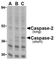 Description
Left: Western blot analysis of caspase-2 in Ramos cells with caspase-2 antibody at (A) 0.5, (B) 1, and (C) 2 µg/ml.
Below: Immunocytochemistry of caspase-2 in A-20 cells with caspase-2 antibody at 2 µg/ml.
Other Product Images
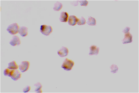 Source : Caspase-2 antibody was raised against a 16 amino acid peptide from the amino-terminus of human Caspase-2.
Purification : Affinity chromatography purified via peptide column
Clonality and Clone : This is a polyclonal antibody.
Host : Caspase-2 antibody was raised in rabbit. Please use anti-rabbit secondary antibodies.
Immunogen : Murine Casp-2 / ICH-1 / Nedd2 (N-Terminus) Peptide (Cat. No. 3447P)
Application : Caspase-2 antibody can be used for the detection of caspase-2 by Western blot at 1 µg/ml.Depending on cell lines or tissues used, other cleavage products may be observed.
Tested Application(s) : E, WB, ICC
Buffer : Antibody is supplied in PBS containing 0.02% sodium azide.
Blocking Peptide : Cat. No. 3447P - Casp-2 Peptide
Long-Term Storage : Caspase-2 antibody can be stored at 4ºC, stable for one year. As with all antibodies care should be taken to avoid repeated freeze thaw cycles. Antibodies should not be exposed to prolonged high temperatures.
Positive Control
1.Cat. No. 1225 - Ramos Cell Lysate
2.Cat. No. 1288 - A20 Cell Lysate
Species Reactivity :H, M
GI Number : 83300977
Accession Number : P42575
Short Description : (NT) a caspase induced by ER stress and DNA damage
References
1.Martinon F and Tschopp J. Inflammatory caspases: linking an intracellular innate immune system to autoinflammatory diseases. Cell 2004; 117:561-74.
2.Zhivotovsky B and Orrenius S. Caspase-2 funtion in response to DNA damage. Biochim. Biophys. Res. Comm. 2005; 331:859-67.
3.Kuida K, Lippke JA, Ku G, et al. Altered cytokine export and apoptosis in mice deficient in interleukin-1 beta converting enzyme. Science 1995; 267:2000-3.
4.Gracie JA, Robertson SE, and McInnes IB. Interleukin-18. J. Leukoc. Biol. 2003; 73:213-224.
Catalog# : 3447
Caspases are a family of cysteine proteases that can be divided into the apoptotic and inflammatory caspase subfamilies. Unlike the apoptotic caspases, members of the inflammatory subfamily are generally not involved in cell death but are associated with the immune response to microbial pathogens (reviewed in 1 and 2). Members of this subfamily include caspase-1, -4, -5, and -12 and can activate proinflammatory cytokines such as IL-1beta and IL-18 (3,4). Although phylogenetically similar to this subfamily, Caspase-2 is thought to be involved in stress-induced apoptosis (5). Caspase-2 has two major isoforms; overexpression on the long form results in apoptosis while that of the short form suppresses cell death (6).
Additional Names : Caspase-2 (NT), ICH-1, Nedd2
 Description
Left: Western blot analysis of caspase-2 in Ramos cells with caspase-2 antibody at (A) 0.5, (B) 1, and (C) 2 µg/ml.
Below: Immunocytochemistry of caspase-2 in A-20 cells with caspase-2 antibody at 2 µg/ml.
Other Product Images
 Source : Caspase-2 antibody was raised against a 16 amino acid peptide from the amino-terminus of human Caspase-2.
Purification : Affinity chromatography purified via peptide column
Clonality and Clone : This is a polyclonal antibody.
Host : Caspase-2 antibody was raised in rabbit. Please use anti-rabbit secondary antibodies.
Immunogen : Murine Casp-2 / ICH-1 / Nedd2 (N-Terminus) Peptide (Cat. No. 3447P)
Application : Caspase-2 antibody can be used for the detection of caspase-2 by Western blot at 1 µg/ml.Depending on cell lines or tissues used, other cleavage products may be observed.
Tested Application(s) : E, WB, ICC
Buffer : Antibody is supplied in PBS containing 0.02% sodium azide.
Blocking Peptide : Cat. No. 3447P - Casp-2 Peptide
Long-Term Storage : Caspase-2 antibody can be stored at 4ºC, stable for one year. As with all antibodies care should be taken to avoid repeated freeze thaw cycles. Antibodies should not be exposed to prolonged high temperatures.
Positive Control
1.Cat. No. 1225 - Ramos Cell Lysate
2.Cat. No. 1288 - A20 Cell Lysate
Species Reactivity :H, M
GI Number : 83300977
Accession Number : P42575
Short Description : (NT) a caspase induced by ER stress and DNA damage
References
1.Martinon F and Tschopp J. Inflammatory caspases: linking an intracellular innate immune system to autoinflammatory diseases. Cell 2004; 117:561-74.
2.Zhivotovsky B and Orrenius S. Caspase-2 funtion in response to DNA damage. Biochim. Biophys. Res. Comm. 2005; 331:859-67.
3.Kuida K, Lippke JA, Ku G, et al. Altered cytokine export and apoptosis in mice deficient in interleukin-1 beta converting enzyme. Science 1995; 267:2000-3.
4.Gracie JA, Robertson SE, and McInnes IB. Interleukin-18. J. Leukoc. Biol. 2003; 73:213-224.
Subscribe to:
Posts (Atom)
