Bio-Synthesis offers conjugation of peptides to a variety of immunogenic carrier proteins, and gel, for use as an immunogen or for affinity purification. A variety of immunogenic carrier proteins such as Keyhole Limpet Hemocyanin (KLH) and Diphtheria toxoid have been use in conjugation with several conjugation chemistries. We also offer coupling of peptides to various types of gel to create an immunosorbent, for positive or negative selection of macromolecular binding entries, such as antibodies.
Serum production packages also available with large selection of host animals, click here to explore more on our custom peptide antibody production.
Carrier: Keyhole Limpet Hemocyanin (KLH), Bovine Serum Albumin (BSA), Rabbit Serum Albumin (RSA), Ovalbumin, Thyroglobulin, or Human Gamma Globulin (HGG). Other proteins are available upon request.
Cross-Links: MBS (and other bi-functional linkers), which couple through sulfhydryl groups, and glutaraldehyde, which couples primarily through alpha, sigma & epsilon amino groups
* Amino Acid Analysis of Conjugates available upon request 
References:
[1] Lee, A.C., Powell, J.E., Tregear, G.W., Niall, H.D. and Stevens, V.C. (1980) A method for preparing b-hCG COOH peptide-carrier conjugates of predictable composition. Mol. Immunol. 17; 749-756.
[2] Posnett, D., McGrath, H., and Tam, J.P. A novel method for producing anti-peptide antibodies
, The Journal of Biological Chemistry, 263, 1719-1725 (1988),
[3] Tam, J.P., Synthetic peptide vaccine design: synthesis and properties of a high-density multiple antigenic peptide system, Proc. Natl. Acad, Sci., 85, 5409-5413 (1988).
[4] Wang, C. Y., Looney, D.J., Li, M.L., Walfield, A.M., Ye, J., Hosein, B., Tam, J.P. and Wong-Staal, F., Long-term high-titer neutralizing activity induced by octameric synthetic HIV antigen, Science, 254, 285-288. (1991)
ST3gal6 Antibody
Catalog# : 5169
Sialyltransferases catalyze the transfer of sialic acid from cytidine 5-prime monophospho-N-acetylneuraminic acid (CMP-NeuAc) to terminal positions of glycoprotein and glycolipid carbohydrate groups. Terminal NeuAc residues are key determinants of carbohydrate structures, such as the sialyl-Lewis X determinants, and are widely distributed in many cell types. However, cancer cells often express more heavily sialylated glycans on their cell surface and this feature sometimes correlates with invasiveness. In contrast, expression of ST3gal6, a member of the sialyltransferase family that sialylates type II lactosamine structures on glycoproteins and glycolipids, was found to be significantly decreased by hypermethylation of the gene in gastrointestinal cancer. At least three isoforms of ST3gal6 are known to exist.
Additional Names : ST3gal6, Alpha2,3-sialyltransferase 6, ST3Gal VI, siat10
 Description
Left: Western blot analysis of ST3gal6 in HeLa cell lysate with ST3gal6 antibody at (A) 1 and (B) 2 µg/ml.
Below: Immunocytochemistry of ST3gal6 in HeLa cells with ST3gal6 antibody at 5 μg/ml.
Other Product Images
 Source : ST3gal6 antibody was raised against a 16 amino acid peptide from near the carboxy terminus of human ST3gal6.
Purification : Affinity chromatography purified via peptide column
Clonality and Clone : This is a polyclonal antibody.
Host : ST3gal6 antibody was raised in rabbit. Please use anti-rabbit secondary antibodies.
Application : ST3gal6 antibody can be used for detection of ST3gal6 by Western blot at 1 - 2 µg/ml.
Tested Application(s) : E, WB
Buffer : Antibody is supplied in PBS containing 0.02% sodium azide.
Blocking Peptide : Cat.No. 5169P - ST3gal6 Peptide
Long-Term Storage : ST3gal6 antibody can be stored at 4ºC, stable for one year. As with all antibodies care should be taken to avoid repeated freeze thaw cycles. Antibodies should not be exposed to prolonged high temperatures.
Positive Control
1. Cat. No. 1201 - HeLa Cell Lysate
Species Reactivity :H, M, R
GI Number : 5174697
Accession Number : NP_006091
Short Description : Alpha2,3-sialyltransferase 6
References
1. Dall’Olio F and Chiricolo M. Sialyltransferases in cancer. Glycoconj. J. 2001; 18:841-50.
2. Okajima T, Fukumoto S, Miyazaki H, et al. Molecular cloning of a novel alpha2,3-sialyltransferase (ST3Gal VI) that sialylates type II lactosamine structures on glycoproteins and glycolipids. J. Biol. Chem. 1999; 274:11479-86.
3. Kawamura YI, Toyota M, Kawashima R, et al. DNA hypermethylation contributes to incomplete synthesis of carbohydrate determinants in gastrointestinal cancer. Gasatroenterology 2008; 135:142-51.
PALB2 Antibody
Catalog# : 5275
The breast cancer early onset proteins BRCA1 and BRCA2 are central to the repair of DNA damage by homologous recombination and are strongly associated with inherited breast and ovarian cancer. The protein PALB2 is required for the localization of BRCA2 to sites of DNA damage and like BRCA1 and 2 is a breast cancer susceptibility gene. PALB2 is thought to function through directly binding to BRCA1, which allows the PALB2 protein to organize BRCA2 and the recombinase RAD51 at the site of DNA damage. Analysis of the BRCA1-PALB2-BRCA2-RAD51network may allow predictions to be made of the responsiveness of a particular tumor to therapeutic treatments. At least four isoforms of PALB2 are known to exist.
Additional Names : PALB2,Partner and localizer of BRCA2, FANCN
 Description
Left: Western blot analysis of PALB2 in SK-N-SH cell lysate with PALB2 antibody at (A) 1 and (B) 2 µg/ml.
Source : PALB2 antibody was raised against a 15 amino acid peptide from near the carboxy terminus of human PALB2.
Purification : Affinity chromatography purified via peptide column
Clonality and Clone : This is a polyclonal antibody.
Host : PALB2 antibody was raised in rabbit. Please use anti-rabbit secondary antibodies.
Application : PALB2 antibody can be used for detection of PALB2 by Western blot at 1 - 2 µg/ml.
Tested Application(s) : E, WB
Buffer : Antibody is supplied in PBS containing 0.02% sodium azide.
Blocking Peptide : Cat.No. 5275P - PALB2 Peptide
Long-Term Storage : PALB2 antibody can be stored at 4ºC, stable for one year. As with all antibodies care should be taken to avoid repeated freeze thaw cycles. Antibodies should not be exposed to prolonged high temperatures.
Positive Control
1. Cat. No. 1220 - SK-N-SH Cell Lysate
Species Reactivity :H, M, R
GI Number : 74727919
Accession Number : Q86YC2
Short Description : Partner and localizer of BRCA2
References
1. Greenberg RA. Recognition of DNA double strand breaks by the BRCA1 tumor suppressor network. Chromosoma 2008; 117:305-17.
2. Xia B, Sheng Q, Nakanishi K, et al. Control of BRCA2 cellular and clinical functions by a nuclear partner, PALB Mol. Cell 2007; 22:719-29.
3. Rahman N, Seal S, Thompson D, et al. PALB2, which encodes a BRCA2-interacting protein, is a breast cancer susceptibility gene. Nat. Genet. 2007; 39:165-7.
4. Zhang F, Fan Q, Ren K, et al. PALB2 functionally connects the breast cancer susceptibility proteins BRCA1 and BRCA2. Mol. Cancer. Res. 2009; 7:1110-8.
OTUD4 Antibody
Catalog# : 5075
OTUD4, also known as HIV-1 induced protein HIN-1, is a member of the OTU (ovarian tumor) domain containing cysteine protease superfamily, in which the OUT domain generally confers deubiquitinase activity. At least three isoforms of OTUD4 are known to exist, and the smallest of these isoforms are only expressed in HIV-1-infected cells (provided by RefSeq). This antibody is predicted to not cross-react with other members of the OTUD family.
Additional Names : OTUD4, OTU domain containing 4, HIN1, DUBA6, HSHIN1
 Description
Left: Western blot analysis of OTUD4 in Daudi cell lysate with OTUD4 antibody at (A) 0.25 and (B) 0.5 µg/ml.
Below: Immunocytochemistry of OTUD4 in Daudi cells with OTUD4 antibody at 2.5 μg/ml.
Other Product Images
 Source : OTUD4 antibody was raised against an 18 amino acid peptide near the carboxy terminus of human OTUD4.
Purification : Affinity chromatography purified via peptide column
Clonality and Clone : This is a polyclonal antibody.
Host : OTUD4 antibody was raised in rabbit. Please use anti-rabbit secondary antibodies.
Application : OTUD4 antibody can be used for detection of OTUD4 by Western blot at 0.25 – 0.5 µg/ml.
Tested Application(s) : E, WB
Buffer : Antibody is supplied in PBS containing 0.02% sodium azide.
Blocking Peptide : Cat.No. 5075P - OTUD4 Peptide
Long-Term Storage : OTUD4 antibody can be stored at 4ºC, stable for one year. As with all antibodies care should be taken to avoid repeated freeze thaw cycles. Antibodies should not be exposed to prolonged high temperatures.
Positive Control
1. Cat. No. 1224 - Daudi Cell Lysate
Species Reactivity :H, M, R
GI Number : 156630992
Accession Number : NP_001096123
Short Description : OTU domain containing 4
References
1. Borodovsky A, Ovaa H, Kolli N, et al. Chemistry-based functional genomics reveals novel members of the deubiquitinating enzyme family. Chem. Biol. 2002; 10:1149-59.
OCC-1 Antibody
Catalog# : 4987
OCC-1 (Overexpressed in colon carcinoma-1) was initially identified as a novel human cDNA overexpressed in a colon carcinoma cell line. Elevated levels of OCC-1 mRNA were observed in three of eight colon carcinomas, suggesting that OCC-1 may be a hallmark of only a subset of colon carcinomas. It is highly expressed in skeletal muscle, kidney, placenta, and pancreas tissues, with low to no expression in heart, brain, lung and liver tissues. At least two different OCC-1 mRNA species are known to exist. In vitro transcription and translation of the OCC-1 cDNA yields polypeptides less than 9 kDa. Despite this, OCC-1 from cell lines is detected at ~28 kDa in SDS-PAGE.
Additional Names : OCC-1 (CT), Overexpressed in colon carcinoma-1
 Description
Left: Western blot analysis of OCC-1 in 293 cell lysate with OCC-1 antibody at (A) 2 µg/ml and (B) 4 µg/ml.
Below: Immunohistochemistry of OCC-1 in human spleen tissue with OCC-1 antibody at 2.5 µg/ml.
Other Product Images
 Source : OCC-1 antibody was raised against a 7 amino acid peptide near the carboxy terminus of human OCC-1.
Purification : Affinity chromatography purified via peptide column
Clonality and Clone : This is a polyclonal antibody.
Host : OCC-1 antibody was raised in rabbit. Please use anti-rabbit secondary antibodies.
Application : OCC-1 antibody can be used for detection of OCC-1 by Western blot at 2 – 4 µg/ml.
Tested Application(s) : E, WB, IHC
Buffer : Antibody is supplied in PBS containing 0.02% sodium azide.
Blocking Peptide : Cat.No. 4987P - OCC-1 Peptide
Long-Term Storage : OCC-1 antibody can be stored at 4ºC, stable for one year. As with all antibodies care should be taken to avoid repeated freeze thaw cycles. Antibodies should not be exposed to prolonged high temperatures.
Positive Control
1. Cat. No. 1210 - 293 Cell Lysate
Species Reactivity :H
GI Number : 112180576
Accession Number : AAH61920
Short Description : (CT) Overexpressed in colon carcinoma-1
References
1. Pibouin L, Villaudy J, Ferbus D, et al. Cloning of the mRNA of overexpression in colon-carcinoma-1: a sequence overexpressed in a subset of colon carcinomas. Cancer Genet. Cytogenet. 2002; 133:55-60.
OCC-1 Antibody
Catalog# : 4997
OCC-1 (Overexpressed in colon carcinoma-1) was initially identified as a novel human cDNA overexpressed in a colon carcinoma cell line. Elevated levels of OCC-1 mRNA were observed in three of eight colon carcinomas, suggesting that OCC-1 may be a hallmark of only a subset of colon carcinomas. It is highly expressed in skeletal muscle, kidney, placenta, and pancreas tissues, with low to no expression in heart, brain, lung and liver tissues. At least two different OCC-1 mRNA species are known to exist. In vitro transcription and translation of the OCC-1 cDNA yields polypeptides less than 9 kDa. Despite this, OCC-1 from cell lines is detected at ~28 kDa in SDS-PAGE.
Additional Names : OCC-1 (NT), Overexpressed in colon carcinoma-1
 Description
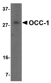
Left: Western blot analysis of OCC-1 in human spleen tissue lysate with OCC-1 antibody at 2 µg/ml.
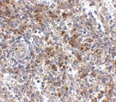
Below: Immunohistochemistry of OCC-1 in human spleen tissue with OCC-1 antibody at 2.5 μg/ml.
Other Product Images
 Source : OCC-1 antibody was raised against a 16 amino acid peptide near the amino terminus of human OCC-1.
Purification : Affinity chromatography purified via peptide column
Clonality and Clone : This is a polyclonal antibody.
Host : OCC-1 antibody was raised in rabbit. Please use anti-rabbit secondary antibodies.
Application : OCC-1 antibody can be used for detection of OCC-1 by Western blot at 2 – 4 µg/ml.
Tested Application(s) : E, WB
Buffer : Antibody is supplied in PBS containing 0.02% sodium azide.
Blocking Peptide : Cat.No. 4997P - OCC-1 Peptide
Long-Term Storage : OCC-1 antibody can be stored at 4ºC, stable for one year. As with all antibodies care should be taken to avoid repeated freeze thaw cycles. Antibodies should not be exposed to prolonged high temperatures.
Positive Control
1. Cat. No. 1210 - 293 Cell Lysate
Species Reactivity :H
GI Number : 112180576
Accession Number : AAH61920
Short Description : (NT) Overexpressed in colon carcinoma-1
References
1. Pibouin L, Villaudy J, Ferbus D, et al. Cloning of the mRNA of overexpression in colon-carcinoma-1: a sequence overexpressed in a subset of colon carcinomas. Cancer Genet. Cytogenet. 2002; 133:55-60.
Bio-Synthesis prides itself in helping scientists worldwide choose the most efficient and economical process for their custom peptide synthesis project. We continue to build on over 20 years of experience in technology development and contract manufacturing services with the capacity to produce peptide quantities from milligrams to kilograms. Our production method include Boc, CBZ and Fmoc, solid phase synthesis to liquid phase chemistry or the combinations of both...
For large scale synthesis, call 800-227-0627 or write to us at biosyn@biosyn.com.
General Features:
* High Control Production Process
Our synthesis reagents used to produce the oligomer that are carefully checked to ensure quality and reproducibility.
* Dedicated Staff and Facilities
We have a strict Quality Management System to ensure all production is handled with the utmost care.
We believe in building strong relationships with our customers and encourage dialogue to ensure that correct synthesis specifications are achieved.
* Fexible Tailor-Made Solutions
With over 20 years of experience in custom synthesis, the knowledge and expertise of our chemists allow us the flexibility to tailor-make our customer's precise requirements.
Synthesis Capabilities:
- Scale range from mg to kg
- Dedicated synthesis apparatus as requested
- Extensive project portfolios of synthesis modifications
Quality Control and Assurance:
- Purification by several preparative HPLC lines
- Dedicated purification column as requested
- Characterization by MALDI-TOF mass spectrometry
MACC1 Antibody
Catalog# : 5197
Metastasis associated in colon cancer 1 (MACC1) is a key regulator of the hepatocyte growth factor (HGF)-HGF receptor (MET) pathway, which is involved in cellular growth, epithelial-mesenchymal transition, angiogenesis, cell motility, invasiveness, and metastasis. MACC1 protein consists of four domains: ZU5, SH3, and two C-terminal death domains (DD). Expression of MACC1 was found significantly upregulated in malignant tissues (colon cancer of all stages as well as liver and lung metastases) compared to normal tissues or adenomas. MACC1 represents an early and crucial prognostic indicator for colon cancer metastasis that is independent of age, sex, tumor infiltration, nodal status, and lymph vessel invasion. Besides its involvement in signal transduction with the MET receptor, MACC1 also links MET signaling and apoptosis. MACC1 may also be an important therapeutic target for colorectal cancer treatment. At least two isoforms of MACC1 are known to exist.
Additional Names : MACC1 (IN), Metastasis associated in colon cancer 1
 Description
Left: Western blot analysis of MACC1 in mouse liver tissue lysate with MACC1 antibody at (A) 1 and (B) 2 µg/ml.
Below: Immunohistochemistry of MACC1 in human liver tissue with MACC1 antibody at 2.5 μg/ml.
Other Product Images
 Source : MACC1 antibody was raised against a 17 amino acid peptide near the center of human MACC1.
Purification : Affinity chromatography purified via peptide column
Clonality and Clone : This is a polyclonal antibody.
Host : MACC1 antibody was raised in rabbit. Please use anti-rabbit secondary antibodies.
Application : MACC1 antibody can be used for detection of MACC1 by Western blot at 1-2 µg/ml.
Tested Application(s) : E, WB
Buffer : Antibody is supplied in PBS containing 0.02% sodium azide.
Blocking Peptide : Cat.No. 5197P - MACC1 Peptide
Long-Term Storage : MACC1 antibody can be stored at 4ºC, stable for one year. As with all antibodies care should be taken to avoid repeated freeze thaw cycles. Antibodies should not be exposed to prolonged high temperatures.
Positive Control
1. Cat. No. 1404 - Mouse Liver Tissue Lysate
Species Reactivity :H, M
GI Number : 32997046
Accession Number : AJ313524
Short Description : (IN) Metastasis associated in colon cancer 1
References
1. Stein U, Walther W, Arlt F, et al. MACC1, a newly identified key regulator of HGF-MET signaling, predicts colon cancer metastasis. Nat. Med. 2009; 15:59-67.
2. Boardman LA. Overexpression of MACC1 leads to downstream activation of HGF/MET and potentiates metastasis and recurrence of colorectal cancer. Genome Med. 2009; 1:36.
3. Stein U, Smith J, Walther W, et al. MACC1 controls Met: what a difference an Sp1 site makes. Cell Cycle 2009; 8:2467-9.
4. Arlt F and Stein U. Colon cancer metastasis: MACC1 and Met as metastatic pacemakers. Int. J. Biochem. Cell Biol. 2009; epub.
JMJD2A Antibody
Catalog# : 5373
Members of the Jumonji domain 2 (JMJD2) family contain a JmjN domain, a JmjC domain, a JD2H domain, two TUDOR domains, and two PHD-type zinc fingers. The first member of this group, JMJD2A, is widely expressed in human tissues and cell lines and functions as a trimethylation-specific demethylase, converting specific trimethylated histone residues to the dimethylated form, and as a transcriptional repressor. JMJD2A can also form a complex with the androgen receptor (AR), a transcription factor that is pivotal for the development of prostate cancer. Overexpression of JMJD2A stimulates AR function and this stimulation is dependent on JMJD2A catalytic activity, suggesting that JMJD2A might be a critical protein with roles in cell proliferation and oncogenesis.
Additional Names : JMJD2A, Jumonji domain-containing protein 2A, Lysine-specific demethylase 4A, KDM4A
 Description
Left: Western blot analysis of JMJD2A in rat liver tissue lysate with JMJD2A antibody at (A) 1 and (B) 2 µg/ml.
Source : JMJD2A antibody was raised against a 14 amino acid peptide from near the center of human JMJD2A.
Purification : Affinity chromatography purified via peptide column
Clonality and Clone : Polyclonal
Host : JMJD2A antibody was raised in rabbit. Please use anti-rabbit secondary antibodies.
Application : JMJD2A antibody can be used for detection of JMJD2A by Western blot at 1 - 2 µg/ml.
Tested Application(s) : E, WB
Buffer : Antibody is supplied in PBS containing 0.02% sodium azide.
Blocking Peptide : Cat.No. 5373P - JMJD2A Peptide
Long-Term Storage : JMJD2A antibody can be stored at 4ºC, stable for one year. As with all antibodies care should be taken to avoid repeated freeze thaw cycles. Antibodies should not be exposed to prolonged high temperatures.
Positive Control
1. Cat. No. 1464 - Rat Liver Tissue Lysate
Species Reactivity :H, M, R
GI Number : 55665236
Accession Number : CAH71021
Short Description : Lysine-specific demethylase 4A
References
1. Gray SG, Iglesias AH, Lizcano F, et al. Functional characterization of JMJD2A, a histone deacetylase- and retinoblastoma-binding protein. J. Biol. Chem. 2005; 31:28507-18.
2. Shin S and Janknecht R. Activation of androgen receptor by histone demethylases JMJD2A and JMJD2D. Biochem. Biophys. Res. Comm. 2007; 359:742-6
JMJD1B Antibody
Catalog# : 5369
The jumonji domain containing 1B protein (JMJD1B) was originally discovered as a candidate for the myeloid leukemia tumor suppressor gene. Besides CD34+ cells and acute myeloid leukemia (AML) cell lines, JMJD1B mRNA is highly expressed in liver, heart, kidney, skeletal muscle, and placenta tissues. The JMJD1B gene is localized to a region of chromosome 5q31, which is frequently deleted in myeloid leukemias and myelodysplasias and expression of JMJD1B in a del(5q) cell line results in suppression of clonogenic growth suggesting that JMJD1B may function as a tumor suppressor. In contrast, JMJD1B gene copy number and mRNA expression level was increased in several non-small cell lung cancers indicating that the role of JMJD1B in cancer formation and progression is more complex than originally postulated. At least three isoforms of JMJD1B are known to exist. This antibody will not cross-react with JMJD1A or JMJD1C.
Additional Names : JMJD1B,5qNCA, Lysine-specific demethylase 3B, KDM3B
 Description
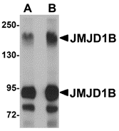
Left: Western blot analysis of JMJD1B in rat liver tissue lysate with JMJD1B antibody at (A) 1 and (B) 2 µg/ml.
Source : JMJD1B antibody was raised against a 20 amino acid peptide from near the center of human JMJD1B.
Purification : Affinity chromatography purified via peptide column
Clonality and Clone : Polyclonal
Host : JMJD1B antibody was raised in rabbit. Please use anti-rabbit secondary antibodies.
Application : JMJD1B antibody can be used for detection of JMJD1B by Western blot at 1 - 2 µg/ml.
Tested Application(s) : E, WB
Buffer : Antibody is supplied in PBS containing 0.02% sodium azide.
Blocking Peptide : Cat.No. 5369P - JMJD1B Peptide
Long-Term Storage : JMJD1B antibody can be stored at 4ºC, stable for one year. As with all antibodies care should be taken to avoid repeated freeze thaw cycles. Antibodies should not be exposed to prolonged high temperatures.
Positive Control
1. Cat. No. 1464 - Rat Liver Tissue Lysate
Species Reactivity :H, M, R
GI Number : 119582545
Accession Number : EAW62141
Short Description : Lysine-specific demethylase 3B
References
1. Lai F, Godley LA, Fernald AA, et al. cDNA cloning and genomic structure of three genes localized to human chromosome band 5q31 encoding potential nuclear proteins. Genomics 2000; 70:123-30.
2. Hu Z, Gomes I, Horrigan SK, et al. A novel nuclear protein, 5qNCA (LOC51780) is a candidate for the myeloid leukemia tumor suppressor gene on chromosome band q31. Oncogene 2001; 20:6946-54.
3. Baik S-H, Jee B-K, Choi J-S, et al. DNA profiling by array comparative genomic hybridization (CGH) of peripheral blood mononuclear cells (PBMC) and tumor tissue cell in non-small cell lung cancer (NSCLC). Mol. Biol. Rep. 2009; 36:1767-78.


