Tuesday, May 31, 2011
DNA Testing Centers
Bio-Synthesis, an international provider of DNA testing services including: government agencies, private institutions, or individuals who need DNA testing for various reasons. We offer comprehensive services such as paternity, maternity and other kinship identification. Our facilities are operated under AABB accreditation guidelines for all your private or legal DNA tests.
DNA Testing Centers
A genealogical DNA test examines the nucleotides at specific locations on a person's DNA for genetic genealogy purposes. The test results are meant to have no informative medical value and do not determine specific genetic diseases or disorders (see possible exceptions in Medical information below); they are intended only to give genealogical information. Genealogical DNA tests generally involve comparing the results of living individuals as opposed to obtaining samples from deceased people.
Mitochondrial DNA (mtDNA) testing
A person's maternal ancestry can be traced using his or her mitochondrial DNA (mtDNA). The DNA in the human mitochondria is passed down by the mother unchanged. One exception, which was linked to infertility, has been shown. Additionally, some people cite paternal mtDNA transmission as invalidating mtDNA testing, but this is not considered problematic in scholarly population genetics studies or genetic genealogy.
DNA Paternity Testing
Lowest price/highest accuracy guarantee! - paternity test prices. See our secret paternity tests (using a toothbrush, ear wax, etc.)
For more information visit: www.biosyn.com
ACVR1 Antibody
Catalog#:4791
Activins are dimeric growth and differentiation factors which belong to the transforming growth factor-beta (TGF-beta) superfamily of structurally related signaling proteins. Activins signal through a heteromeric complex of receptor serine kinases which include at least two type I and two type II receptors. Unlike ACVR1B and ACVR1C, ACVR1, also known as activin receptor-like kinase 2 (ALK2), can not transduce activin-mediated signaling, but will transduce BMP and Mullerian inhibiting substance (MIS) group signaling. It is thought that ACVR1 also inhibits activin signaling by blocking the binding of activin to its type II receptor. Recent studies indicate that genetic variation in ACVR1 is associated with polycystic ovary syndrome, suggesting that ACVR1 signaling contributes to disturbed folliculogenesis in these patients. At least four isoforms of ACVR1 are known to exist. This antibody is predicted to have no cross-reactivity to ACVR1B or ACVR1C.
Additional Names: ACVR1,Activin A receptor type IA, Activin receptor-like kinase 2, Activin receptor type 1A precursor, ACTR-IA, ACVRLK2, ALK2, ALK-2
 Description
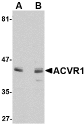
Left: Western blot analysis of ACVR1 in A549 cell lysate with ACVR1 antibody at 1 µg/ml in (A) the absence and (B) the presence of blocking peptide.
Source:ACVR1 antibody was raised against a 14 amino acid peptide near the amino terminus of the human ACVR1.
Purification: Affinity chromatography purified via peptide column
Clonality and Clone: This is a polyclonal antibody.
Host: ACVR1 antibody was raised in rabbit.
Please use anti-rabbit secondary antibodies.
Application: ACVR1 antibody can be used for detection of ACVR1 by Western blot at 1 – 2 µg/ml.
Tested Application(s): E, WB
Buffer: Antibody is supplied in PBS containing 0.02% sodium azide.
Blocking Peptide:Cat.No. 4791P - ACVR1 Peptide
Long-Term Storage: ACVR1 antibody can be stored at 4ºC, stable for one year. As with all antibodies care should be taken to avoid repeated freeze thaw cycles. Antibodies should not be exposed to prolonged high temperatures.
Positive Control:
1. Cat. No. 1203 - A549 Cell Lysate
Species Reactivity: H, M
GI Number: 4501895
Accession Number: NP_001096
Short Description: Activin receptor-like kinase 2
References
1. Tsuchida K, Sawchenko PN, Nishikawa S, et al. Molecular cloning of a novel type I receptor serine/threonine kinase for the TGF beta superfamily from rat brain. Mol. Cell. Neurosci. 1996; 76:467–78.
2. ten Dijke P, Yamashita H, Sampath TK, et al. Identification of type I receptors for osteogenic protein-1 and bone morphogenetic protein-4. J. Biol. Chem. 1994; 269:16985-8.
3. Clarke TR, Hoshiya Y, Yi SE, et al. Mullerian inhibiting substance signaling uses a BMP-like pathway mediated by ALK2 and induces Smad6 expression. Mol. Endocrinol. 2001; 15:946-59.
4. Renlund N, O’Neill FH, Zhang L, et al. Activin receptor-like kinase-2 inhibits activin signaling by blocking the binding of activin to its type II receptor. J. Endocrinol. 2007; 195:95-103.
DescriptionLeft: Western blot analysis of ACVR1 in A549 cell lysate with ACVR1 antibody at 1 µg/ml in (A) the absence and (B) the presence of blocking peptide.
Source:ACVR1 antibody was raised against a 14 amino acid peptide near the amino terminus of the human ACVR1.
Purification: Affinity chromatography purified via peptide column
Clonality and Clone: This is a polyclonal antibody.
Host: ACVR1 antibody was raised in rabbit.
Please use anti-rabbit secondary antibodies.
Application: ACVR1 antibody can be used for detection of ACVR1 by Western blot at 1 – 2 µg/ml.
Tested Application(s): E, WB
Buffer: Antibody is supplied in PBS containing 0.02% sodium azide.
Blocking Peptide:Cat.No. 4791P - ACVR1 Peptide
Long-Term Storage: ACVR1 antibody can be stored at 4ºC, stable for one year. As with all antibodies care should be taken to avoid repeated freeze thaw cycles. Antibodies should not be exposed to prolonged high temperatures.
Positive Control:
1. Cat. No. 1203 - A549 Cell Lysate
Species Reactivity: H, M
GI Number: 4501895
Accession Number: NP_001096
Short Description: Activin receptor-like kinase 2
References
1. Tsuchida K, Sawchenko PN, Nishikawa S, et al. Molecular cloning of a novel type I receptor serine/threonine kinase for the TGF beta superfamily from rat brain. Mol. Cell. Neurosci. 1996; 76:467–78.
2. ten Dijke P, Yamashita H, Sampath TK, et al. Identification of type I receptors for osteogenic protein-1 and bone morphogenetic protein-4. J. Biol. Chem. 1994; 269:16985-8.
3. Clarke TR, Hoshiya Y, Yi SE, et al. Mullerian inhibiting substance signaling uses a BMP-like pathway mediated by ALK2 and induces Smad6 expression. Mol. Endocrinol. 2001; 15:946-59.
4. Renlund N, O’Neill FH, Zhang L, et al. Activin receptor-like kinase-2 inhibits activin signaling by blocking the binding of activin to its type II receptor. J. Endocrinol. 2007; 195:95-103.
Aak1 Antibody
Catalog#:4841
AP2-associated protein kinase 1 (Aak1) is a member of the Ark1/Prk1 subfamily of Ser/Thr protein kinases that are thought to regulate endocytosis by phosphorylating the accessory endocytic components. Aak1 interacts with and phosphorylates the mu2 subunit of the AP-2 complex, which promotes binding of the AP-2 to tyrosine based (Yxxf) internalization motif-containing receptors and subsequent receptor endocytosis. At least two isoforms of Aak1 are known to exist; the longer isoform contains an extended carboxy-terminus that contains an additional clathrin-binding domain. Overexpression of this long isoform or Aak1 depletion by RNA interference impairs transferrin recycling from the early/sorting endosome, suggesting that Aak1 functions at multiple steps of the endosomal pathway by regulating transferrin internalization and its recycling back to the plasma membrane.
Additional Names: Aak1 (NT), AP2-associated protein kinase 1
Description
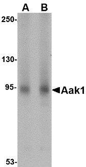
Left: Western blot analysis of Aak1 in A-20 lysate with Aak1 antibody at (A) 1 and (B) 2 µg/ml.
Below: Immunocytochemistry of Aak1 in A-20 cells with Aak1 antibody at 5 µg/ml.
Other Product Images
 Source:Aak1 antibody was raised against a 20 amino acid peptide near the amino terminus of the human Aak1.
Purification: Affinity chromatography purified via peptide column
Clonality and Clone: This is a polyclonal antibody.
Host: Aak1 antibody was raised in rabbit.
Please use anti-rabbit secondary antibodies.
Application: Aak1 antibody can be used for detection of Aak1 by Western blot at 1 – 2 µg/ml.
Tested Application(s): E, WB, ICC
Buffer: Antibody is supplied in PBS containing 0.02% sodium azide.
Blocking Peptide:Cat.No. 4841P - Aak1 Peptide
Long-Term Storage: Aak1 antibody can be stored at 4ºC, stable for one year. As with all antibodies care should be taken to avoid repeated freeze thaw cycles. Antibodies should not be exposed to prolonged high temperatures.
Positive Control:
1. Cat. No. 1466 - Rat Spleen Tissue Lysate
Species Reactivity: H, M, R
GI Number: 148277037
Accession Number: NP_055726
Short Description: (NT) AP2-associated protein kinase 1
References
1. Connor SD and Schmid SL. Identification of an adaptor-associated kinase, AAK1, as a regulator of clathrin-mediated endocytosis. J. Cell Biol. 2002; 156:921-9.
2. Smythe E and Ayscough KR. The Ark1/Prk1 family of protein kinases. Regulators of endocytosis and the actin skeleton. EMBO Rep. 2003; 4:246-51.
3. Ricotta D, Connor SD, Schmid SL, et al. Phosphorylation of the AP2 m2 subunit by AAK1 mediates high affinity binding to membrane protein sorting signals. J. Cell Biol. 2002; 156:791-5.
4. Connor SD and Henderson DM. A novel AAK1 splice variant functions at multiple steps of the endocytic pathway. Mol. Biol. Cell 2007; 18:2698-706.
Description
Left: Western blot analysis of Aak1 in A-20 lysate with Aak1 antibody at (A) 1 and (B) 2 µg/ml.
Below: Immunocytochemistry of Aak1 in A-20 cells with Aak1 antibody at 5 µg/ml.
Other Product Images
Source:Aak1 antibody was raised against a 20 amino acid peptide near the amino terminus of the human Aak1.Purification: Affinity chromatography purified via peptide column
Clonality and Clone: This is a polyclonal antibody.
Host: Aak1 antibody was raised in rabbit.
Please use anti-rabbit secondary antibodies.
Application: Aak1 antibody can be used for detection of Aak1 by Western blot at 1 – 2 µg/ml.
Tested Application(s): E, WB, ICC
Buffer: Antibody is supplied in PBS containing 0.02% sodium azide.
Blocking Peptide:Cat.No. 4841P - Aak1 Peptide
Long-Term Storage: Aak1 antibody can be stored at 4ºC, stable for one year. As with all antibodies care should be taken to avoid repeated freeze thaw cycles. Antibodies should not be exposed to prolonged high temperatures.
Positive Control:
1. Cat. No. 1466 - Rat Spleen Tissue Lysate
Species Reactivity: H, M, R
GI Number: 148277037
Accession Number: NP_055726
Short Description: (NT) AP2-associated protein kinase 1
References
1. Connor SD and Schmid SL. Identification of an adaptor-associated kinase, AAK1, as a regulator of clathrin-mediated endocytosis. J. Cell Biol. 2002; 156:921-9.
2. Smythe E and Ayscough KR. The Ark1/Prk1 family of protein kinases. Regulators of endocytosis and the actin skeleton. EMBO Rep. 2003; 4:246-51.
3. Ricotta D, Connor SD, Schmid SL, et al. Phosphorylation of the AP2 m2 subunit by AAK1 mediates high affinity binding to membrane protein sorting signals. J. Cell Biol. 2002; 156:791-5.
4. Connor SD and Henderson DM. A novel AAK1 splice variant functions at multiple steps of the endocytic pathway. Mol. Biol. Cell 2007; 18:2698-706.
Aak1 Antibody
Catalog#:4831
AP2-associated protein kinase 1 (Aak1) is a member of the Ark1/Prk1 subfamily of Ser/Thr protein kinases that are thought to regulate endocytosis by phosphorylating the accessory endocytic components. Aak1 interacts with and phosphorylates the mu2 subunit of the AP-2 complex, which promotes binding of the AP-2 to tyrosine based (Yxxf) internalization motif-containing receptors and subsequent receptor endocytosis. At least two isoforms of Aak1 are known to exist; the longer isoform contains an extended carboxy-terminus that contains an additional clathrin-binding domain. Overexpression of this long isoform or Aak1 depletion by RNA interference impairs transferrin recycling from the early/sorting endosome, suggesting that Aak1 functions at multiple steps of the endosomal pathway by regulating transferrin internalization and its recycling back to the plasma membrane.
Additional Names: Aak1 (CT), AP2-associated protein kinase 1
Description
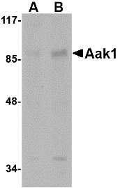
Left: Western blot analysis of Aak1 in A-20 lysate with Aak1 antibody at (A) 1 and (B) 2 µg/m
Source:Aak1 antibody was raised against a 18 amino acid peptide near the carboxy terminus of the human Aak1.
Purification: Affinity chromatography purified via peptide column
Clonality and Clone: This is a polyclonal antibody.
Host: Aak1 antibody was raised in rabbit.
Please use anti-rabbit secondary antibodies.
Application: Aak1 antibody can be used for detection of Aak1 by Western blot at 1 – 2 µg/ml.
Tested Application(s): E, WB
Buffer: Antibody is supplied in PBS containing 0.02% sodium azide.
Blocking Peptide:Cat.No. 4831P - Aak1 Peptide
Long-Term Storage: Aak1 antibody can be stored at 4ºC, stable for one year. As with all antibodies care should be taken to avoid repeated freeze thaw cycles. Antibodies should not be exposed to prolonged high temperatures.
Positive Control:
1. Cat. No. 1288 - A20 Cell Lysate
Species Reactivity: H, M, R
GI Number: 148277037
Accession Number: NP_055726
Short Description: (CT) AP2-associated protein kinase 1
References
1. Connor SD and Schmid SL. Identification of an adaptor-associated kinase, AAK1, as a regulator of clathrin-mediated endocytosis. J. Cell Biol. 2002; 156:921-9.
2. Smythe E and Ayscough KR. The Ark1/Prk1 family of protein kinases. Regulators of endocytosis and the actin skeleton. EMBO Rep. 2003; 4:246-51.
3. Ricotta D, Connor SD, Schmid SL, et al. Phosphorylation of the AP2 m2 subunit by AAK1 mediates high affinity binding to membrane protein sorting signals. J. Cell Biol. 2002; 156:791-5.
4. Connor SD and Henderson DM. A novel AAK1 splice variant functions at multiple steps of the endocytic pathway. Mol. Biol. Cell 2007; 18:2698-706.
4E-BP1 Antibody
Catalog#:3513
The translation of mRNA in eukaryotic cells is regulated by the presence of amino acids through multiple mechanisms (reviewed in 1). One such mechanism involves the evolutionarily conserved serine/threonine kinase TOR (Target of rapamycin, also known as mTOR), which regulates cell growth and cell cycle through its ability to integrate signals from nutrient levels and growth factors (reviewed in 2). One downstream target of TOR is the eukaryotic initiation factor 4E binding protein 1 (4E-BP1) whose phosphorylation prevents its association with eIF4E, preferentially stimulating translation of mRNAs containing long, highly structured 5’-UTRs (1). Rapamycin inhibits TOR resulting in reduced cell growth and reduced rates of cell cycle and cell proliferation (reviewed in 3), at least in part by inhibiting the activity of TOR towards 4E-BP1.
Additional Names: 4E-BP1, initiation factor 4E binding protein 1, sample
 Description
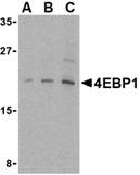
Left: Western blot analysis of 4E-BP1 in 3T3 cell lysate with 4E-BP1 antibody at (A) 2.5, (B) 5 and (C) 10 µg/ml.
Below: Immunocytochemistry of 4E-BP1 in 3T3 cells with 4E-BP1 antibody at 2 µg/ml.
Other Product Images
Source:4E-BP1 antibody was raised against a 14 amino acid peptide from near the carboxy-terminus of human 4E-BP1.
Purification: Affinity chromatography purified via peptide column
Clonality and Clone: This is a polyclonal antibody.
Host: 4E-BP1 antibody was raised in rabbit.
Please use anti-rabbit secondary antibodies.
Immunogen: Human 4E-BP1 / initiation factor 4E binding protein 1 Peptide (Cat. No. 3513P)
Application: 4E-BP1 antibody can be used for the detection of 4E-BP1 by Western blot at 2.5 – 5 µg/ml.
Tested Application(s): E, WB, ICC
Buffer: Antibody is supplied in PBS containing 0.02% sodium azide.
Blocking Peptide:Cat. No. 3513P - 4E-BP1 Peptide
Long-Term Storage: 4E-BP1 antibody can be stored at 4ºC, stable for one year. As with all antibodies care should be taken to avoid repeated freeze thaw cycles. Antibodies should not be exposed to prolonged high temperatures.
Positive Control:
1. Cat. No. 1212 - 3T3 Cell Lysate
Species Reactivity: H, M
GI Number: 4758258
Accession Number: NP_004086
Short Description: a eukaryotic translation initiation factor
References
1. Shah OJ, Anthony JC, Kimball SR, et al. 4E-BP1 and S6K1: translational integration sites for nutritional and hormonal information in muscle. Am. J. Physiol. Endocrinol. Metab. 2000; 279:E715-29.
2. Shamji AF, Ngheim P, and Schreiber SL. Integration of growth factor and nutrient signaling: implications for cancer biology. Mol. Cell 2003; 12:271-80.
Monday, May 30, 2011
Protein RNA
Bio-Synthesis Inc. - the world leader in custom services, with the industry growing so rapidly, there are “young” companies sprouting all over the place. Many of these same companies are not successful and do not last very long. Over two decades of the highest quality of products, coupled with fast turnaround time and affordability has enabled BSI to stand in no one else’s shadow. Go with a company you know you can trust.
DNA-RNA-Protein
DNA carries the genetic information of a cell and consists of thousands of genes. Each gene serves as a recipe on how to build a protein molecule. Proteins perform important tasks for the cell functions or serve as building blocks. The flow of information from the genes determines the protein composition and thereby the functions of the cell.
RNA-Binding Proteins
RNA-binding proteins are typically cytoplasmic and nuclear proteins that associate with (bind) double-strand or single-strand RNAs through RNA recognition motif (RRM). RNA-binding proteins may regulate the translation of RNA, and post-transcriptional events, such as RNA splicing, editing.
RNA Research Analysis
Today RNA biology is becoming important in all areas of biomedical research. This is extremely useful for researchers who design molecules for RNA interference, and need to statistically test complex structure hypotheses.
Small Interfering RNA
Small interfering RNA (siRNA), sometimes known as short interfering RNA or silencing RNA, are a class of 20-25 nucleotide-long double-stranded RNA molecules that play a variety of roles in biology. Most notably, siRNA is involved in the RNA interference (RNAi) pathway where the siRNA interferes with the expression of a specific gene.
RNAi Pathway
In addition to their role in the RNAi pathway, siRNAs also act in RNAi-related pathways, e.g. as an antiviral mechanism or in shaping the chromatin structure of a genome; the complexity of these pathways is only now being elucidated.
siRNAs
siRNAs were first discovered by David Baulcombe's group in Norwich, England, as part of post-transcriptional gene silencing (PTGS) in plants, and published their findings in Science in a paper titled "A species of small antisense RNA in posttranscriptional gene silencing in plants". Shortly thereafter, in 2001, synthetic siRNAs were then shown to be able to induce RNAi in mammalian cells by Thomas Tuschl and colleagues in a paper published in Nature. This discovery led to a surge in interest in harnessing RNAi for biomedical research and drug development.
For more information visit : www.biosyn.com
SARS Spike Antibody
Catalog#:3225
A novel coronavirus has recently been identified as the causative agent of SARS (Severe Acute Respiratory Syndrome) (1-2). Coronaviruses are a major cause of upper respiratory diseases in humans (3). The genomes of these viruses are positive-stranded RNA approximately 27-31kb in length. SARS infection can be mediated by the binding of the viral spike protein, a glycosylated 139 kDa protein and the major surface antigen of the virus, to the angiotensin-converting enzyme 2 (ACE2) on target cells. This binding can be blocked by a soluble form of ACE2 (4).
Additional Names: SARS Spike (IN3), SARS
Source:SARS Spike antibody was raised against a synthetic peptide corresponding to amino acids near the center of the SARS Spike glycoprotein.
Purification: Affinity chromatography purified via peptide column
Clonality and Clone: This is a polyclonal antibody.
Host: SARS Spike antibody was raised in rabbit.
Please use anti-rabbit secondary antibodies.
Immunogen: Human SARS Spike (Intermediate Domain 3) Peptide (Cat. No. 3225P)
Application: SARS Spike (IN3) antibody can be used for the detection of SARS Spike protein in ELISA. It will detect 10 ng of free peptide at 1 µg/ml.Other applications are pending.
Tested Application(s): E
Buffer: Antibody is supplied in PBS containing 0.02% sodium azide.
Blocking Peptide:Cat. No. 3225P - SARS Spike (IN3) Peptide
Long-Term Storage: SARS Spike antibody can be stored at 4ºC, stable for one year. As with all antibodies care should be taken to avoid repeated freeze thaw cycles. Antibodies should not be exposed to prolonged high temperatures.
Species Reactivity: V
GI Number: 30173397
Accession Number: P59594
Short Description: (IN3) SARS virus membrane protein
References
1. Marra MA, Jones SJ, Astell CR, et al. The Genome sequence of the SARS-associated corona virus. Science 2003;300:1399-404.
2. Rota PA, Oberste MS, Monroe SS, et al. Characterization of a novel coronavirus associated with severe acute respiratory syndrome. Science 2003;300:1394-9.
3. Navas-Nartin SR and Weiss S. Coronavirus replication and pathogenesis: Implications for the recent outbreak of severe acute respiratory syndrome (SARS), and the challenge for vaccine development. J Neurovirol. 2004;10:75-85.
4. Li W, Moore MJ, Vasileva N, et al. Angiotensin-converting enzyme 2 is a functional receptor for the SARS coronavirus. Nature 2003;426:450-4.
SARS Spike Antibody
Catalog#:3223
A novel coronavirus has recently been identified as the causative agent of SARS (Severe Acute Respiratory Syndrome) (1-2). Coronaviruses are a major cause of upper respiratory diseases in humans (3). The genomes of these viruses are positive-stranded RNA approximately 27-31kb in length. SARS infection can be mediated by the binding of the viral spike protein, a glycosylated 139 kDa protein and the major surface antigen of the virus, to the angiotensin-converting enzyme 2 (ACE2) on target cells. This binding can be blocked by a soluble form of ACE2 (4).
Additional Names: SARS Spike (IN2), SARS
Source:SARS Spike antibody was raised against a synthetic peptide
Purification: Affinity chromatography purified via peptide column
Clonality and Clone: This is a polyclonal antibody.
Host: SARS Spike antibody was raised in rabbit.
Please use anti-rabbit secondary antibodies.
Immunogen: Human SARS Spike (Intermediate Domain 2) Peptide (Cat. No. 3223P)
Application: SARS Spike antibody can be used for the detection of SARS Spike protein in ELISA. It will detect 10 ng of free peptide at 1 µg/ml.Other applications are pending.
Tested Application(s): E
Buffer: Antibody is supplied in PBS containing 0.02% sodium azide.
Blocking Peptide:Cat. No. 3223P - SARS Spike (IN2) Peptide
Long-Term Storage: SARS Spike antibody can be stored at 4ºC, stable for one year. As with all antibodies care should be taken to avoid repeated freeze thaw cycles. Antibodies should not be exposed to prolonged high temperatures.
Species Reactivity: V
GI Number: 30173397
Accession Number: P59594
Short Description: (IN2) SARS virus membrane protein
References
1. Marra MA, Jones SJ, Astell CR, et al. The Genome sequence of the SARS-associated corona virus. Science 2003;300:1399-404.
2. Rota PA, Oberste MS, Monroe SS, et al. Characterization of a novel coronavirus associated with severe acute respiratory syndrome. Science 2003;300:1394-9.
3. Navas-Nartin SR and Weiss S. Coronavirus replication and pathogenesis: Implications for the recent outbreak of severe acute respiratory syndrome (SARS), and the challenge for vaccine development. J Neurovirol. 2004;10:75-85.
4. Li W, Moore MJ, Vasileva N, et al. Angiotensin-converting enzyme 2 is a functional receptor for the SARS coronavirus. Nature 2003;426:450-4.
corresponding to amino acids near the center of the SARS Spike glycoprotein.
SARS Spike Antibody
Catalog#:3221
A novel coronavirus has recently been identified as the causative agent of SARS (Severe Acute Respiratory Syndrome) (1-2). Coronaviruses are a major cause of upper respiratory diseases in humans (3). The genomes of these viruses are positive-stranded RNA approximately 27-31kb in length. SARS infection can be mediated by the binding of the viral spike protein, a glycosylated 139 kDa protein and the major surface antigen of the virus, to the angiotensin-converting enzyme 2 (ACE2) on target cells. This binding can be blocked by a soluble form of ACE2 (4).
Additional Names: SARS Spike (IN1), SARS
Source:SARS Spike antibody was raised against a synthetic peptide corresponding to amino acids near the center of the SARS Spike glycoprotein.
Purification: Affinity chromatography purified via peptide column
Clonality and Clone: This is a polyclonal antibody.
Host: SARS Spike antibody was raised in rabbit.
Please use anti-rabbit secondary antibodies.
Immunogen: Human SARS Spike (Intermediate Domain 1) Peptide (Cat. No. 3221P)
Application: SARS Spike antibody can be used for the detection of SARS Spike protein in ELISA. It will detect 10 ng of free peptide at 1 µg/ml.Other applications are pending.
Tested Application(s): E
Buffer: Antibody is supplied in PBS containing 0.02% sodium azide.
Blocking Peptide:Cat. No. 3221P - SARS Spike (IN1) Peptide
Long-Term Storage: SARS Spike antibody can be stored at 4ºC, stable for one year. As with all antibodies care should be taken to avoid repeated freeze thaw cycles. Antibodies should not be exposed to prolonged high temperatures.
Species Reactivity: V
GI Number: 30173397
Accession Number: P59594
Short Description: (IN1) SARS virus membrane protein
References
1. Marra MA, Jones SJ, Astell CR, et al. The Genome sequence of the SARS-associated corona virus. Science 2003;300:1399-404.
2. Rota PA, Oberste MS, Monroe SS, et al. Characterization of a novel coronavirus associated with severe acute respiratory syndrome. Science 2003;300:1394-9.
3. Navas-Nartin SR and Weiss S. Coronavirus replication and pathogenesis: Implications for the recent outbreak of severe acute respiratory syndrome (SARS), and the challenge for vaccine development. J Neurovirol. 2004;10:75-85.
4. Li W, Moore MJ, Vasileva N, et al. Angiotensin-converting enzyme 2 is a functional receptor for the SARS coronavirus. Nature 2003;426:450-4.
Friday, May 27, 2011
SIRNA Synthesis
Small interfering RNA (siRNA), sometimes known as short interfering RNA or silencing RNA, are a class of 20-25 nucleotide-long double-stranded RNA molecules that play a variety of roles in biology. Most notably, siRNA is involved in the RNA interference (RNAi) pathway where the siRNA interferes with the expression of a specific gene. In addition to their role in the RNAi pathway, siRNAs also act in RNAi-related pathways, e.g. as an antiviral mechanism or in shaping the chromatin structure of a genome; the complexity of these pathways is only now being elucidated.
siRNAs
siRNAs were first discovered by David Baulcombe's group in Norwich, England, as part of post-transcriptional gene silencing (PTGS) in plants, and published their findings in Science in a paper titled "A species of small antisense RNA in posttranscriptional gene silencing in plants". Shortly thereafter, in 2001, synthetic siRNAs were then shown to be able to induce RNAi in mammalian cells by Thomas Tuschl and colleagues in a paper published in Nature. This discovery led to a surge in interest in harnessing RNAi for biomedical research and drug development.
RNA Interference
RNA Interference Home has everything you need for your RNAi and siRNA research. You will find information here and links on RNAi protocols, an RNAi Forum, and RNAi bioinformatic software for design of RNAi and SiRNA inhibitory molecules.
SARS Spike Antibody
Catalog# :3219
A novel coronavirus has recently been identified as the causative agent of SARS (Severe Acute Respiratory Syndrome) (1-2). Coronaviruses are a major cause of upper respiratory diseases in humans (3). The genomes of these viruses are positive-stranded RNA approximately 27-31kb in length. SARS infection can be mediated by the binding of the viral spike protein, a glycosylated 139 kDa protein and the major surface antigen of the virus, to the angiotensin-converting enzyme 2 (ACE2) on target cells. This binding can be blocked by a soluble form of ACE2 (4).
Additional Names : SARS Spike (NT), SARS
Source :SARS Spike antibody was raised against a synthetic peptide corresponding to amino acids at the amino-terminus of the SARS Spike glycoprotein.
Purification : Affinity chromatography purified via peptide column
Clonality and Clone : This is a polyclonal antibody.
Host : SARS Spike antibody was raised in rabbit.
Please use anti-rabbit secondary antibodies.
Immunogen : Human SARS Spike (N-Terminus) Peptide (Cat. No. 3219P)
Application : SARS Spike antibody can be used for the detection of SARS Spike protein in ELISA. It will detect 10 ng of free peptide at 1 µg/ml.Other applications are pending.
Tested Application(s) : E
Buffer : Antibody is supplied in PBS containing 0.02% sodium azide.
Blocking Peptide :Cat. No. 3219P - SARS Spike Peptide
Long-Term Storage : SARS Spike antibody can be stored at 4ºC, stable for one year. As with all antibodies care should be taken to avoid repeated freeze thaw cycles. Antibodies should not be exposed to prolonged high temperatures.
Species Reactivity : V
GI Number : 30173397
Accession Number : P59594
Short Description : (NT) SARS virus membrane protein
References
1. Marra MA, Jones SJ, Astell CR, et al. The Genome sequence of the SARS-associated corona virus. Science 2003;300:1399-404.
2. Rota PA, Oberste MS, Monroe SS, et al. Characterization of a novel coronavirus associated with severe acute respiratory syndrome. Science 2003;300:1394-9.
3. Navas-Nartin SR and Weiss S. Coronavirus replication and pathogenesis: Implications for the recent outbreak of severe acute respiratory syndrome (SARS), and the challenge for vaccine development. J Neurovirol. 2004;10:75-85.
4. Li W, Moore MJ, Vasileva N, et al. Angiotensin-converting enzyme 2 is a functional receptor for the SARS coronavirus. Nature 2003;426:450-4.
SARS Spike Antibody
Catalog# :3525
A novel coronavirus has recently been identified as the causative agent of SARS (Severe Acute Respiratory Syndrome). Coronaviruses are a major cause of upper respiratory diseases in humans. The genomes of these viruses are positive-stranded RNA approximately 27-31kb in length. SARS infection can be mediated by the binding of the viral spike protein, a glycosylated 139 kDa protein and the major surface antigen of the virus, to the angiotensin-converting enzyme 2 (ACE2) on target cells. This binding can be blocked by a soluble form of ACE2.
Additional Names : SARS Spike (CT), SARS
Source :SARS Spike antibody was raised against a synthetic peptide
Purification : Affinity chromatography purified via peptide column
Clonality and Clone : This is a polyclonal antibody.
Host : SARS Spike antibody was raised in rabbit.
Please use anti-rabbit secondary antibodies.
Immunogen : Human SARS Spike (C-Terminus) Peptide (Cat. No. 3525P)
Application : SARS Spike antibody can be used for the detection of SARS Spike protein in ELISA. It will detect 10 ng of free peptide at 1 µg/ml.Other applications are pending.
Tested Application(s) : E
Buffer : Antibody is supplied in PBS containing 0.02% sodium azide.
Blocking Peptide :Cat. No. 3525P - SARS Spike Peptide
Long-Term Storage : SARS Spike antibody can be stored at 4ºC, stable for one year. As with all antibodies care should be taken to avoid repeated freeze thaw cycles. Antibodies should not be exposed to prolonged high temperatures.
Species Reactivity : V
GI Number : 30173397
Accession Number : P59594
Short Description : (CT) SARS virus membrane protein
References
1. Marra MA, Jones SJ, Astell CR, et al. The Genome sequence of the SARS-associated corona virus. Science 2003;300:1399-404.
2. Rota PA, Oberste MS, Monroe SS, et al. Characterization of a novel coronavirus associated with severe acute respiratory syndrome. Science 2003;300:1394-9.
3. Navas-Nartin SR and Weiss S. Coronavirus replication and pathogenesis: Implications for the recent outbreak of severe acute respiratory syndrome (SARS), and the challenge for vaccine development. J Neurovirol. 2004;10:75-85.
4. Li W, Moore MJ, Vasileva N, et al. Angiotensin-converting enzyme 2 is a functional receptor for the SARS coronavirus. Nature 2003;426:450-4.
corresponding to amino acids at the carboxy-terminus of the SARS Spike glycoprotein.
SARS Matrix Antibody
Catalog# :3529
A novel coronavirus has recently been identified as the causative agent of SARS (Severe Acute Respiratory Syndrome). Coronaviruses are a major cause of upper respiratory diseases in humans. The genomes of these viruses are positive-stranded RNA approximately 27-31kb in length. The M protein (Membrane protein, Matrix protein) is one of the major structural viral proteins. It is an integral membrane protein involved in the budding of the viral particles and interacts with S (Spike) protein and the nucleocapsid protein.
Additional Names : SARS Matrix (CT), SARS, SARS M
Source :SARS matrix antibody was raised against a synthetic peptide
Purification : Affinity chromatography purified via peptide column
Clonality and Clone : This is a polyclonal antibody.
Host : SARS Matrix antibody was raised in rabbit.
Please use anti-rabbit secondary antibodies.
Immunogen : Human SARS Matrix (C-Terminus) Peptide (Cat. No. 3529P)
Application : SARS matrix antibody can be used for the detection of SARS matrix protein in ELISA. It will detect 10 ng of free peptide at 1 µg/ml.Other applications are pending.
Tested Application(s) : E
Buffer : Antibody is supplied in PBS containing 0.02% sodium azide.
Blocking Peptide :Cat. No. 3529P - SARS Matrix Peptide
Long-Term Storage : SARS Matrix antibody can be stored at 4ºC, stable for one year. As with all antibodies care should be taken to avoid repeated freeze thaw cycles. Antibodies should not be exposed to prolonged high temperatures.
Species Reactivity : V
GI Number : 30173398
Accession Number : P59596
Short Description : (CT) SARS virus matrix protein
References
1. Marra MA, Jones SJ, Astell CR, et al. The Genome sequence of the SARS-associated corona virus. Science 2003;300:1399-404.
2. Rota PA, Oberste MS, Monroe SS, et al. Characterization of a novel coronavirus associated with severe acute respiratory syndrome. Science 2003;300:1394-9.
3. Navas-Nartin SR and Weiss S. Coronavirus replication and pathogenesis: Implications for the recent outbreak of severe acute respiratory syndrome (SARS), and the challenge for vaccine development. J Neurovirol. 2004;10:75-85.
4. Opstelten DJ, Raamsman MJ, Wolfs K, et al. Envelope glycoprotein interactions in coronavirus assembly. J Cell Biol. 1995;131:339-49.
corresponding to amino acids at the carboxy-terminus of the SARS matrix protein.
Thursday, May 26, 2011
Genetics RNA
With over 20 years experience in custom synthesis for the biomedical research communities, BSI has developed the expertise to deliver custom synthesized RNA with quality that meets all your RNAi, siRNA, shRNA and other RNA projects.
Synthesis
Synthesis of RNA is usually catalyzed by an enzyme—RNA polymerase—using DNA as a template, a process known as transcription. Initiation of transcription begins with the binding of the enzyme to a promoter sequence in the DNA (usually found "upstream" of a gene). The DNA double helix is unwound by the helicase activity of the enzyme.
Chimera (genetics)
A chimera is an animal that has two or more different populations of genetically distinct cells that originated in different zygotes; if the different cells emerged from the same zygote, it is called a mosaicism. Chimerism is rare in human beings: there have been only about 40 reported cases.
DNA and Chromosomes
The molecular basis for genes is deoxyribonucleic acid (DNA). DNA is composed of a chain of nucleotides, of which there are four types: adenine (A), cytosine (C), guanine (G), and thymine (T). Genetic information exists in the sequence of these nucleotides, and genes exist as stretches of sequence along the DNA chain. Viruses are the only exception to this rule—sometimes viruses use the very similar molecule RNA instead of DNA as their genetic material.
SARS Matrix Antibody
Catalog# :3527
A novel coronavirus has recently been identified as the causative agent of SARS (Severe Acute Respiratory Syndrome). Coronaviruses are a major cause of upper respiratory diseases in humans. The genomes of these viruses are positive-stranded RNA approximately 27-31kb in length. The M protein (Membrane protein, Matrix protein) is one of the major structural viral proteins. It is an integral membrane protein involved in the budding of the viral particles and interacts with S (Spike) protein and the nucleocapsid protein.
Additional Names : SARS Matrix (NT), SARS, SARS M
Source :SARS matrix antibody was raised against a synthetic peptide
Purification : Affinity chromatography purified via peptide column
Clonality and Clone : This is a polyclonal antibody.
Host : SARS Matrix antibody was raised in rabbit.
Please use anti-rabbit secondary antibodies.
Immunogen : Murine SARS Matrix (N-Terminus) Peptide (Cat. No. 3527P)
Application : SARS matrix antibody can be used for the detection of SARS matrix protein in ELISA. It will detect 10 ng of free peptide at 1 µg/ml.Other applications are pending.
Tested Application(s) : E
Buffer : Antibody is supplied in PBS containing 0.02% sodium azide.
Blocking Peptide :Cat. No. 3527P - SARS Matrix Peptide
Long-Term Storage : SARS Matrix antibody can be stored at 4ºC, stable for one year. As with all antibodies care should be taken to avoid repeated freeze thaw cycles. Antibodies should not be exposed to prolonged high temperatures.
Species Reactivity : V
GI Number : 30173398
Accession Number : P59596
Short Description : (NT) SARS virus matrix protein
References
1. Marra MA, Jones SJ, Astell CR, et al. The Genome sequence of the SARS-associated corona virus. Science 2003;300:1399-404.
2. Rota PA, Oberste MS, Monroe SS, et al. Characterization of a novel coronavirus associated with severe acute respiratory syndrome. Science 2003;300:1394-9.
3. Navas-Nartin SR and Weiss S. Coronavirus replication and pathogenesis: Implications for the recent outbreak of severe acute respiratory syndrome (SARS), and the challenge for vaccine development. J Neurovirol. 2004;10:75-85.
4. Opstelten DJ, Raamsman MJ, Wolfs K, et al. Envelope glycoprotein interactions in coronavirus assembly. J Cell Biol. 1995;131:339-49.
corresponding to amino acids at the amino-terminus of the SARS matrix protein.
SARS Envelope Antibody
Catalog# :3533
A novel coronavirus has recently been identified as the causative agent of SARS (Severe Acute Respiratory Syndrome). Coronaviruses are a major cause of upper respiratory diseases in humans. The genomes of these viruses are positive-stranded RNA approximately 27-31kb in length. SARS infection can be mediated by the binding of the viral spike protein, a glycosylated 139 kDa protein and the major surface antigen of the virus, to the angiotensin-converting enzyme 2 (ACE2) on target cells. This binding can be blocked by a soluble form of ACE2 (4).
Additional Names : SARS Envelope (CT), SARS, SARS E, SARS Env
Source :SARS envelope antibody was raised against a synthetic peptide
Purification : Affinity chromatography purified via peptide column
Clonality and Clone : This is a polyclonal antibody.
Host : SARS Envelope antibody was raised in rabbit.
Please use anti-rabbit secondary antibodies.
Immunogen : Human SARS Envelope (C-Terminus) Peptide (Cat. No. 3533P)
Application : SARS envelope antibody can be used for the detection of SARS envelope protein in ELISA. It will detect 10 ng of free peptide at 1 µg/ml.Other applications are pending.
Tested Application(s) : E
Buffer : Antibody is supplied in PBS containing 0.02% sodium azide.
Blocking Peptide :Cat. No. 3533P - SARS Envelope Peptide
Long-Term Storage : SARS Envelope antibody can be stored at 4ºC, stable for one year. As with all antibodies care should be taken to avoid repeated freeze thaw cycles. Antibodies should not be exposed to prolonged high temperatures.
Species Reactivity : V
GI Number : 30173401
Accession Number : P59637
Short Description : (CT) SARS virus envelope protein
References
1. Marra MA, Jones SJ, Astell CR, et al. The Genome sequence of the SARS-associated corona virus. Science 2003;300:1399-404.
2. Rota PA, Oberste MS, Monroe SS, et al. Characterization of a novel coronavirus associated with severe acute respiratory syndrome. Science 2003;300:1394-9.
3. Navas-Nartin SR and Weiss S. Coronavirus replication and pathogenesis: Implications for the recent outbreak of severe acute respiratory syndrome (SARS), and the challenge for vaccine development. J Neurovirol. 2004;10:75-85.
4. Arbely E, Khattari Z, Brotons G, et al. A highly unusual palindromic transmembrane helical hairpin formed by SARS coronavirus E protein. J Mol. Biol. 2004;3414:769-79.
corresponding to amino acids at the carboxy-terminus of the SARS envelope protein.
SARS Envelope Antibody
Catalog# :3531
A novel coronavirus has recently been identified as the causative agent of SARS (Severe Acute Respiratory Syndrome). Coronaviruses are a major cause of upper respiratory diseases in humans. The genomes of these viruses are positive-stranded RNA approximately 27-31kb in length. SARS infection can be mediated by the binding of the viral spike protein, a glycosylated 139 kDa protein and the major surface antigen of the virus, to the angiotensin-converting enzyme 2 (ACE2) on target cells. This binding can be blocked by a soluble form of ACE2 (4).
Additional Names : SARS Envelope (NT), SARS, SARS E, SARS Env
Source :SARS envelope antibody was raised against a synthetic peptide corresponding to amino acids at the amino-terminus of the SARS envelope protein.
Purification : Affinity chromatography purified via peptide column
Clonality and Clone : This is a polyclonal antibody.
Host : SARS Envelope antibody was raised in rabbit.
Please use anti-rabbit secondary antibodies.
Immunogen : Human SARS Envelope (N-Terminus) Peptide (Cat. No. 3531P)
Application : SARS envelope antibody can be used for the detection of SARS envelope protein in ELISA. It will detect 10 ng of free peptide at 1 µg/ml.Other applications are pending.
Tested Application(s) : E
Buffer : Antibody is supplied in PBS containing 0.02% sodium azide.
Blocking Peptide :Cat. No. 3531P - SARS Envelope Peptide
Long-Term Storage : SARS Envelope antibody can be stored at 4ºC, stable for one year. As with all antibodies care should be taken to avoid repeated freeze thaw cycles. Antibodies should not be exposed to prolonged high temperatures.
Species Reactivity : V
GI Number : 30173401
Accession Number : P59637
Short Description : (NT) SARS virus envelope protein
References
1. Marra MA, Jones SJ, Astell CR, et al. The Genome sequence of the SARS-associated corona virus. Science 2003;300:1399-404.
2. Rota PA, Oberste MS, Monroe SS, et al. Characterization of a novel coronavirus associated with severe acute respiratory syndrome. Science 2003;300:1394-9.
3. Navas-Nartin SR and Weiss S. Coronavirus replication and pathogenesis: Implications for the recent outbreak of severe acute respiratory syndrome (SARS), and the challenge for vaccine development. J Neurovirol. 2004;10:75-85.
4. Arbely E, Khattari Z, Brotons G, et al. A highly unusual palindromic transmembrane helical hairpin formed by SARS coronavirus E protein. J Mol. Biol. 2004;3414:769-79.
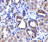
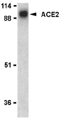
ACE2 Antibody
Catalog# :3229
Angiotensin-converting enzyme 2 (ACE2) plays a central role in vascular, renal, and myocardial physiology (1-2). In contrast to its homolog ACE, ACE2 expression is restricted to heart, kidney, and testis. Recently. ACE2 has also been shown to be a functional receptor of the SARS coronavirus (3). The normal function of ACE2 is to convert the inactive vasoconstrictor angiotensin I (AngI) to Ang1-9 and the active form AngII to Ang1-7, unlike ACE, which converts AngI to AngII. While the role of these vasoactive peptides is not well understood, lack of ACE2 expression in ace2-/ace2- mice leads to severely reduced cardiac contractility, indicating its importance in regulating heart function (4).
Source :ACE2 antibody was raised against a synthetic peptide corresponding to amino acids near the center of human ACE2.
Purification : Affinity chromatography purified via peptide column
Clonality and Clone : This is a polyclonal antibody.
Host : ACE2 antibody was raised in rabbit.
Please use anti-rabbit secondary antibodies.
Immunogen : Human ACE2 (Intermediate Domain 2) Peptide (Cat. No. 3229P)
Application : ACE2 antibody can be used for the detection of ACE2 by Western blot at 0.5 to 2 µg/ml.A 90 kDa band can be detected. Anti-ACE2 has no cross response to ACE1.
Tested Application(s) : E, WB, IHC
Buffer : Antibody is supplied in PBS containing 0.02% sodium azide.
Blocking Peptide :Cat. No. 3229P - ACE2 (IN2) Peptide
Long-Term Storage : ACE2 antibody can be stored at 4ºC, stable for one year. As with all antibodies care should be taken to avoid repeated freeze thaw cycles. Antibodies should not be exposed to prolonged high temperatures.
Positive Control :
1. Cat. No. 1305 - Human Kidney Tissue Lysate
Species Reactivity : H
GI Number : 11225609
Accession Number : NP_068576
Short Description : (IN3) Angiotensin-converting enzyme 2, SARS receptor
References
1. Donoghue M, Hsieh F, Baronas E, et al. A novel angiotensin-converting enzyme-related carboxypeptidase (ACE2) converts angiotension I to angiotension 1-9. Circ. Res. 2000;87:1-9.
2. Tipnis SR, Hooper NM, Hyde R, et al. A human homolog of angiotensin-converting enzyme. Cloning and functional expression as a captopril-insensitive carboxypeptidase. J Biol. Chem. 2000;275:33238-43.
3. Li W, Moore MJ, Vasileva N, et al. Angiotensin-converting enzyme 2 is a functional receptor for the SARS coronavirus. Nature 2003;426:450-4.
4. Crackower MA, Sarao R, Oudit GY, et al. Angiotensin-converting enzyme 2 is an essential regulator of heart function. Nature 2002;417:822-8.
Wednesday, May 25, 2011
HIV Epitope
For over 20 years, BioSynthesis has provided custom peptide production services and stock HIV peptides. Our unique and comprehensive experience has enable us in providing chemically synthesized peptides ranging from highly challenging ultra-pure peptidomimetics and long peptides to complex peptide libraries consisting of thousands of fully characterized peptides to highly regulated multiple kilogram per batch GMP peptides. Our goal is to be your single peptide partner at every stage in the peptide drug development continuum from design of peptide libraries, through to GMP manufacturing of active pharmaceutical ingredients (APIs) and commercialization. Our consistency, viability, and delivery are guaranteed.
Epitope
An Epitope is the part of a macromolecule that is recognized by the immune system, specifically by antibodies, or T cells. Although epitope are usually thought to be derived from non self proteins, sequences derived from the host that can be recognized are also classified as epitope.
Epitope Mapping
It is the process of matching surface proteins to antibodies that will bond to them. Epitope mapping is the process of identification and characterization of the minimum molecular structures that are able to be recognized by the Immune System elements, mainly T and B cells. A collection of in vivo and in vitro methodologies are used for epitope mapping.
Antibodies
Antibodies (also known as immunoglobulins are proteins that are found in blood or other bodily fluids of vertebrates, and are used by the immune system to identify and neutralize foreign objects, such as bacteria and viruses.
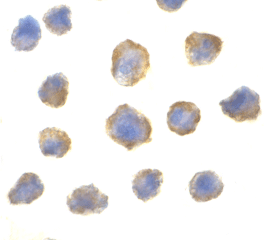
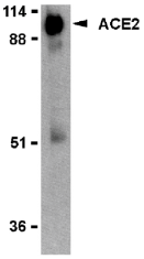
ACE2 Antibody
Catalog# :3227
Angiotensin-converting enzyme 2 (ACE2) plays a central role in vascular, renal, and myocardial physiology (1-2). In contrast to its homolog ACE, ACE2 expression is restricted to heart, kidney, and testis. Recently. ACE2 has also been shown to be a functional receptor of the SARS coronavirus (3). The normal function of ACE2 is to convert the inactive vasoconstrictor angiotensin I (AngI) to Ang1-9 and the active form AngII to Ang1-7, unlike ACE, which converts AngI to AngII. While the role of these vasoactive peptides is not well understood, lack of ACE2 expression in ace2-/ace2- mice leads to severely reduced cardiac contractility, indicating its importance in regulating heart function (4).
Additional Names : ACE2 (NT), ACE2
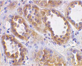
Description
Left: Western blot analysis of ACE2 in human kidney lysate with ACE2 antibody at 1 µg/ml.
Below: Immunohistochemical staining of human kidney cells using ACE2 antibody at 2 µg/ml.
Other Product Images
Description
Left: Western blot analysis of ACE2 in human kidney lysate with ACE2 antibody at 1 µg/ml.
Below: Immunohistochemical staining of human kidney cells using ACE2 antibody at 2 µg/ml.
Other Product Images
 Source :ACE2 antibody was raised against a synthetic peptide corresponding to amino acids near the N-terminus of human ACE2.
Purification : Affinity chromatography purified via peptide column
Clonality and Clone : This is a polyclonal antibody.
Host : ACE2 antibody was raised in rabbit.
Please use anti-rabbit secondary antibodies.
Immunogen : Human ACE2 (N-Terminus) Peptide (Cat. No. 3227P)
Application : ACE2 antibody can be used for the detection of ACE2 by Western blot at 0.5 to 2 µg/ml.A 90 kDa band can be detected. Anti-ACE2 has no cross response to ACE1.
Tested Application(s) : E, WB, IHC
Buffer : Antibody is supplied in PBS containing 0.02% sodium azide.
Blocking Peptide :Cat. No. 3227P - ACE2 Peptide
Long-Term Storage : ACE2 antibody can be stored at 4ºC, stable for one year. As with all antibodies care should be taken to avoid repeated freeze thaw cycles. Antibodies should not be exposed to prolonged high temperatures.
Positive Control :
1. Cat. No. 1305 - Human Kidney Tissue Lysate
Species Reactivity : H
GI Number : 11225609
Accession Number : NP_068576
Short Description : (NT) Angiotensin-converting enzyme 2, SARS receptor
References
1. Donoghue M, Hsieh F, Baronas E, et al. A novel angiotensin-converting enzyme-related carboxypeptidase (ACE2) converts angiotension I to angiotension 1-9. Circ. Res. 2000;87:1-9.
2. Tipnis SR, Hooper NM, Hyde R, et al. A human homolog of angiotensin-converting enzyme. Cloning and functional expression as a captopril-insensitive carboxypeptidase. J Biol. Chem. 2000;275:33238-43.
3. Li W, Moore MJ, Vasileva N, et al. Angiotensin-converting enzyme 2 is a functional receptor for the SARS coronavirus. Nature 2003;426:450-4.
4. Crackower MA, Sarao R, Oudit GY, et al. Angiotensin-converting enzyme 2 is an essential regulator of heart function. Nature 2002;417:822-8.
Subscribe to:
Posts (Atom)